

Abstract
Reptiles are very likely to have the same pain experience as mammals, as the structures involved with nociception and processing are homologous. However, there is a big difference in expressive behaviours of pain between reptiles and mammals. This makes pain assessment challenging. Difficulty in pain assessment, along with other factors, makes poor pain management a welfare issue in the veterinary and the wider reptile - keeping context. Research in reptile pain is very limited so far; this article aims to provide some basic information on assessing pain in reptiles.
The author would like to thank David Stokes for his help with this article.
This tortoise was hospitalised following a cystotomy at a previous veterinary practice and clinically deteriorating. He had a CT that showed intracoelomic gas and effusion, sediment in the bladder, and a surgical site secondary infection (already reported by bloods/swabs of the surgical site) visualised on the images.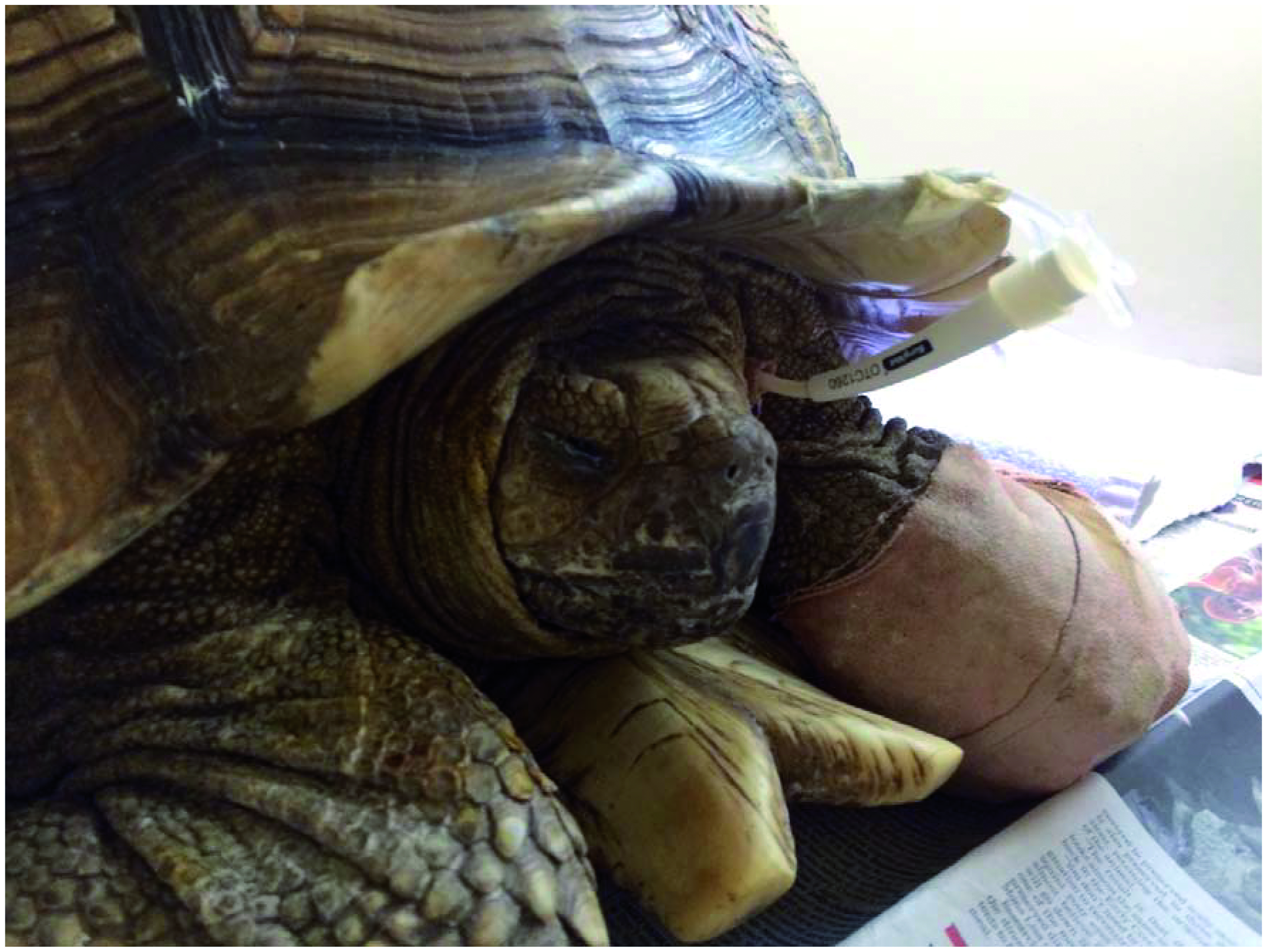
The tortoise was severely dehydrated on bloodwork and clinical exam, evident from the dull corneas and crusting around the eyes, mouth and nares, and showing signs of pain and illness with closed eyes and very little movement. He was started on IM morphine injections. He was placed on a thick bed in case there was pain or discomfort associated with the surgical site.
Bearded dragon with follicular stasis. Notice the swollen/gravid abdomen, the half - closed eyes and the dark colour changes. There is likely to be a degree of abdominal discomfort with this condition.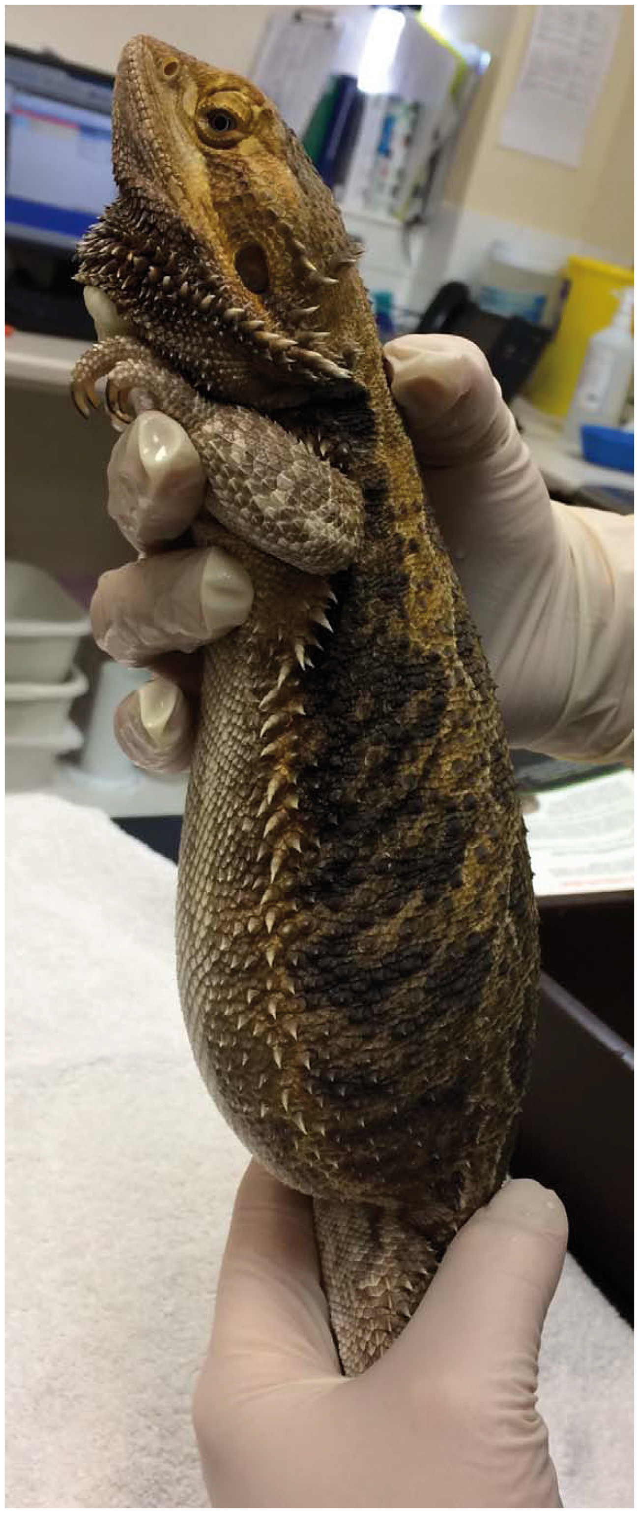
In this photo you can see the colour changes to the ventral aspect of her chin, and the inability to support her weight.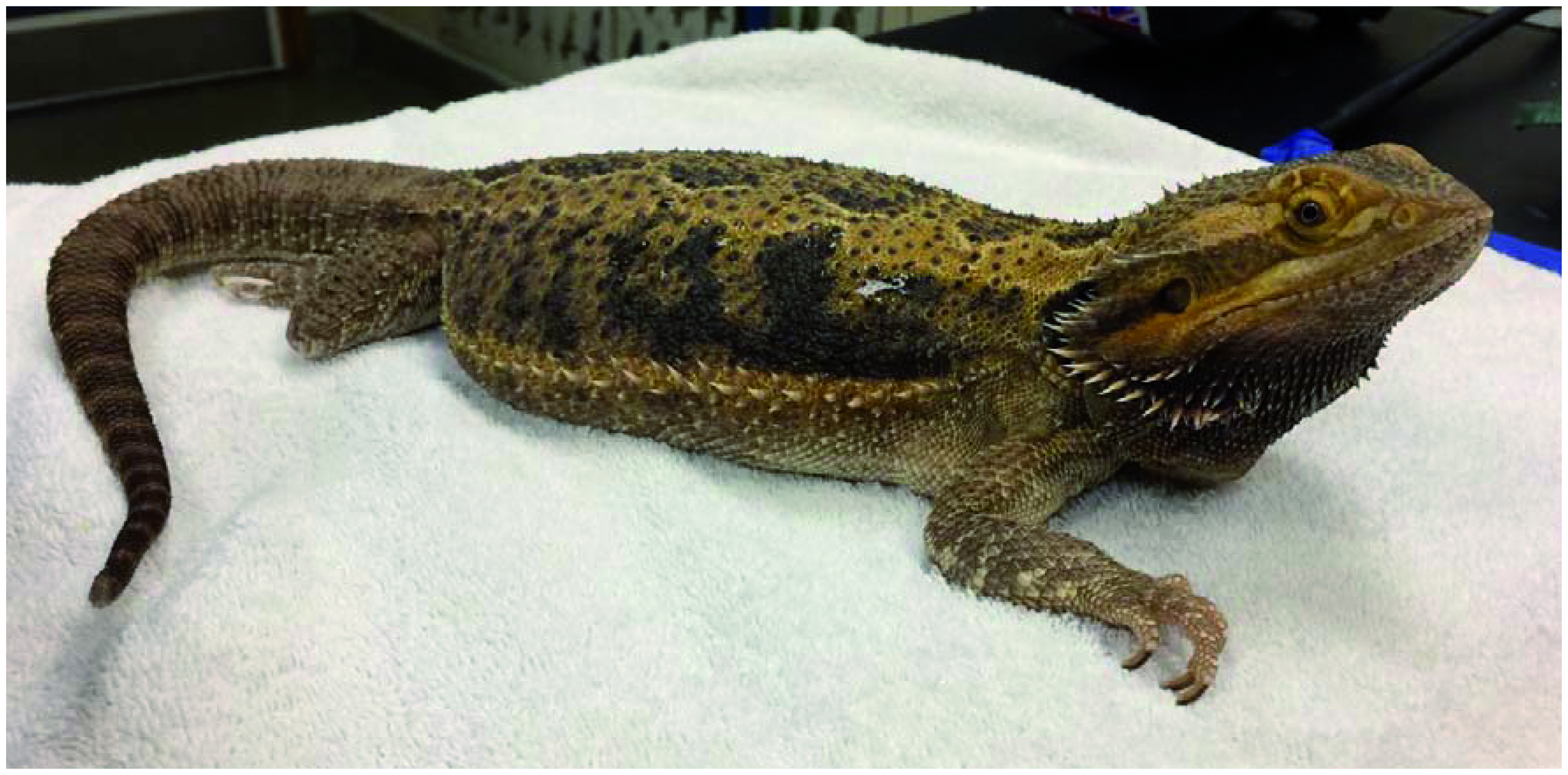
This baby tortoise was attacked by a dog and following flushing and cleaning of the area under sedation, was dressed and bandaged. She was on fluids, antibiotics and morphine IM every 24 h. It was very difficult to tempt this patient to eat or move about, even when tempting to handfeed with tiny pieces of her favourite food. Innappetance can be a non- specific pain behaviour. In this photo she was not attempting to carry her own weight with her legs. Note the dull eyes and demeanour. She was eventually euthanased, due to no clinical improvement.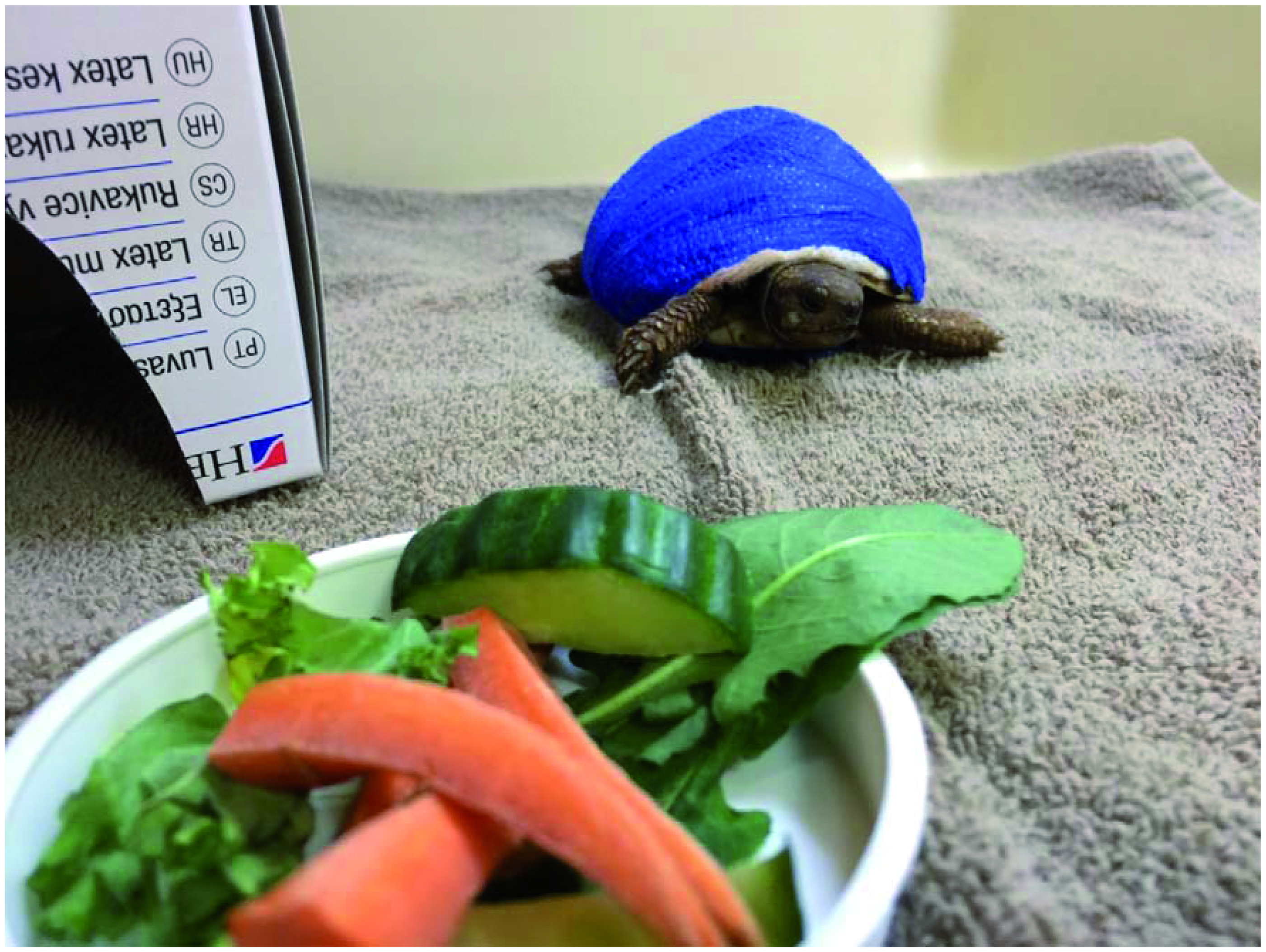
Tortoise with a right hind fracture. This patient was started on morphine IM Q24 h, meloxicam once hydrated, and had a sling support to reduce movement of the fractured leg. Note the half - closed eyes, dull demeanour and withdrawn posture.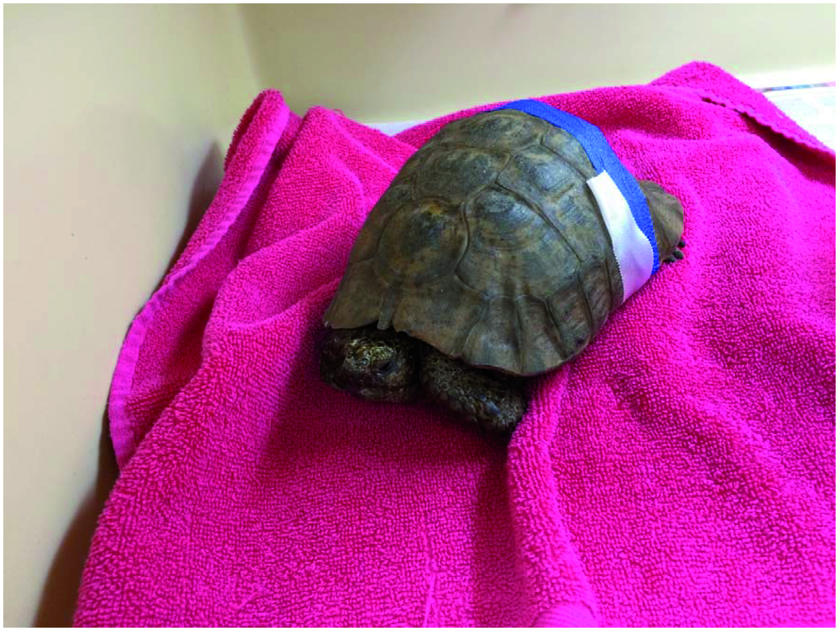
Iguana with MBD: Iguana showing neurological signs and musculoskeletal malformation as a result of metabolic bone disease. Note the twisted appearance to the hindlimb digits and tail. This patient was unable to mobilise properly and was very sick when admitted.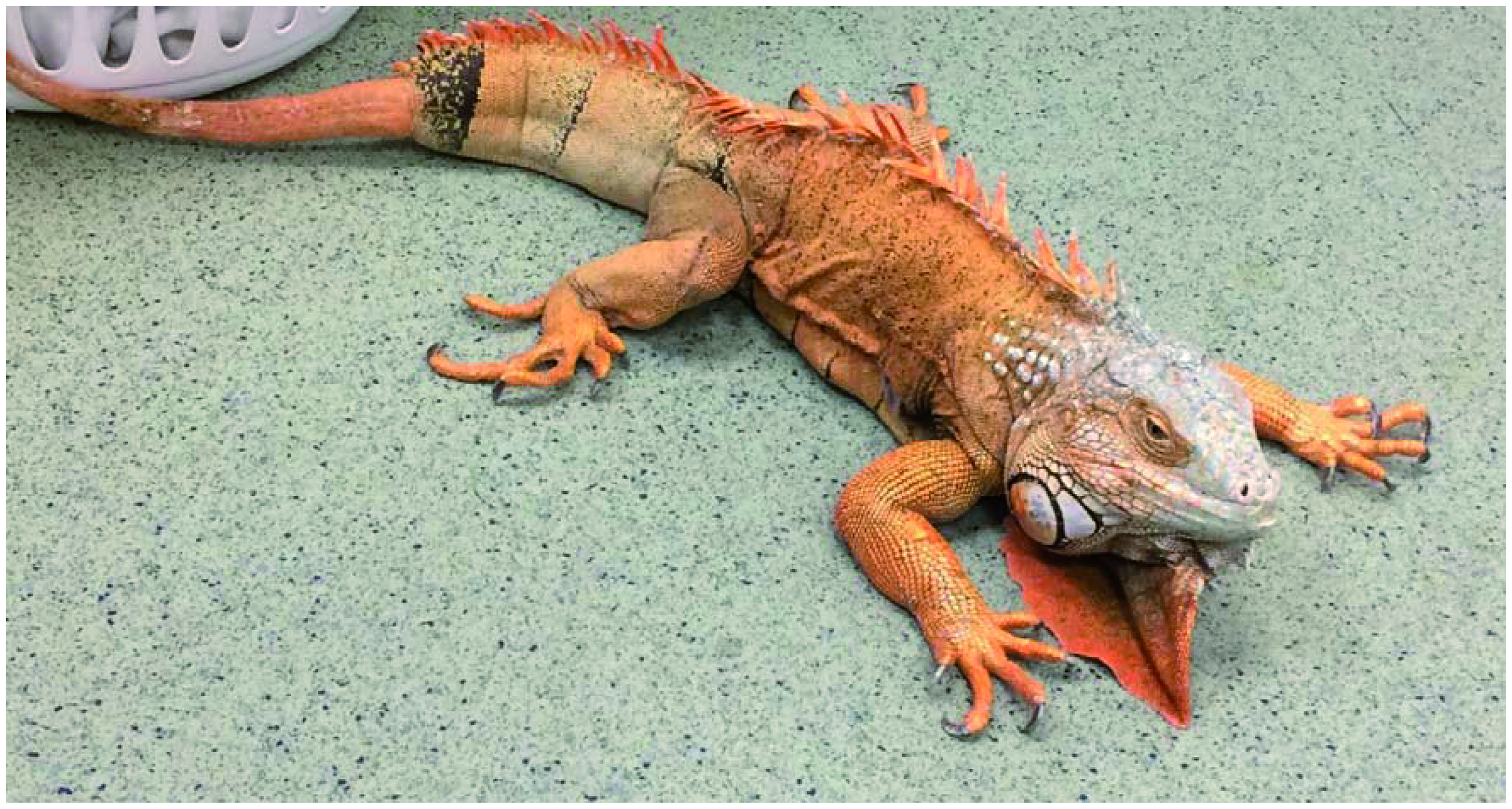
Iguana with MBD: X-ray of the above iguana. Note the reduced bone density in the extremities. There is potentially a degree of bone pain with metabolic bone disease, and muscle ache and strain as muscle is built up to compensate the inadequate bone growth. The treatment plan included injectable tramadol.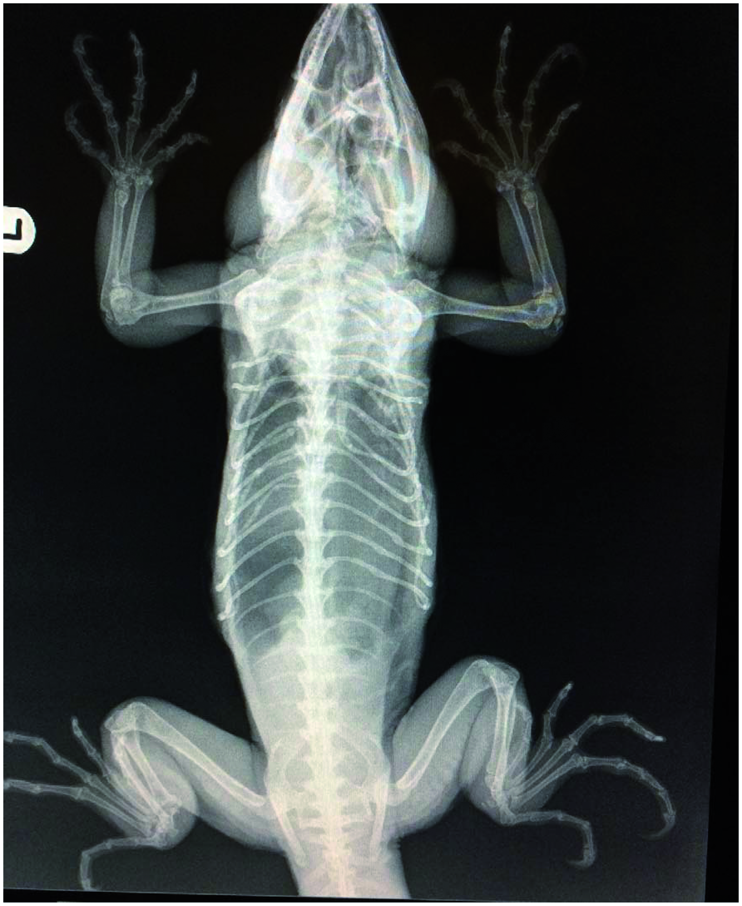
This bearded dragon was suffering from severe oral stomatitis, which had meant he had been unable to eat for some time. Note the poor body condition. The patient has closed eyes and a darkened ventral chin, that could indicate pain and/or general illness. The patient had a swab taken and the gums were cleaned. He was given fluids, and started on antibiotic injections, and IM tramadol injections to be given at home. There is a risk of refeeding syndrome when gavage feeding dehydrated patients such as this, so the decision was to rehydrate before considering feeding.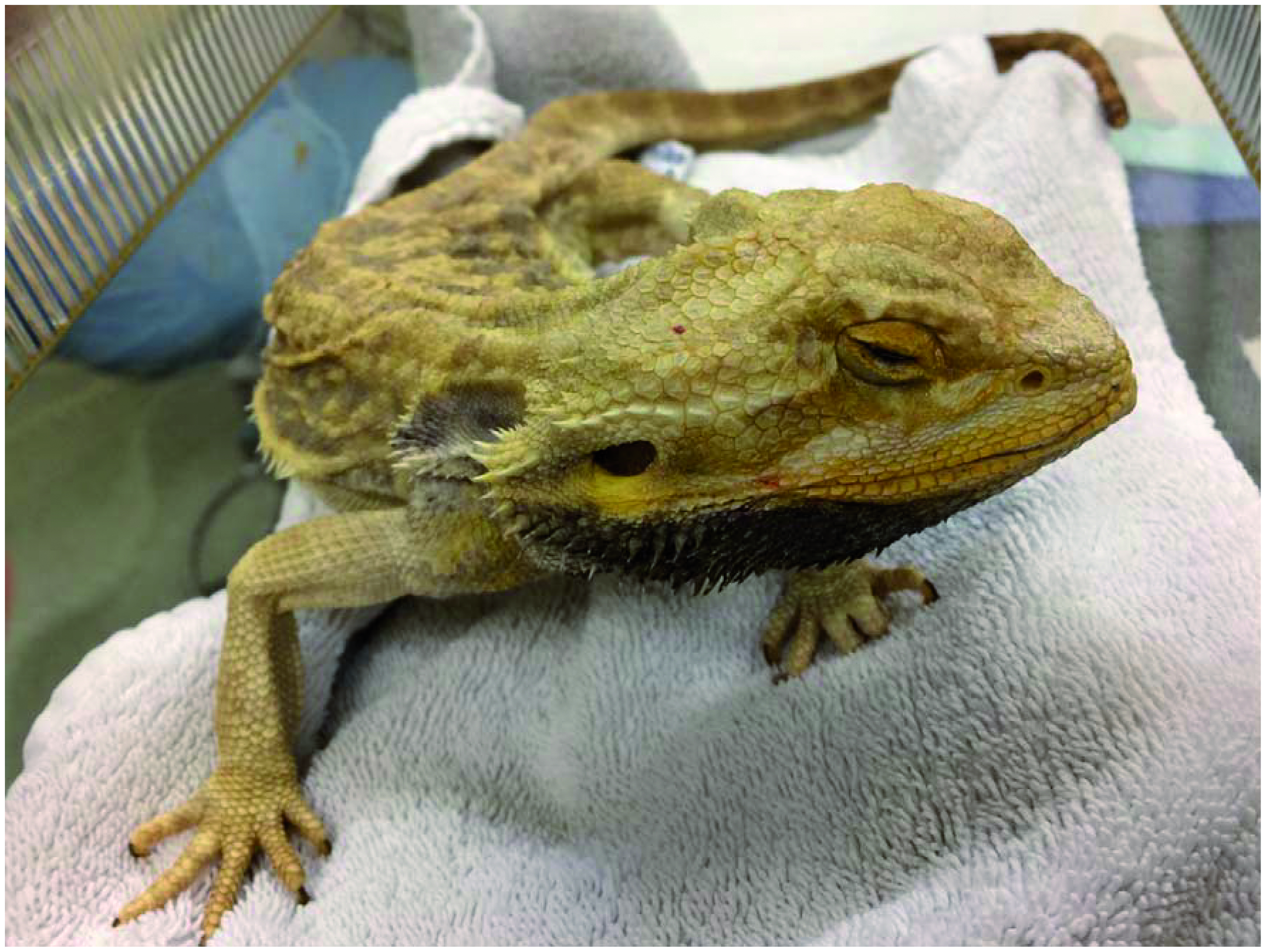
Bearded dragon exhibiting a postural pain behaviour. Note also the dark discolouration to the ventral neck and head. This patient had an intestinal disorder and parasite burden and was possibly suffering from visceral pain or discomfort.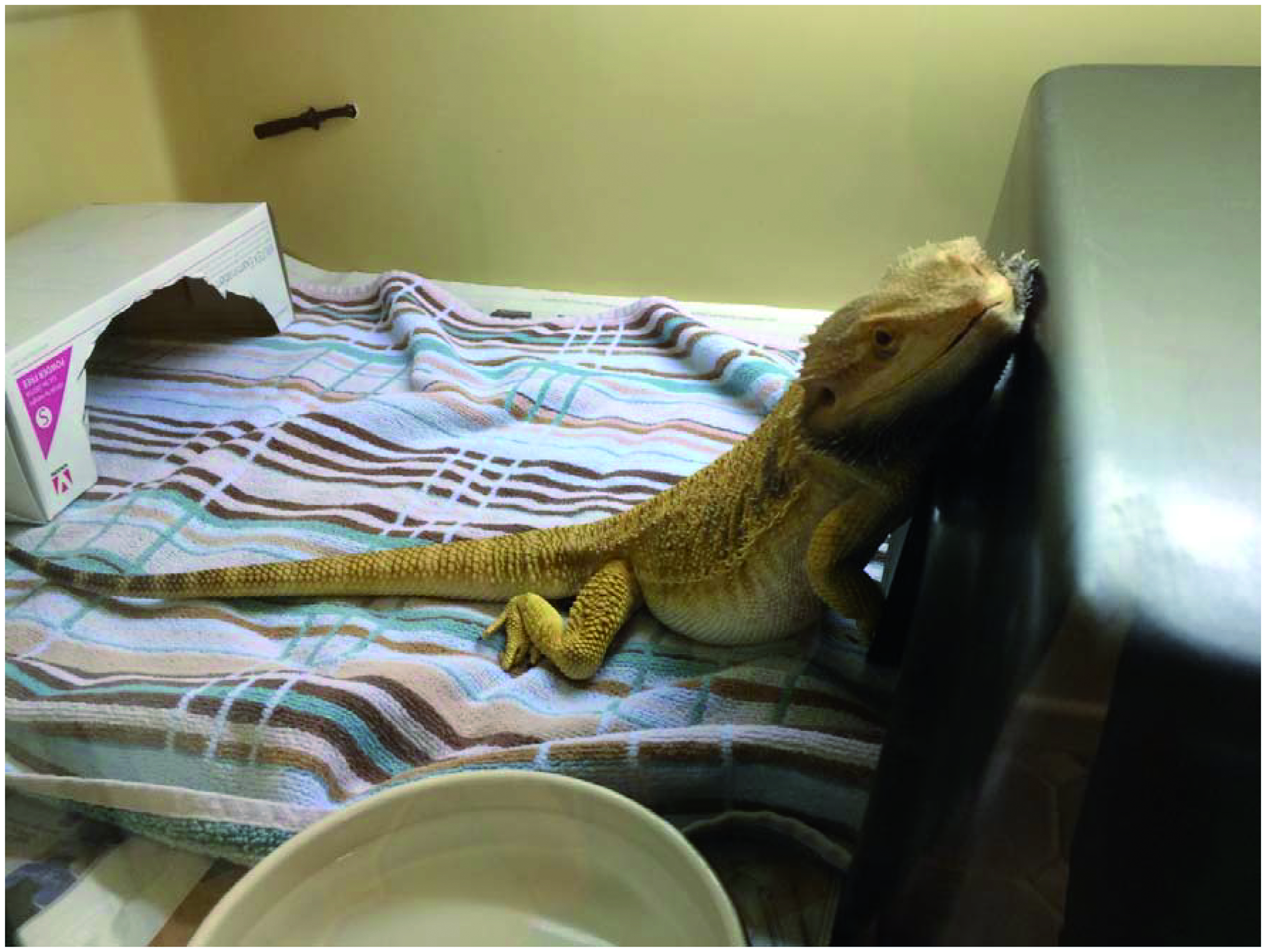
This little leopard gecko had several husbandry related issues, having reproductive disease being a persistent egg layer, osteopenia, severe metabolic bone disease (MBD), suspect hepatic lipidosis, and dysecdysis (difficulty shedding) in all four limbs, with necrosis occurring at the distal ends. She walked on the dorsal aspects of her carpi. Note the abnormal weight bearing on the hind limbs, and inflamed digits. There is likely to be a degree of pain or discomfort associated with all of her conditions, therefore her treatment plan included oral tramadol to take at home.
This leopard gecko presented with tail tip necrosis due to dysecdysis (difficulty shedding). Note the sore forelimb digits. The necrosed tail was removed via manual autotomy, which is removing it along the natural fracture plane that geckos would normally do themselves in the wild if caught by a predator (‘dropping their tail’). This method has better outcomes than surgical amputation. The procedure was done under sevofluorane anaesthesia, with an injection of hydromorphone given IM beforehand.
Shell wound tortoise: This tortoise presented with a wound to her leg and shell that had infected large areas of the ventral shell scutes. Note the dull half- closed eyes and lethargic demeanour.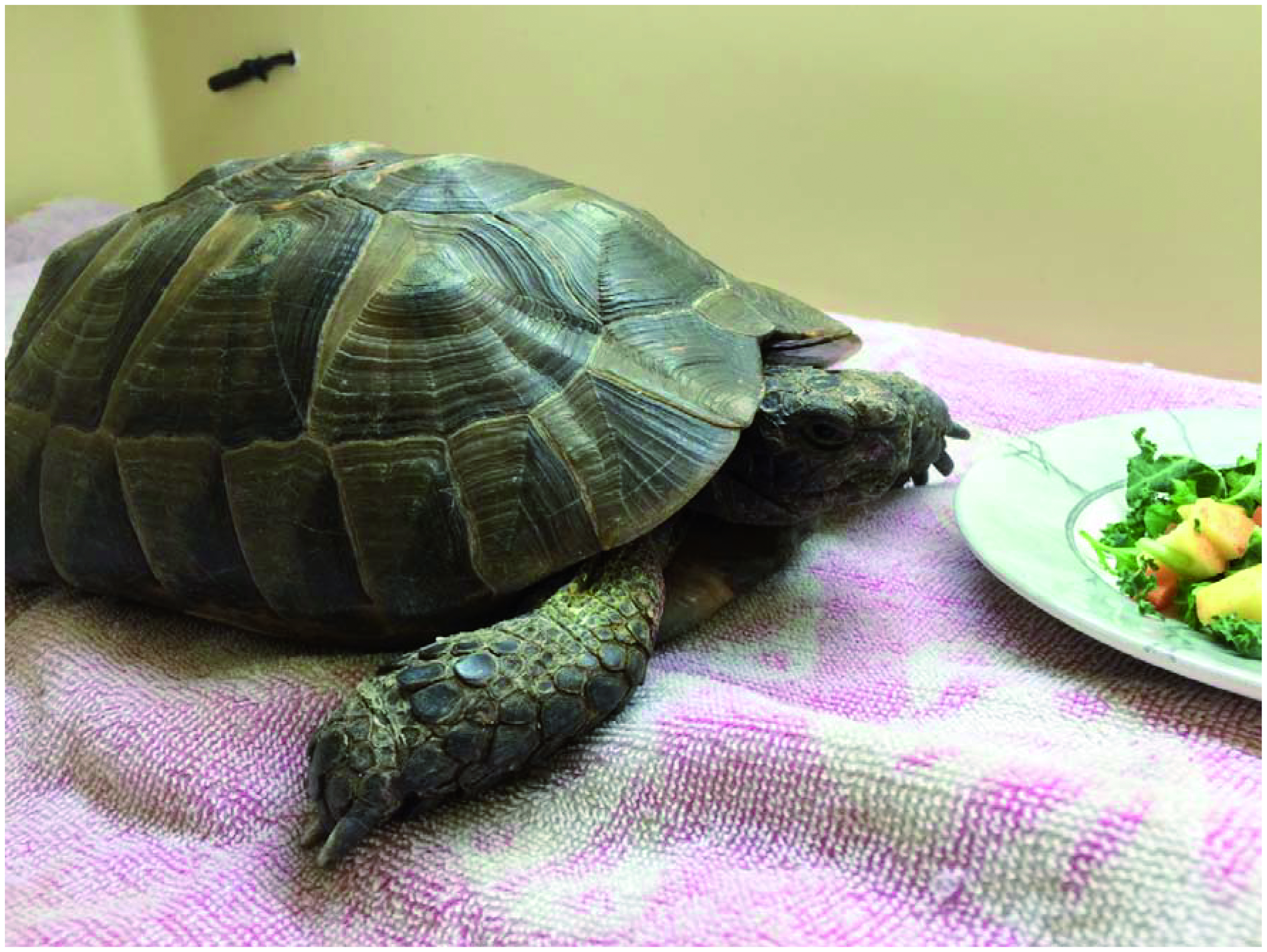
Shell wound tortoise: The same tortoise viewed ventrally. A CT showed severe osteopenia with suspected pathological fractures and possible sequestra, and a small amount of gas present in the coelom (body cavity), and coelomic effusion. Coincidentally the CT also showed multiple follicles in the coelom. The owner opted for treatment over euthanasia and this tortoise received hydromorphone in hospital and went home on oral tramadol.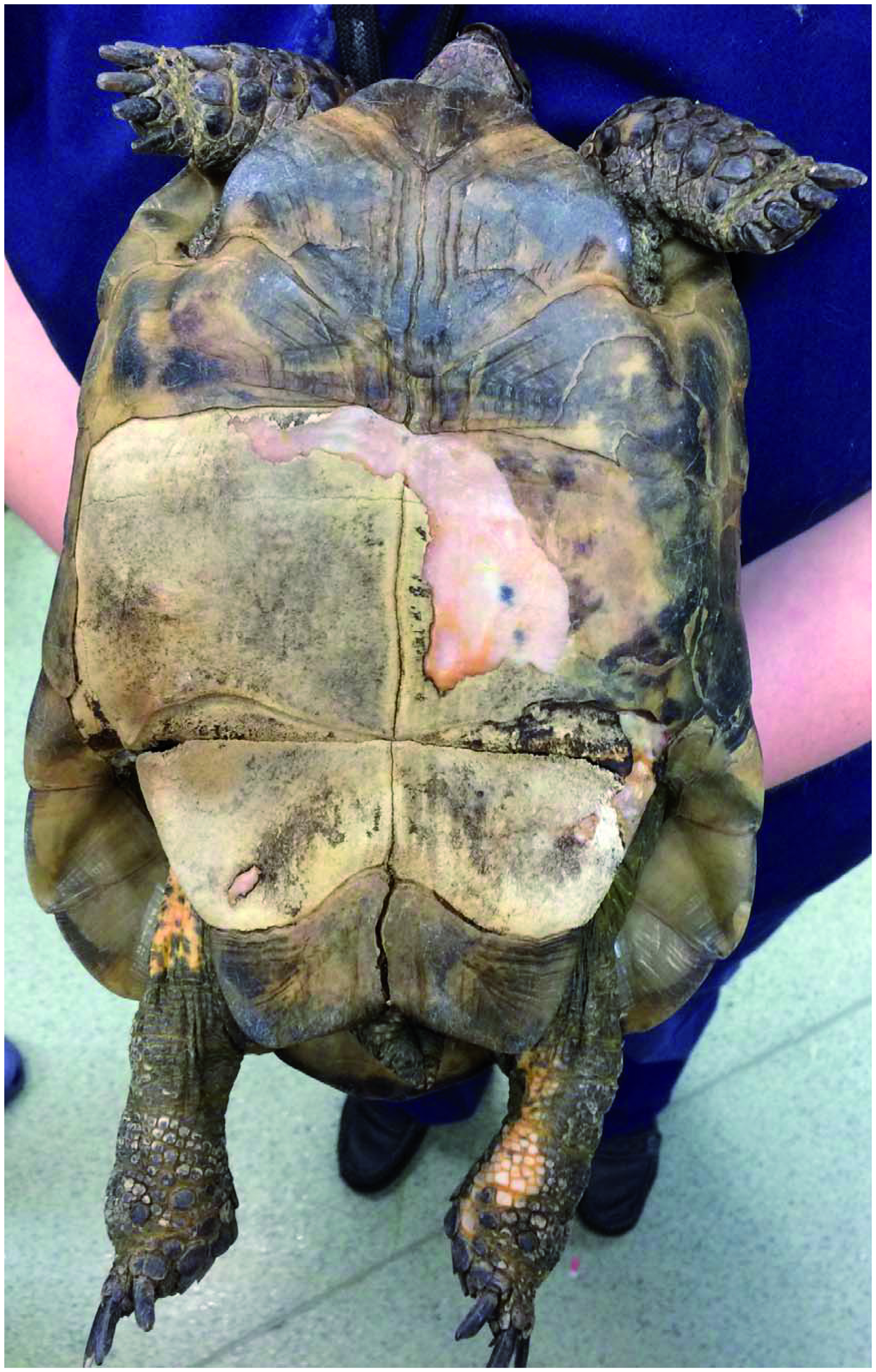
Tortoise (bite wounds): Pictures of a tortoise that presented being chewed by rats due to being ill and weakened by a long period (years) of suboptimal husbandry (kept in the garden).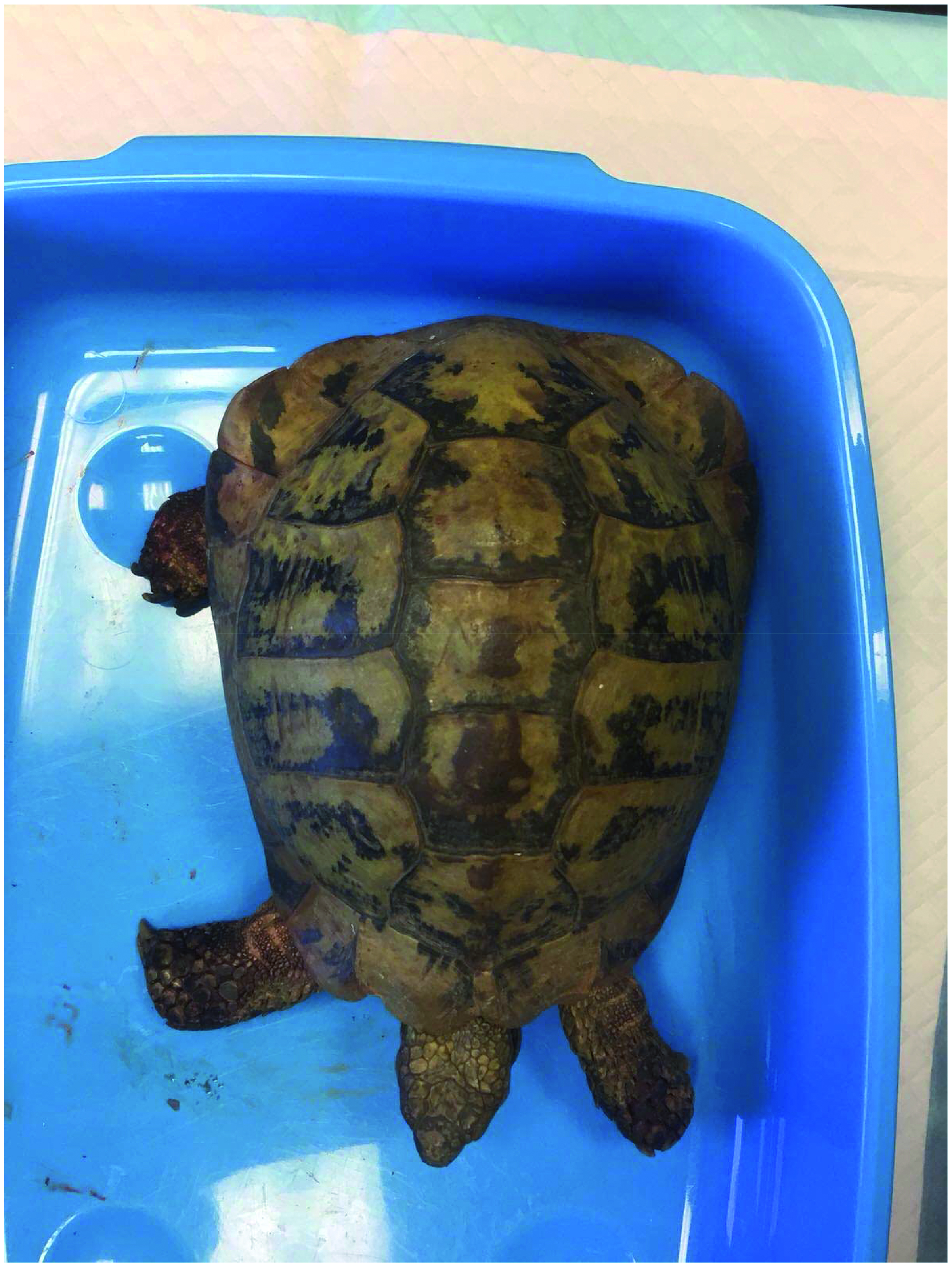
With kind permission from Stacey Vickery
Tortoise (bite wounds)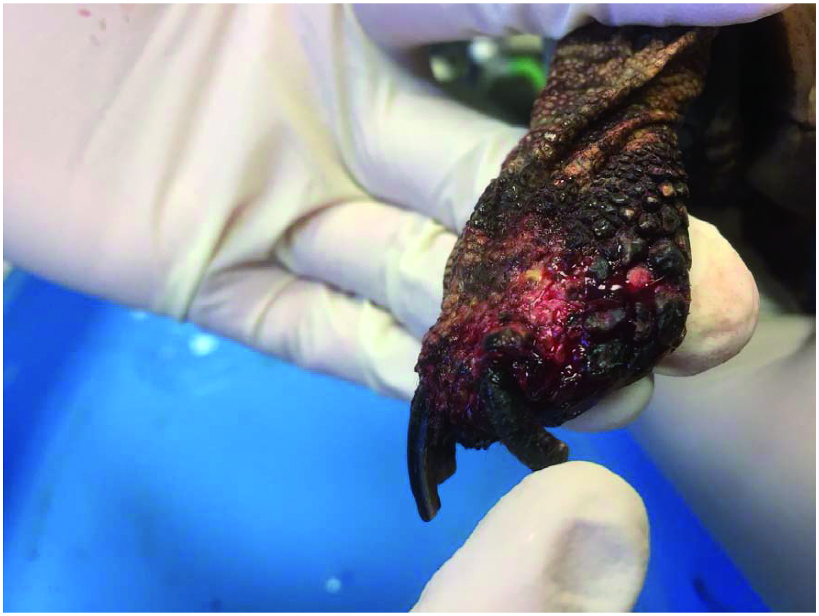
Tortoise (bite wounds): After cleaning the wounds, the patient was sent home on IM tramadol injections, antibiotics, wound cleaning, and advised to keep indoors on padded and clean bedding to reduce further discomfort, before moving to an appropriate indoor set up once healed.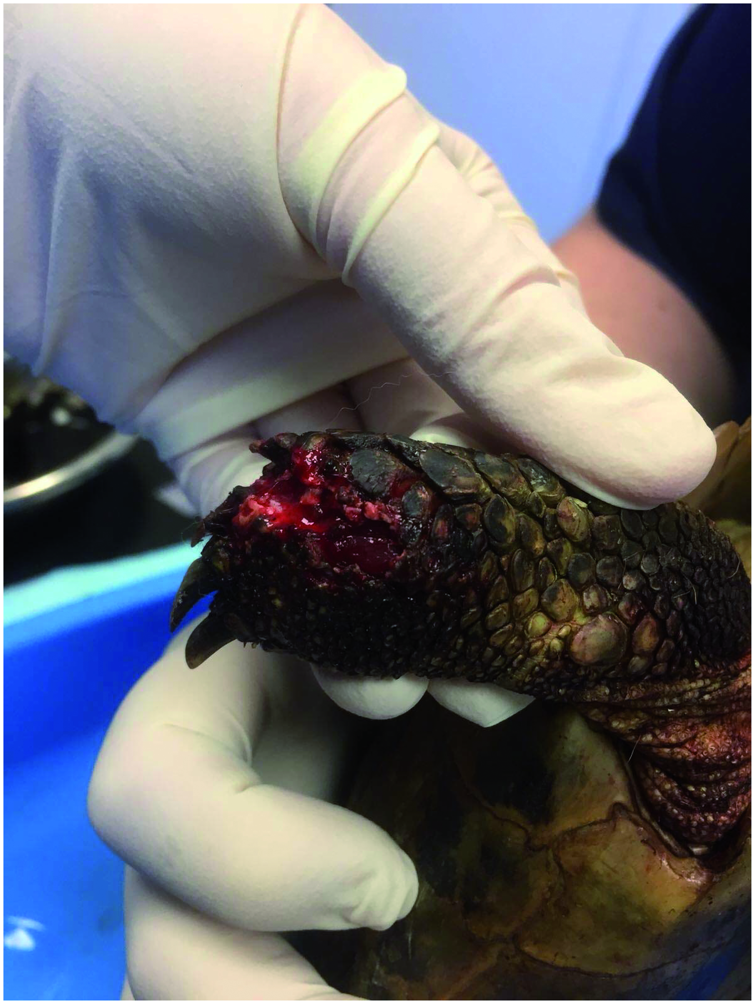
TWith kind permission from Stacey Vickery
Comfortable posture in a bearded dragon, 24 hours post salpingohysterectomy (due to follicular stasis). It has been noted in other sources (Eatwell, Citation2010) that bearded dragons showing abdominal discomfort due to a surgical site will lift the abdomen off the ground to avoid contact with the floor. This patient was deemed very comfortable by the vet on examination, and you can observe that she is not showing any lifting posture. She received hydromorphone on admission the day before, and her premedication included hydromorphone, dexmedetomidine and ketamine.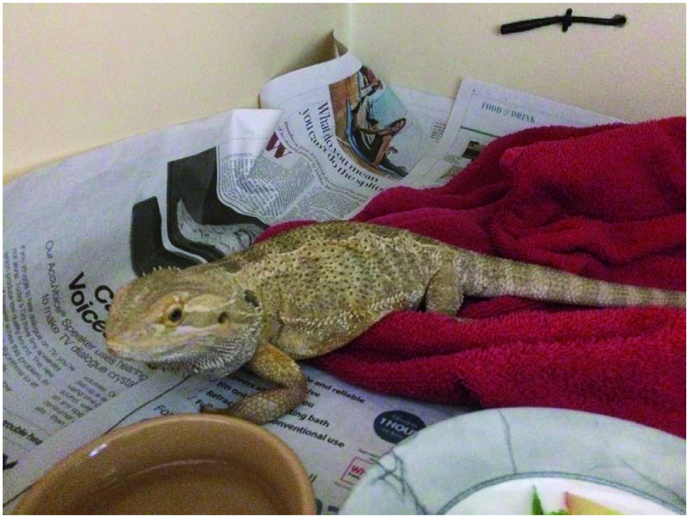
Cloacal prolapse: This tortoise (hiding in its shell) presented with cloacal prolapse which it had for two days, due to a massive parasite burden found by faecal sample.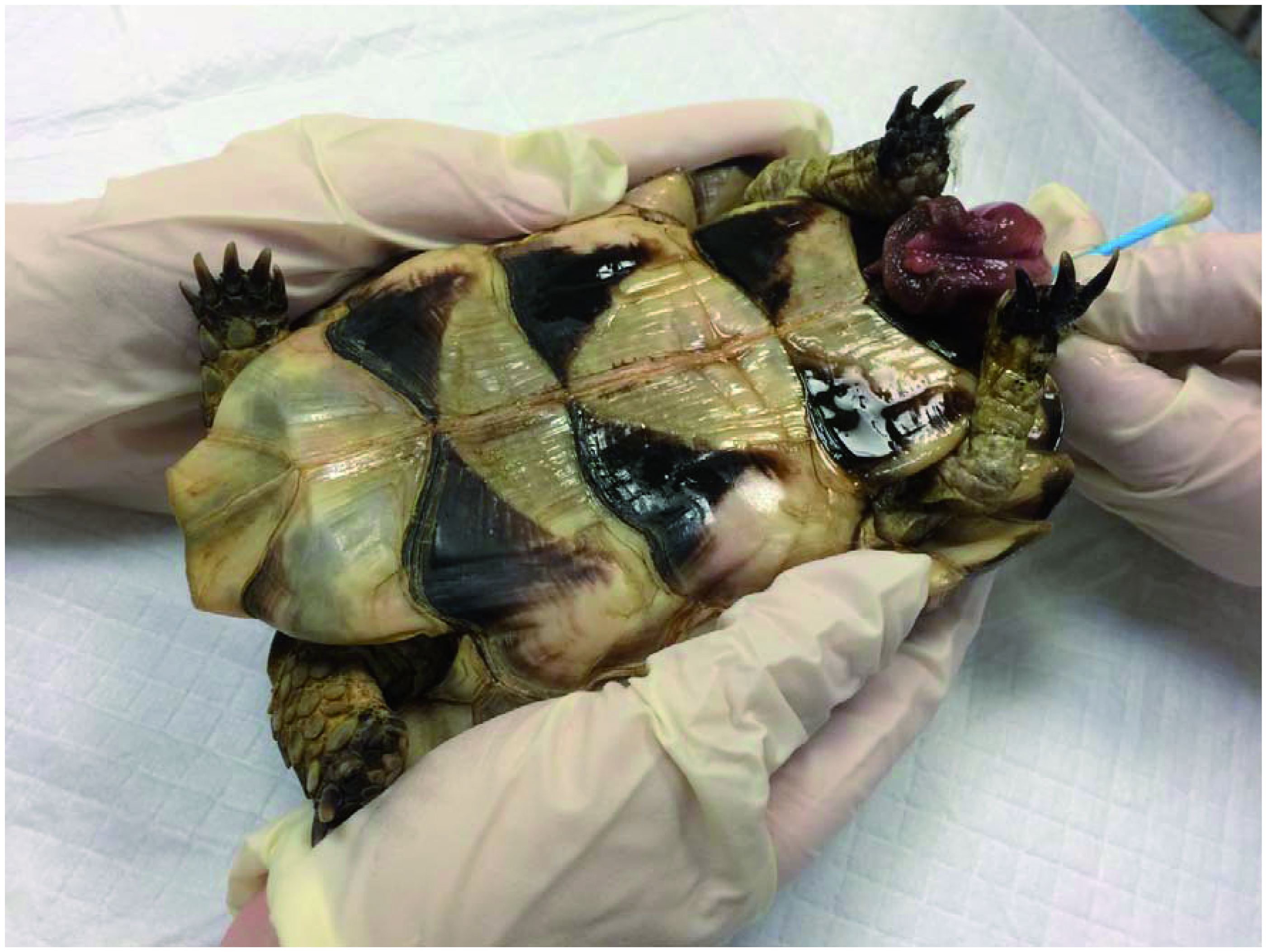
Cloacal prolapse: The patient received hydromorphone and fluids on admission. It was impossible to replace the prolapse due to the extent of the problem, and the owners opted for euthanasia.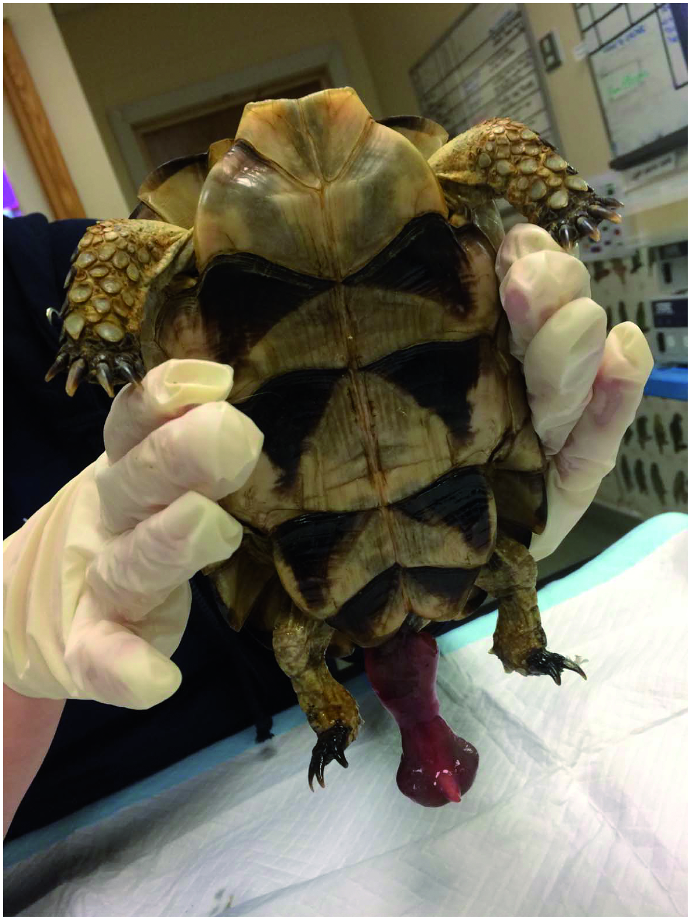
Young tortoise presenting with shell wounds after being attacked by a dog. Note the closed eyes and almost collapsed state. He was administered hydromorphone on admission.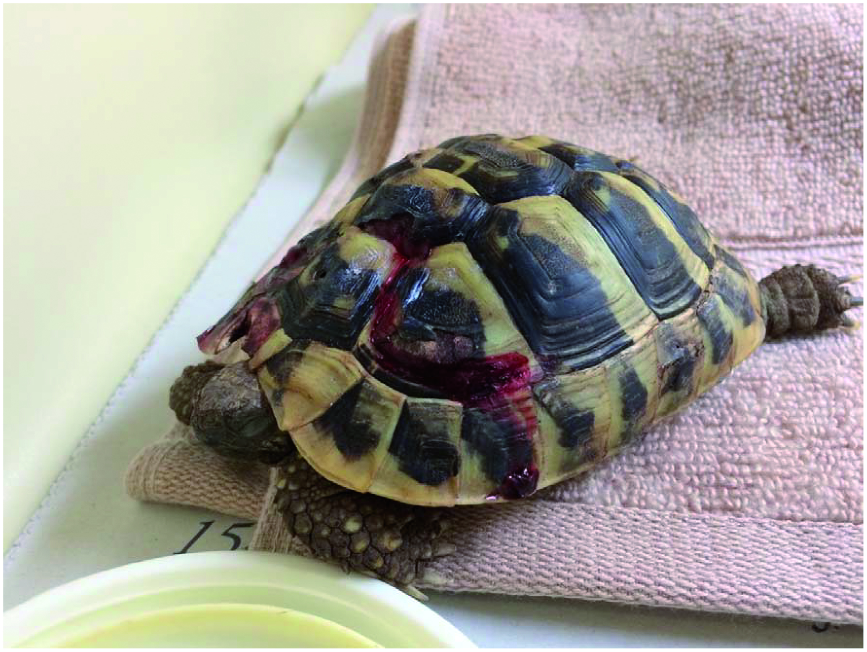
This iguana is 24 h post replacing a cloacal prolapse that was likely due to a reproductive cause. She had hydromorphone on admission and her premedication included hydromorphone, dexmedetomidine and ketamine. It is very difficult to assess post - operative pain in reptiles, but she appeared to be in minimal discomfort. She was posturing the affected area comfortably and was fairly alert and moving around the vivarium reasonably well.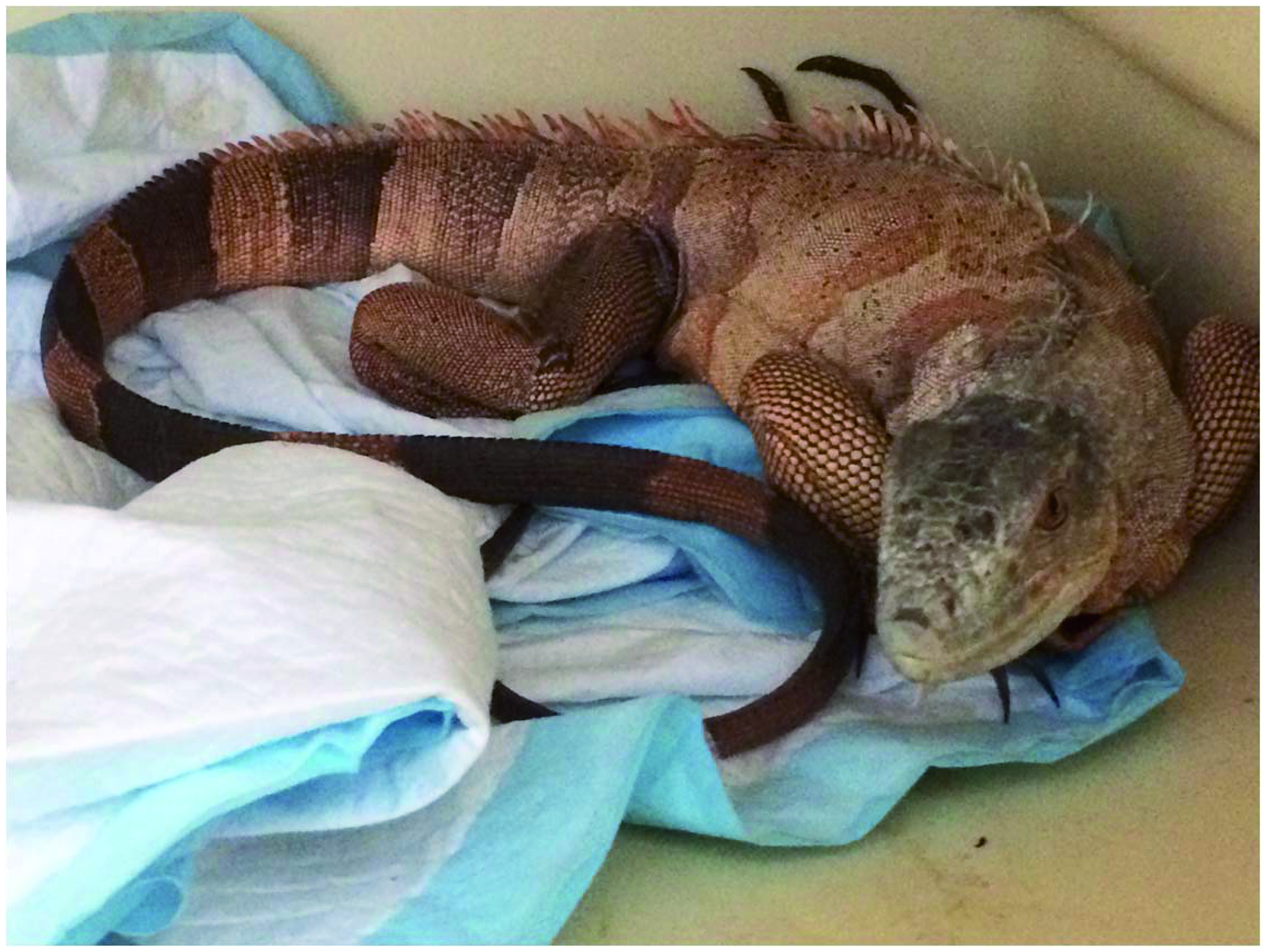
Green tree python: Patient with severe stomatitis. This patient was administered a fentanyl patch.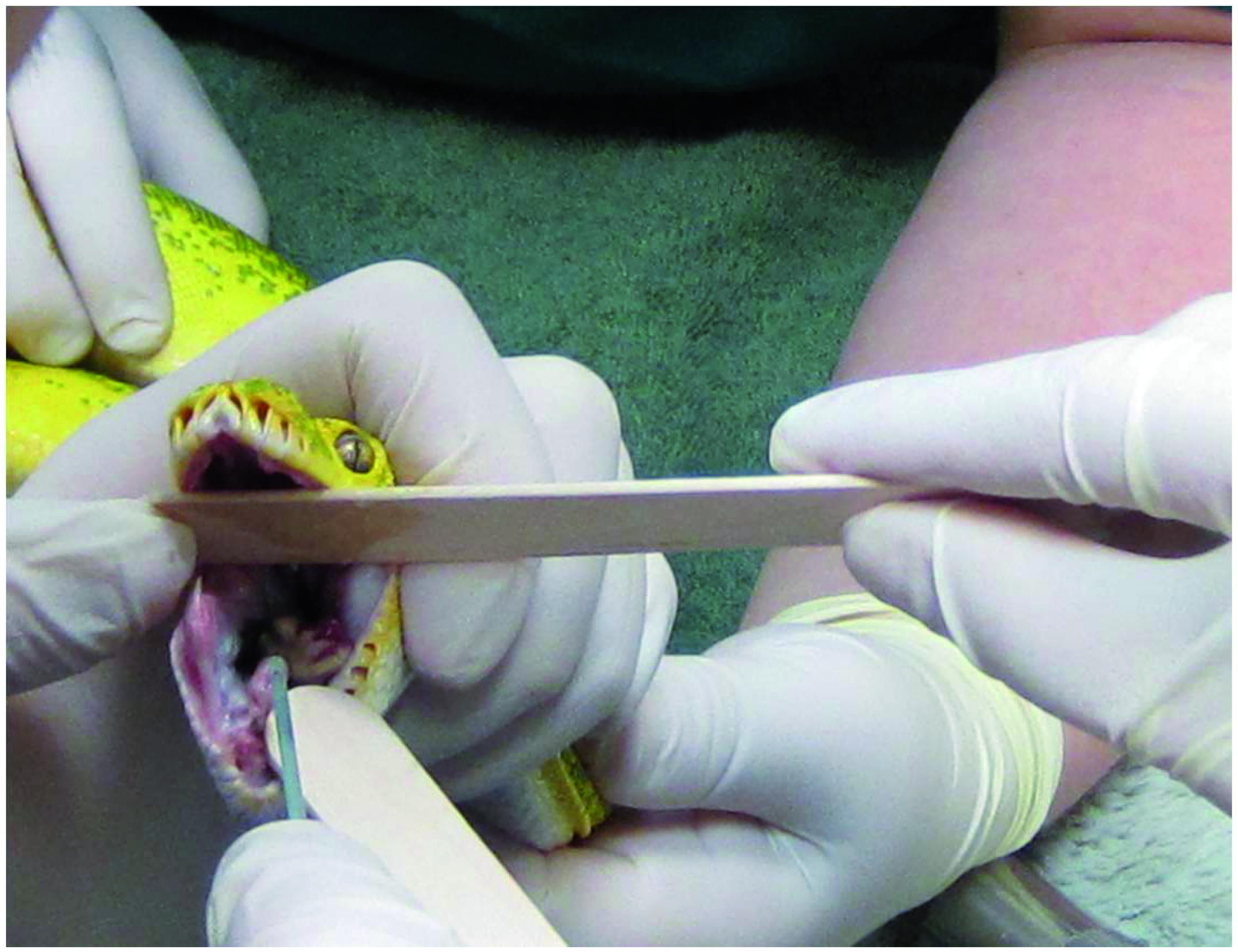
With kind permission from Tariq Abou – Zahr
Green tree python: the fentanyl patch (just visible!) on the afore- mentioned python.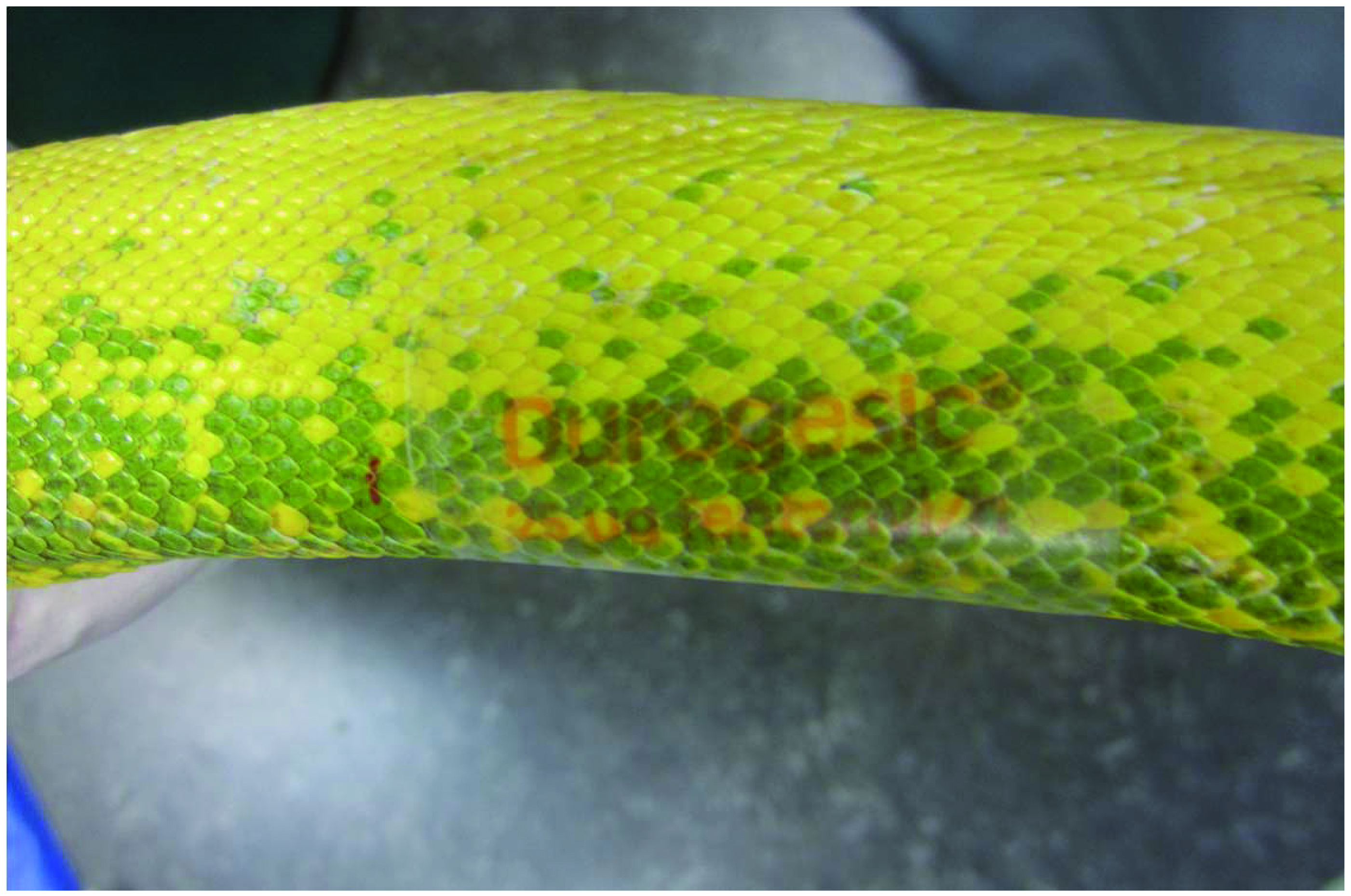
With kind permission from Tariq Abou – Zahr
Albino Burmese python presenting with thermal burns. The patient was on morphine IM Q24 h. The burns were dressed and bandaged.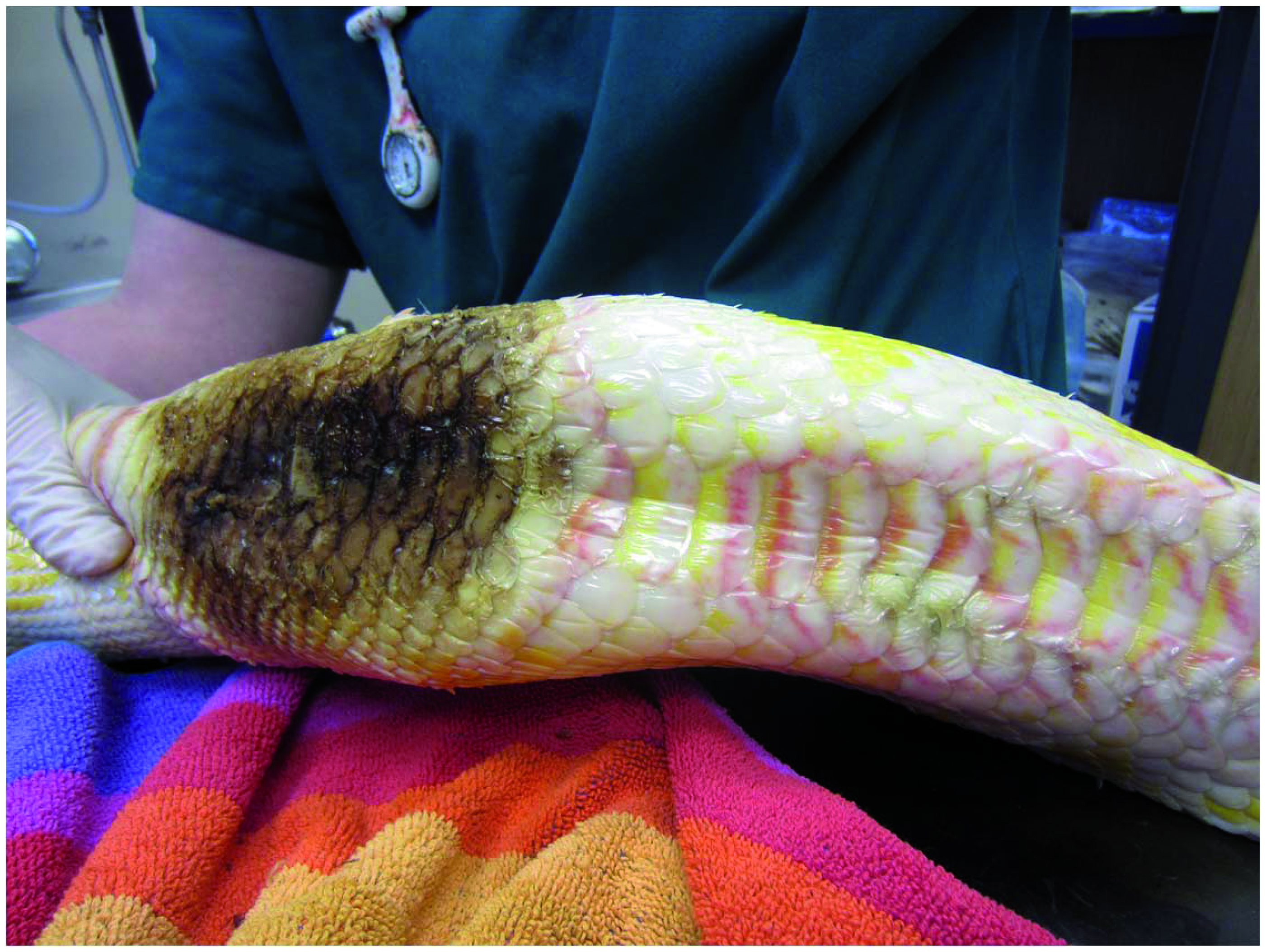
With kind permission from Stacey Vickery and Tariq Abou – Zahr
Young Tokay gecko with a dislocated stifle. The dislocation was resolved and leg splinted under sedation. The patient started on IM tramadol injections.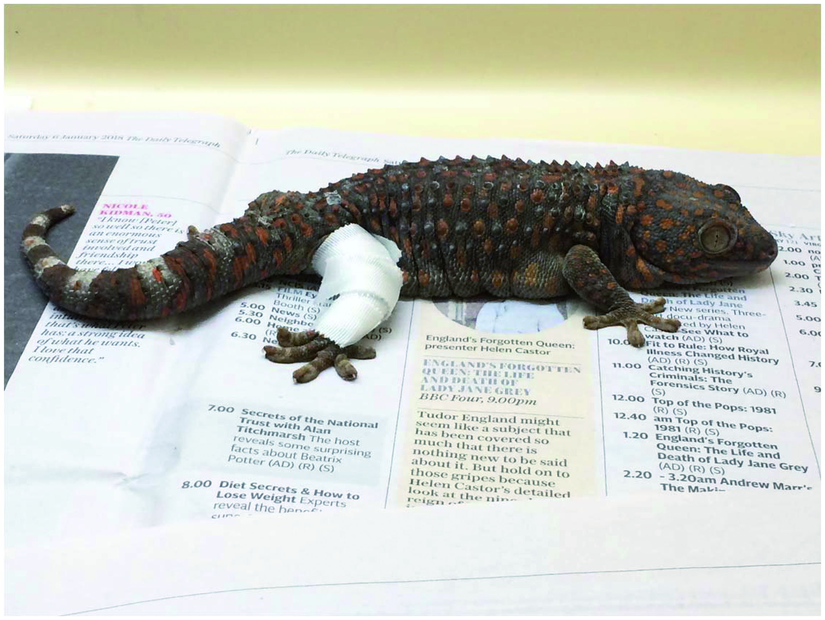
With kind permission from Sara Jones
Bearded dragon 1: Patient presenting with a week - long history of anorexia and lethargy. He also had mites. He was semi – collapsed and dehydrated. Due to cost limitations, bloods and faecal samples were taken only.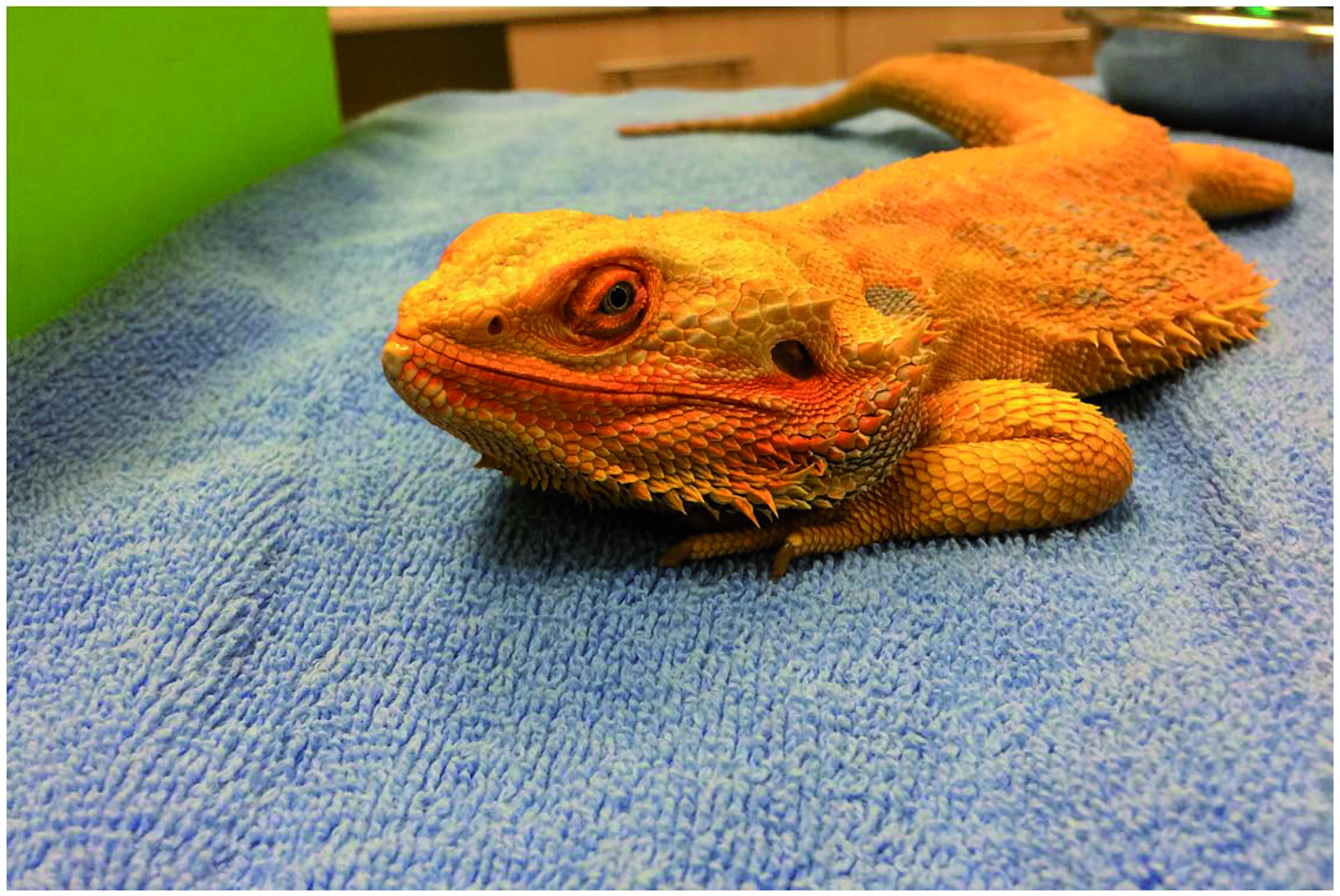
Bearded dragon 2: The patient improved slowly with fluids and supportive care. He was started on IM tramadol just in case there was any pain present (that would be especially difficult to ascertain due to the collapsed state). He was starting to move around his vivarium. Small amounts of gavage feeding commenced on day three.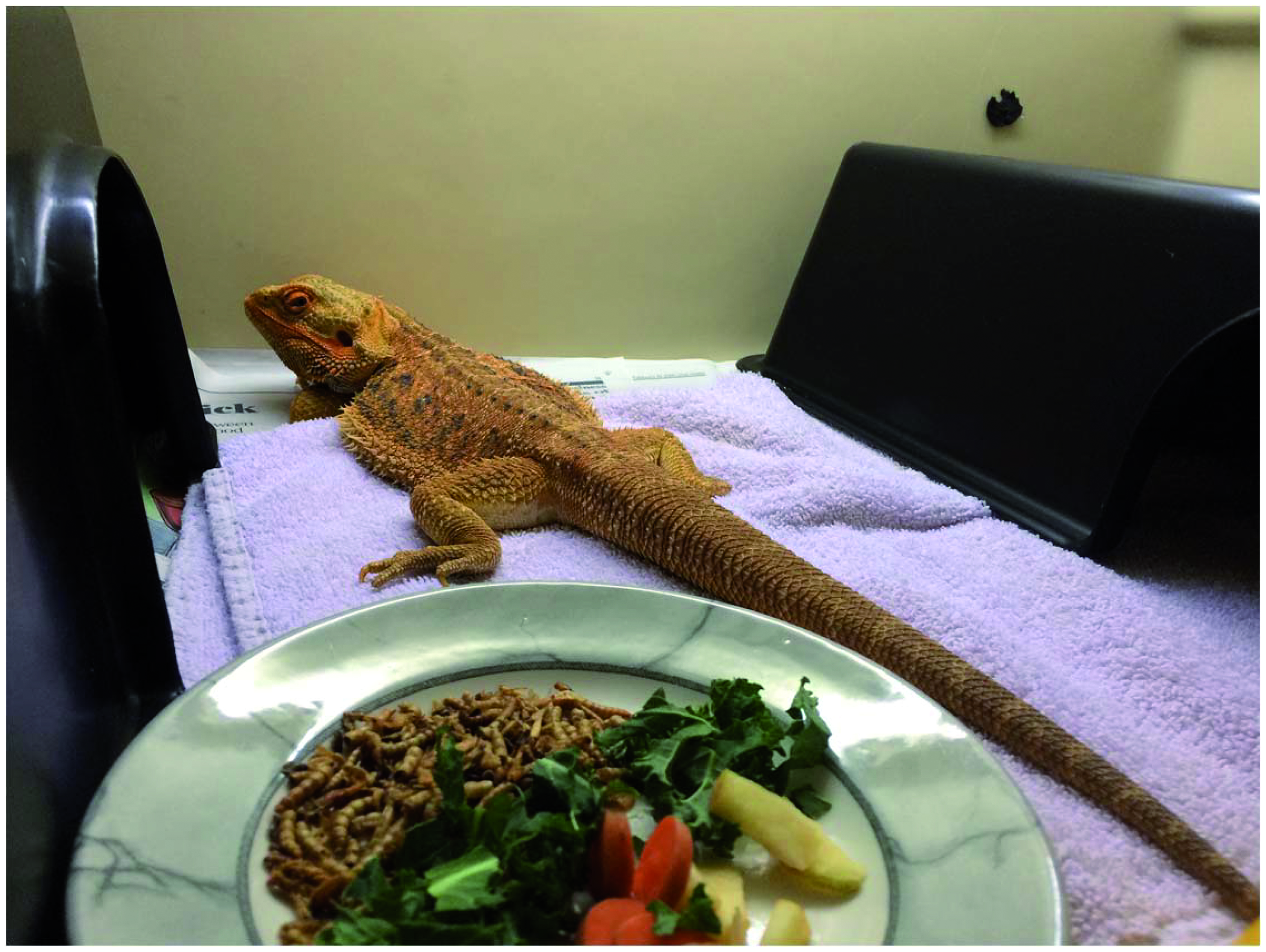
Bearded dragon 3: The patient deteriorated again after four days (note the closed eyes, weak posture and blackened chin). This possibly may have been the illness showing through now that the shock and dehydration had been corrected. The blood results came back showing heterophilia; indicating a possibility for coelomitis among other differentials; the owners opted for no further diagnostics and euthanasia.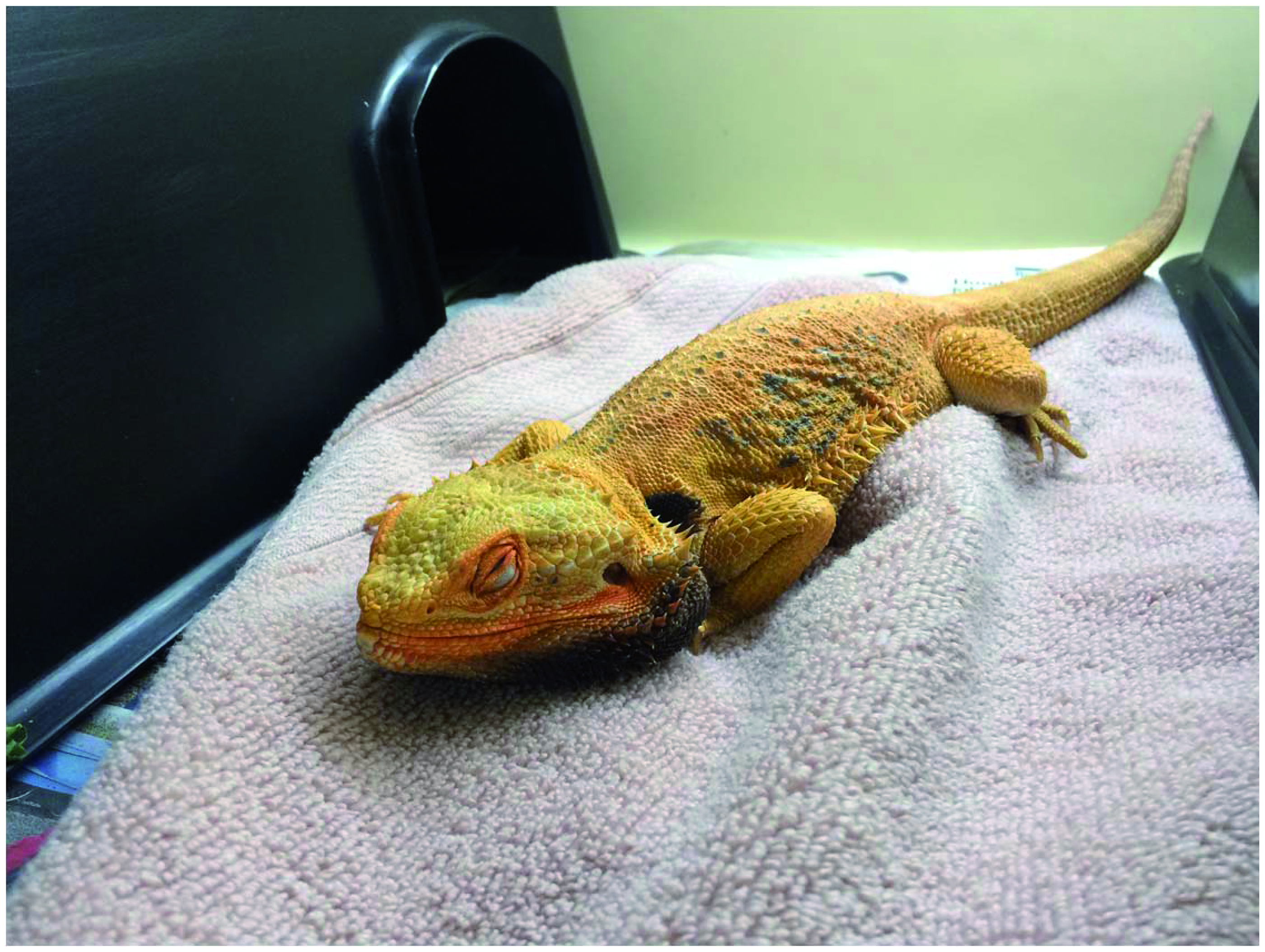
Bearded dragon (eyelid biopsy): This patient had a small biopsy of a mass on his lower eyelid. The patient was administered tramadol as a premedication and IV alfaxan for the anaesthetic.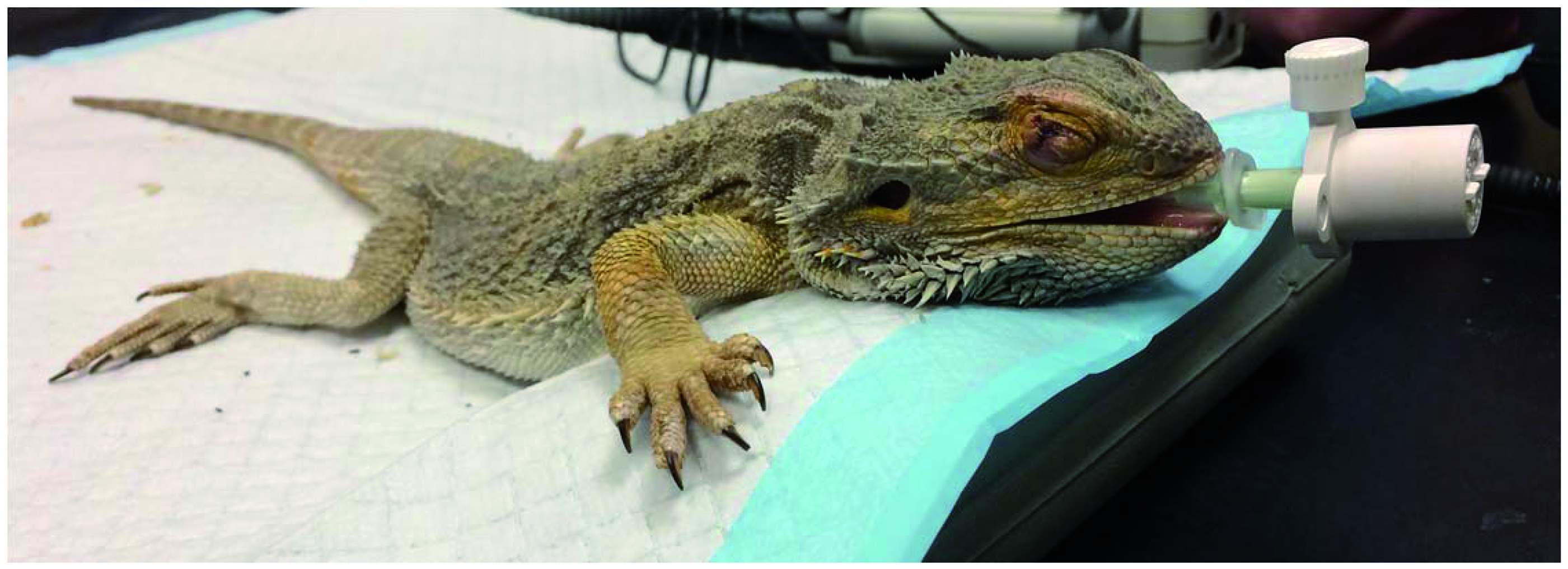
Bearded dragon (eyelid biopsy). Recovering from the surgery. It is very difficult to assess post – operative pain in reptiles due to the long recoveries from anaesthesia (in comparison to birds or mammals). He was also in fairly poor body condition, which can prolong anaesthetic recovery.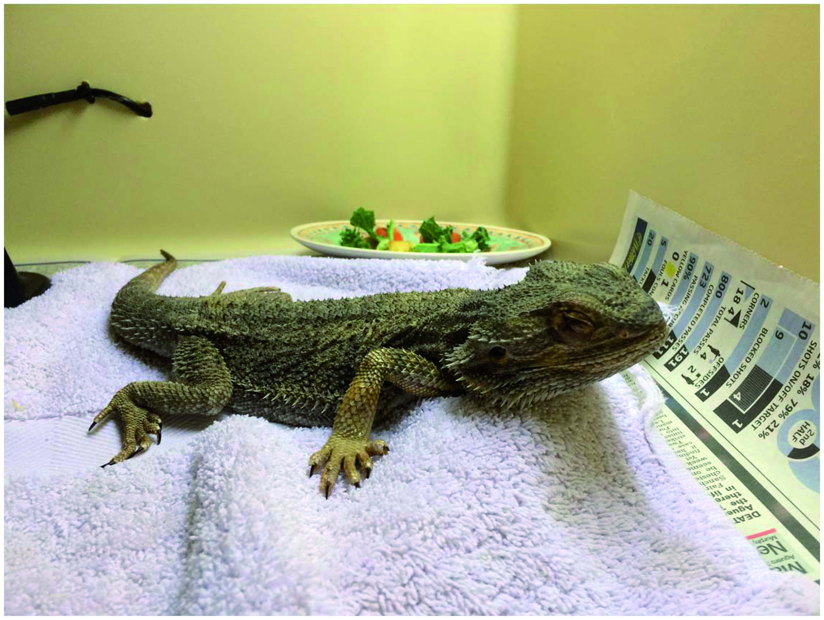
Asian grass lizard presenting with a sore eye. After examination and treatment, the patient went home on oral tramadol. With kind permission from Sara Jones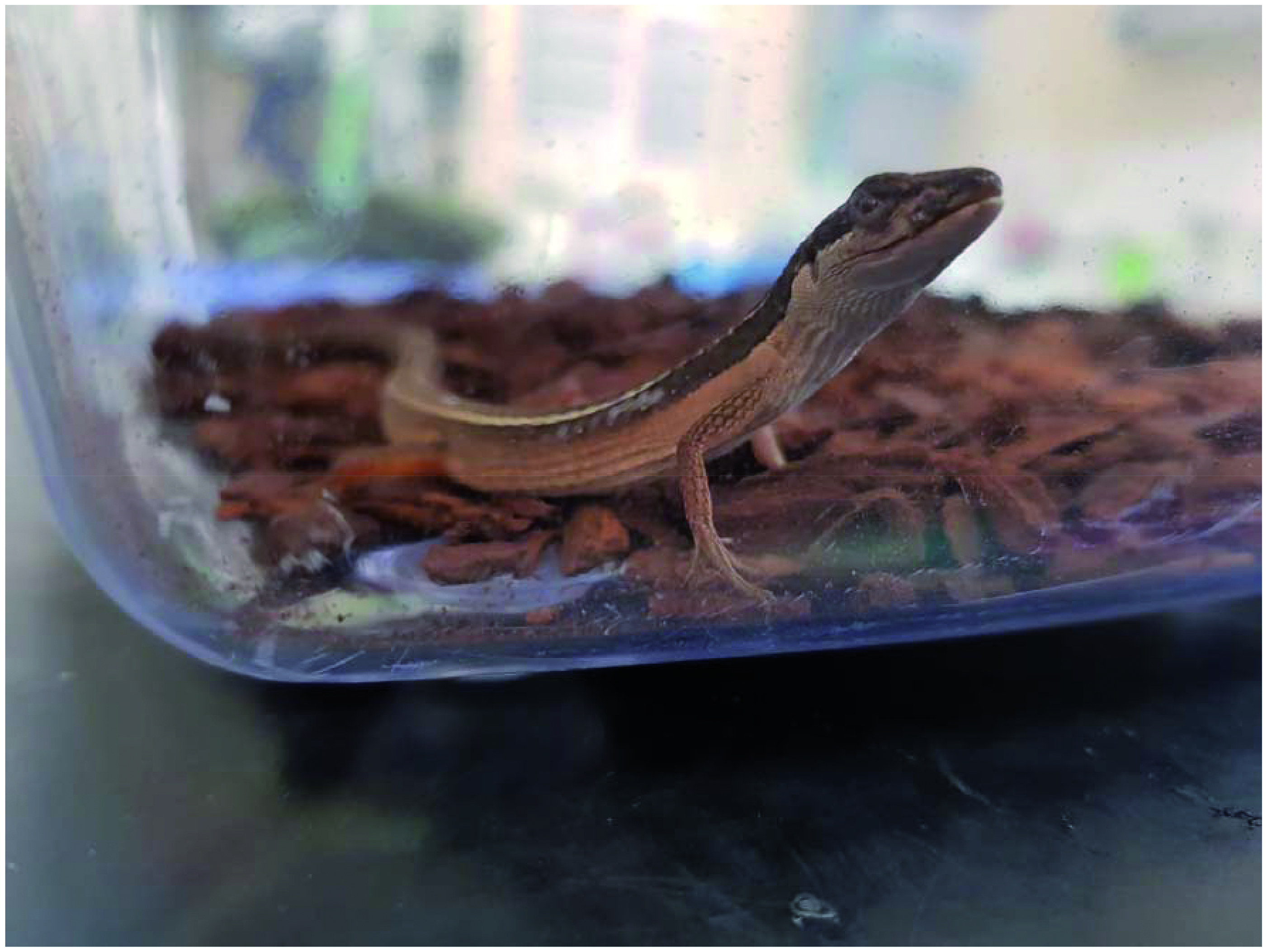
Additional information
Notes on contributors

Aneesa Malik
Aneesa Malik MSc RVN Cert VNES Cert VNECC Aneesa worked in first opinion, emergency, referral, and for the RSPCA. She went on to pursue an interest in animal welfare science, particularly pain assessment and management. Her focus is the factors that affect very different pain management regimes in our veterinary patients, for instance, factors such as the staff involved, the species of animal, or whether patients are ‘owned’, ‘stray’, or wildlife.