
Abstract
The use of hand painting an iris button using oil paint remains the conventional method of artificial eye manufacturing. The authors found that replacing this technique with a digital photograph taken of a patient’s unaffected eye offers several advantages over the conventional method but the process from capture to print must be standardised and colour accurate. The authors of this paper suggest a tried and tested formulated photographic process of capture and printing prior to polymerisation. It discusses issues that can arise and how these can be overcome in order to achieve a high-quality print that can be used to produce a ‘life like’ ocular prosthesis.
Introduction
Trauma is one of the leading causes of eye loss in the UK (Shapira, Worrell, & Ullrich, Citation2020). Increased imperceptibility of an artificial eye may lead to higher patient satisfaction rates (Song, Oh, & Baek, Citation2006; Walshaw et al., Citation2019), and may help reduce the impact on a patient’s mental health from the patients perceived disfigurement following enucleations or eviscerations (McBain et al., Citation2014). To achieve this imperceptibility in the production of an artificial eye, the appearance of key distinguishing features such as iris colour and scleral vasculature must match the patient’s unaffected eye.
The most common method of iris reproduction consists of hand painting using oil paint, a technique undertaken by the National Artificial Eye Service (NAES) and regional maxillofacial prosthetic centres by artistry skilled ocularists. The current continued use of hand paint is down to the ease of adaptation, and ultimate control over final colour through colour mixing, but relies in part on the skill of the Ocularist. Other methods have been tested such as monomer-polymer mixture on an artificial iris (Goiato et al., Citation2010), an inverted painting technique using prefabricated caps as well as photographic reproduction of the patient’s healthy iris (Artopoulou, Montgomery, Wesley, & Lemon, Citation2006; Walshaw, Zoltie, Bartlett, & Gout, Citation2018). Currently no viable alternatives to hand painting have been proved scalable. Current NAES turnaround time for receipt of an artificial eye is six weeks, however patients have reported up to a six month wait (Patient and Public Involvement PPI Meeting, 26th February, 2019).
The authors describe an intricate method of utilising digital photography to reproduce both the iris and sclera in order to achieve a robust method of production that can be standardised, measured, and effectively replicated. This improves upon previous methods of photographic production that focussed on photographing and printing only the iris, and subsequently still required the use of hand painting of the sclera.
Method
Camera equipment & lighting
A Nikon D7500 digital single lens reflex (DSLR) camera fitted with a Nikon AF-S 105mm f2.8G Macro lens was used to capture photographs of the patient’s unaffected eye. To illuminate the eye, three lighting methods were tested; studio lighting, cross polarisation, and a speedlight attached by an off-camera flash cord ().
Figure 1. Illumination techniques for photography of a patients unaffected eye. (A) Two studio lights with softbox attachments (B) Cross Polarisation (C) Speedlight with off-camera remote cord.
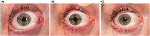
The use of two studio flashes positioned at 45° to the patient was found to create double reflections that covered a considerable amount of the iris. This was partly due to the size of the light source in relation to the subject, but also because of the convex nature of the cornea.
The second method tested was cross polarisation, utilising a Sigma DG140 ring flash with Cross-Polar© filter attachment (IT Logika, UAB). While the flash reflections were eliminated, due to the linear birefringence of the human cornea, visible optical birefringence over the periphery of the limbus rendered the image unusable. A previous study by Knighton and Huang has suggested substantial variation of corneal birefringence amongst individuals (Knighton & Huang, Citation2002) therefore the authors recommend further study into the application of cross polarisation for photography in artificial eye manufacturing.
The third tested method of illumination was the use of a Nikon SC-29 off-camera remote cord to place a Nikon SB-800 flash directly above the lens barrel. This was found to provide the most accurate result, resulting in only a singular flash reflection on the pupil which could be easily removed in post-production.
Colour
A custom white balance was set using a grey card. Following this, a custom DNG profile was created to allow colorimetric interpretation of the raw image data based upon the exact shooting condition and equipment being used, as this can often vary between manufacturer. A photograph of an X-rite ColorChecker passport© was taken and a profile created within the ColorChecker Passport Desktop Application (X-Rite, Citation2020). The profile was stored within Adobe Photoshop for later calibration during post processing.
Camera settings and technique
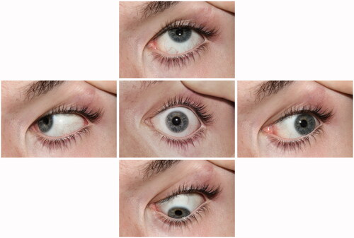
A distance of 1½ ft was set on the lens to ensure the full eye was included in the frame, using a rock a pull technique for focussing. The speedlight was set to ¼ power, and a light metre used to measure the aperture required for exact exposure with the camera set at ISO100 with a shutter speed of 1/125. The light metre recorded an aperture of f/25 which also allowed for a wide depth of field. Five photographs were taken of the eye at primary and secondary positions shown in , commonly used to demonstrate ocular motility (IMI Institute of Medical Illustrators, Citation2008; Miller, Citation2020). This provided a wide view of the scleral vasculature to enable stitching during the postproduction process. A measurement of the iris and pupil size was also taken using a surgical ruler.
Figure 2. Five positions of gaze photographed to provide a wider view of the scleral vasculature.
Postproduction workflow
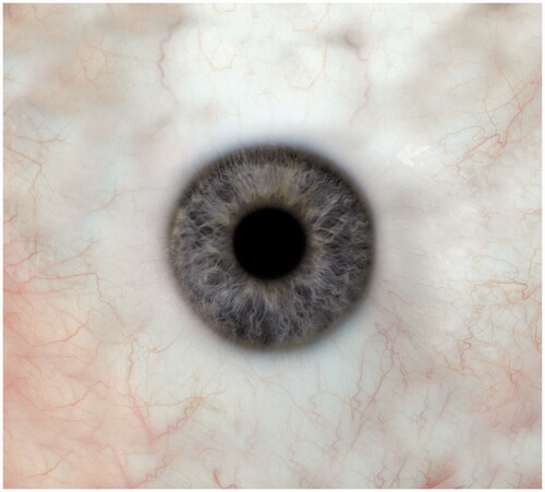
The five images of gaze were imported into Adobe Photoshop, and the camera profile was applied in Adobe RAW. The iris on the photograph of the patient’s normal alignment on forward gaze was used a baseline for merging the wider scleral views. The clone stamp tool was used to cover any areas of missing vasculature. A feathered circular mask tool was utilised to ensure that the iris retained a clear edge, this in turn enabled clean manipulation of the sclera. The result was a wide scleral view which was then resized to match the patient’s recorded iris measurement. The flash reflections were removed using the paint tool. To accommodate for pupil dilation caused by the patient being in low light prior to photography, the pupil size was reduced to a normal pupil range of 3mm using the liquify tool. Slight colour and brightness adjustments were made to accommodate for colour shift during the manufacture process, with 10–30% desaturation and 0.5–0.9 decrease in midtones being applied. The image was then ready for printing (). The image was printed using a custom print profile on to a specialist adhesive paper that can withstand the polymerisation process using a Canon MG5750 inkjet printer and dye-based inks. Once complete, the image was sent to the maxillofacial lab for production and encapsulation.
Figure 3. Wide scleral view containing the five merged positions of gaze.
Results
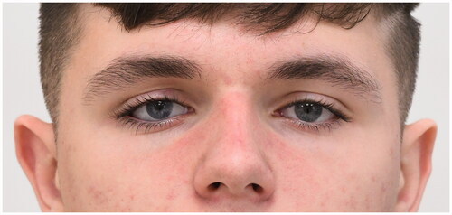
A digital photograph of the eye presents an advantage over hand painting by replicating the iris with minimal colour adjustments required (). The authors were also able to match the characteristics of both the iris and the sclera, improving on previous published methods which relied upon the use of silk thread to create the scleral detail (Goiato et al., Citation2014). The increased iris crypt detail and contrast on the pupil and limbus margin created a more lifelike appearance. Previous patient evaluations undertaken by the authors found that outcomes were favourable in improving the patient’s rehabilitation pathway, quality of life and experience (Gout et al., Citation2020).
Figure 4. Patient wearing the artificial eye produced using the novel digital photographic technique (patient’s right eye).
Hand painted iris colour shifting has previously been found to occur during the polymerisation of the colourless artificial resin which is part of the manufacturing process (Moreno et al., Citation2015). The authors concluded the same with the photographic technique. Slight colour shifts and increased luminance were apparent, with brown eyes having lower values of colour change than blue. The authors found the adaptations required to accommodate for the colour shift during polymerisation require formulisation and recommend further study in this area.
Discussion
Hand painting in artificial eye manufacturing has stood the test of time, with no viable scalable alternative. Oil paints are more convenient to use and offer a far broader range than any other paint medium and this may go some way as to explain their continued use over alternative digital methods (Blake, Citation1997). Other reported reasons include lack of ability to achieve colour accuracy and questions over permanence when using digital photography (NHS Lothian, Citation2020).
As digital photography has evolved, so has colour calibration technology and ink quality. The ability to print an exact colour replica of what was taken in camera has become much simpler. Digital photography arguably provides a more scientifically robust method of production that can be standardised, measured, and effectively reproduced. It is possible that this method is also more cost efficient with a faster production time, however further study is recommended to substantiate this.
Conclusion
Digital photography offers a viable alternative to hand painting an artificial eye, that is accurate and reproduceable. Currently the technique requires the use of specialist photography expertise, equipment and software which may not be readily available for national or regional service providers. A protocoled process such as that described in this paper will allow local or regional medical illustration departments to provide the service which in turn, will enable scalability. Further study is required to investigate permanence of dye-based ink versus oil paint and the colour shift associated with the polymerisation process.
Disclosure statement
No potential conflict of interest was reported by the author(s).
References
- Artopoulou, I.-I., Montgomery, P.C., Wesley, P.J., & Lemon, J.C. (2006). Digital imaging in the fabrication of ocular prostheses. The Journal of Prosthetic Dentistry, 95, 327–330. doi:https://doi.org/10.1016/j.prosdent.2006.01.018
- Blake, W. (1997). Acrylic painting : A complete guide. Mineola, N.Y.: Dover Publications.
- Cross-Polar filter, IG Logika, UAB; Retrieved 19 November, 2020, from https://cross-polar.com/
- Goiato, M.C., Bannwart, L.C., Haddad, M.F., dos Santos, D.M., Pesqueira, A.A., & Miyahara, G.I. (2014). Fabrication techniques for ocular prostheses–An overview. Orbit, 33, 229–233. doi:https://doi.org/10.3109/01676830.2014.881395
- Goiato, M.C., Moreno, A., dos Santos, D.M., de Carvalho Dekon, S.F., Pellizzer, E.P., & Pesqueira, A.A. (2010). Effect of polymerization and accelerated aging on iris color stability of ocular prosthesis. Contact Lens & Anterior Eye, 33, 215–218. doi:https://doi.org/10.1016/j.clae.2010.04.004
- Gout, T., Walshaw, E.G., Zoltie, T., Bartlett, P., Archer, T., Altaie, A., Parmar, J., El-Hindy, N., Chang, B. and Kalantzis, G. (2020). Novel artificial eye service evaluation using patient reported outcome measures. Eye (London, England). [online]; Retrieved 26 Nov, 2020, from https://pubmed.ncbi.nlm.nih.gov/33051621/
- Institute of Medical Illustrators (2008). IMI National Guidelines: Ophthalmic Imaging. [online]; Retrieved 26 Nov, 2020, from https://www.imi.org.uk/wp-content/uploads/2019/01/IMINatGuidelinesOphthalmicImagingFeb_1.pdf
- Knighton, R.W., & Huang, X.R. (2002). Linear birefringence of the central human cornea. Investigative Ophthalmology & Visual Science, 43, 82–86.
- McBain, H.B., Ezra, D.G., Rose, G.E., & Newman, S.P. (2014). The psychosocial impact of living with an ocular prosthesis. Orbit, 33, 39–44. doi:https://doi.org/10.3109/01676830.2013.851251
- Miller, G., 2020 Ocular Motility Photography. [online] Ophthalmic Photographers Society Inc.; Retrieved 23 November, 2020, from https://www.opsweb.org/page/Motility
- Moreno, A., Goiato, M.C., Oliveira, K.F., Iyda, M.G., Haddad, M.F., de Carvalho Dekon, S.F., & dos Santos, D.M. (2015). Color stability of the artificial iris button in an ocular prosthesis before and after acrylic resin polymerization. Contact Lens and Anterior Eye, 38, 414–418. doi:https://doi.org/10.1016/j.clae.2015.05.003
- NHS Lothian (2020). Are Artificial Eyes still Hand-painted|The Artificial Eye Clinic. [online] services.nhslothian.scot; Retrieved 26 Nov, 2020, from https://services.nhslothian.scot/artificialeyes/FAQs/Pages/Why-are-Artificial-Eyes-still-Hand-Painted-in-the-Digital-World.aspx
- Shapira, Y., Worrell, E., Ullrich, K. (2020). UK National Artificial Eye Questionnaire study: Comparisons between cosmetic shell and artificial eye users. Part 1: Demographics, comfort and satisfaction. British Journal of Ophthalmology. doi:https://doi.org/10.1136/bjophthalmol-2020-317015
- Song, J.-S., Oh, J., & Baek, S.H. (2006). A survey of satisfaction in anophthalmic patients wearing ocular prosthesis. Graefe’s Archive for Clinical and Experimental Ophthalmology, 244, 330–335. doi:https://doi.org/10.1007/s00417-005-0037-0
- Walshaw, E., Pamar, J., Gout, T., Bartlett, P., Chang, B., Zoltie, T., & Kalantzis, G. (2019). Personalised ocular prostheses – Patient evaluations of digitally printing prosthetics. British Journal of Oral and Maxillofacial Surgery, 57, e40. doi:https://doi.org/10.1016/j.bjoms.2019.10.109
- Walshaw, E., Zoltie, T., Bartlett, P., & Gout, T. (2018). Manufacture of a high definition ocular prosthesis. British Journal of Oral and Maxillofacial Surgery, 56, 893–894. doi:https://doi.org/10.1016/j.bjoms.2018.08.016
- X-Rite. 2020. Creating DNG Profiles For Adobe Camera Raw. [online]; Retrieved 19 November, 2020, from https://www.xrite.com/service-support/creating_dng_profiles_for_adobe_camera_raw