Abstract
Background Fossa piriformis is considered the correct point of entry for a straight femoral nail. A trochanteric overhang may make the access to fossa piriformis difficult. We investigated the anatomy of the trochanteric region, paying special attention to the entry point for antegrade intramedullary femoral nailing.
Methods and results We studied 100 cadaver specimens. In 63 specimens a shape with a free entry point was found, whereas in 37 cases the entry point was either half or fully covered. In 9 specimens the entry points could not be exactly located from a cranial aspect.
Interpretation The anatomic variations of the trochanteric sometimes make it difficult to identify the correct entry point for an intramedullary nail.
The recommended entry point for antegrade intramedullary femoral nails is determined by the design of the implant. Some nails are to be implanted in the fossa piriformis () and others more laterally at the tip of the greater trochanter (Kempf et al. Citation1985, Harper and Carson Citation1987, Miller et al. Citation1993, Krettek et al. Citation1996, Gausepohl et al. Citation2002, Moein et al. Citation2005).
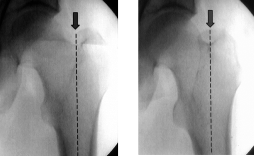
Figure 1. In the AP view, the entry point for the intramedullary femoral nail is exactly within the extended medullary canal line (two different rotational positions are shown).

We examined how frequently the fossa piriformis is covered by the greater trochanter. Due to these anatomic variations, there is a risk that femoral nails be badly positioned.
Methods
We investigated 100 preserved cadaver specimens, mean age 74 (48–90) years at death. The specimens were selected at random and extremities with arthrosis or other pathological changes were excluded from the study. After complete dissection of the bone, the femur was assembled on a reproduction unit in order to take standardized photographs. The femurs were photographed in cranial view (in the exact extension of the medullary canal line and a constant distance of 25 cm) and dorsal view (90° angle to the medullary canal line and constant distance of 25 cm) with constant angles and distances. The relationship between the anatomy of the greater trochanter and the fossa piriformis was subsequently analyzed using these photographs.
Results
We divided our findings into 4 groups.
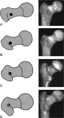
Group 1. In 45 specimens we found the most common shape of the greater trochanter with a free fossa piriformis. The outline of the spine coming to the tip of the greater trochanter just touched or exceeded the entry point by a maximum of ∼2 mm ().
Figure 2. Sketches illustrating the standard entry point in relation to the various shapes of the greater trochanter. A. Group 1: the most common shape, free entry point (45% of cases). B. Group 2: the outline of the spine is projected laterally to the entry point (18% of cases). C. Group 3: the entry point is partially covered (12% of cases). D. Group 4: the entry point is completely covered (25% of cases).

Group 2. In 18 cases the projection of this spine of the greater trochanter was lateral to the entry point (). The shapes of group 1 and 2 would not hinder the implantation of nails in the fossa piriformis. In 37 specimens, the fossa piriformis was exceeded by more than ∼2 mm. This group was subdivided up once again.
Group 3. In 12 cases half of the entry point was covered ().
Group 4. In 25 specimens the entry point could not be exactly located from a cranial view due to the shape of the greater trochanter (). In 9 specimens of this group a prominent gluteal tuberosity—a so-called third trochanter—was observed (Standring et al. Citation2005).
Discussion
Choosing the exact entry point for a femoral nail is important. An incorrect entry point may lead to tensions in the implant-bone interface, inducing additional iatrogenic fractures (Gausepohl et al. Citation2002). The entry point is determined by the design of the implant (Kempf et al. Citation1985, Harper and Carson Citation1987, Miller et al. Citation1993, Krettek et al. Citation1996). Gausepohl et al. (Citation2002) recommended the piriform fossa as the correct entry point for straight nails; bent nails should be implanted more dorsally.
Radiographic control using an image intensifier and digital palpation give the surgeon the correct orientation. The tip of the trochanter and the transition zone to the femoral neck are important landmarks using AP-radiography. These landmarks are difficult to evaluate in axial view and in relation to the trochanteric shape and also the rotational axis of the leg. In proximal femoral shaft fractures, orientation in particular may be even more difficult due to dislocation of the proximal fragment caused by muscular traction. Georgiadis et al. (Citation1996) reported that a trochanteric overhang may result in a much more medial entry point than intended, which may increase the risk of additional fracture or avascular femoral head necrosis.
In our series, the entry point could be reached without problems in two-thirds of the specimens. In one-tenth we faced slight problems, and in one-quarter severe problems in reaching the entry point. Especially in patients with a so-called third trochanter, the entry point is often covered by parts of the greater trochanter. The K-wire for opening of the medullary canal may be misguided by the tip of the trochanter, resulting in a much more medial entry point than intended. Once the leading pin is in the correct position, the opening of the medullary canal by drill—and not by awl—is to be recommended. In this way, troublesome parts of the trochanteric region are resected.
With preoperative CT, special attention should be paid to the scans of the trochanteric region. In order to assure a correct implant position, these anatomic variations should always be kept in mind when doing intramedullary femoral nailing.
We wish to thank Dr Andreas Pichler (Université de Provence, Aix-en-Provence, France) for linguistic revision.
Contributions of authors
WG: reviewed the literature, planned and executed the study and wrote the first draft of the paper. WP: participated in planning and performing the study, and revised the manuscript. HC: commented on and revised the text. NPT: reviewed the anatomical literature and participated in carrying out the analysis. SG: participated in realizing the study. All authors took part in interpretation of the results and prepared the final version of the paper.
- Gausepohl T, Pennig D, Koebke J, Harnoss S. Antegrade femoral nailing: an anatomical determination of the correct entry point. Injury 2002; 33(8)701–5
- Georgiadis G M, Olexa T A, Ebraheim N A. Entry sites for antegrade femoral nailing. Clin Orthop 1996, 330: 281–7
- Harper M C, Carson W L. Curvature of the femur and the proximal entry point for an intramedullary rod. Clin Orthop 1987, 220: 155–61
- Kempf I, Grosse A, Beck G. Closed locked intramedullary nailing. Is application to communited fractures of the femur. J Bone Joint Surg (Am) 1985; 67(5)709–20
- Krettek C, Rudolf J, Schandelmaier P. Unreamed intramedullary nailing of femoral shaft fractures: operative technique and early clinical experience with the standard locking optin. Injury 1996; 27((Suppl 4))233–54
- Miller S D, Burkart B, Damson E. The effect of the entry hole of an intramedullary nail on the strength of the proximal femur. J Bone Joint Surg (Br) 1993; 75(2)202–6
- Moein C M, Verhofstad M H, Bleys R L, van der Werken C. Soft tissue injury related to choice of entry point in antegrade femoral nailing: piriform fossa or greater trochanter tip. Injury 2005; 36(11)1337–42
- Standring S, Ellis H, Healy J, Johnson D, Williams A. Gray's Anatomy 39th. The anatomicaal basis of clinical practice. Elsevier, New York 2005
