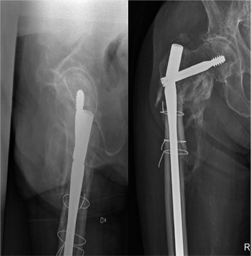
An 80-year-old man sustained a closed displaced comminuted subtrochanteric fracture in a fall accident. The fracture was stabilized with a Long Gamma locking nail (Stryker Trauma, Schönkirchen, Germany). Postoperative radiographs showed good placement of the nail and acceptable fracture alignment, but also significant bone loss beneath the calcar. Postoperatively, the patient was allowed to walk using crutches and partial weight bearing. At 6-month follow-up the patient complained about severe pain and reduced walking ability since several days. Radiographs showed nonunion and breakage of the nail through the hip screw hole (). There was no history of new trauma. The nail was changed to a Trigen trochanteric antegrade nail (Smith & Nephew Orthopedics, Memphis, TN).
Figure 1. The anteroposterior and lateral radiographs taken 6 months after the injury show development of nonunion, varus malalignment, and the broken nail.
Technique ( and )
The patient was placed supine on a fracture table. Using image intensifier guidance, the hip screw was removed. After opening of the intramedullary canal, the proximal part of the broken nail was also removed. The tip of a ball-tipped guide wire (Trigen; Smith & Nephew) was bent slightly in order to be able to change the direction of the guide wire into the medullary canal, when necessary. The guide wire could be gently introduced into the medullary canal through the footprint of the removed proximal nail fragment. Thereafter, the distal locking screws were removed and the guide wire was advanced through the nail until the tip of the guide wire was located distal to the tip of the nail, visible by image intensifier. A 12-mm-long 3.5 AO cortex screw was placed in one of the distal holes using a screwdriver, ensuring that the screw advanced into the contralateral side of the nail hole. This would make the assembly more secure during the extraction of the nail. The guide wire was pulled out until the orb abutted against the screw which took fast hold of the orb. Using the guide wire holder and gentle hammer blows, the nail and the guide wire were pulled out. After the broken nail had been removed, the nonunion site was revised from fibrotic soft tissue and sclerotic fracture site. The hip screw hole was filled with redundant bone. After reaming of the medullary canal, the fracture was stabilized with a 13-mm Trigen trochanteric antegrade nail. An autogenous iliac bone graft was impacted around the fracture. The non-union was healed 4 months after the reoperation.
Figure 2. A. Position of the ball-tipped guide wire. B. Position of the 12-mm-long screw. C. The guide wire is blocked by the screw, and the nail can be removed by gentle hammer blows.
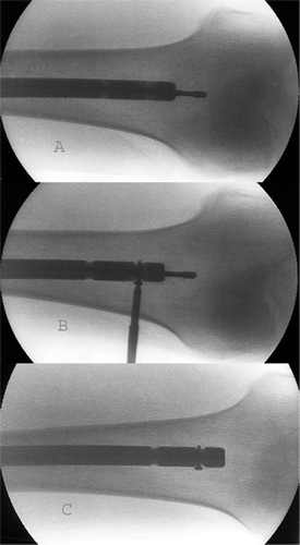
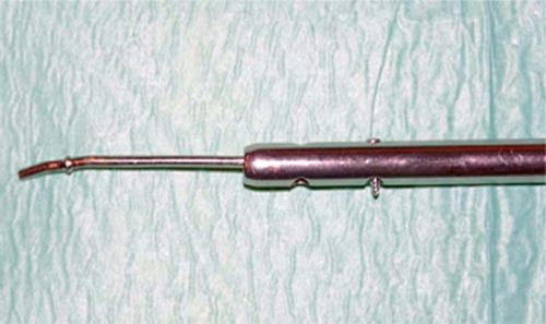
Figure 3. The photograph demonstrates how a ball-tipped guide wire is passed through the nail. A 3.5-mm screw is placed in one of the distal holes which will lock the guide wire during extraction.
Discussion
Our technique was applied for removal of a Long Gamma nail which has an inner canal of 5.7 mm diameter. The Trigen ball-tipped guide wire that was used has a 3-mm-thick wire and 3.5-mm-thick ball. This guide wire could be delivered into the nail without difficulty. (The diameter of the nail's inner canal and the guide wire and the supplementary screw should be counted if a nail other than the Long Gamma nail is subjected to this method.) The head of an AO 3.5-mm cortex screw is 6 mm in diameter; it could pass through the old screw hole from the distal locking screw without difficulty, after it had been cleared of soft tissues.
The standard and the AO-recommended technique involve extraction of the proximal fragment followed by over-reaming of the medullary canal down to the distal nail fragment (Muller et al. Citation1979). An extraction hook is then used to extract the distal nail fragment. However, this technique is time-consuming and the hook tends to come free, thus requiring several re-insertions before full extraction of the nail. To solve this problem, Brewster et al. (Citation1995) modified the AO-recommended technique by using several guide wires impacted around the hook to form a stable construct which could easily be removed as one piece. The technique described by Brewster et al. may be successful, but requires the use of several pieces of equipment and substantial force for removal. Khan et al. (Citation1997) recommended a technique using a hand-reamer and Sivananthan et al. (Citation2000) suggested impaction of a smaller nail into the opening of the distal nail fragment. These methods are not applicable to the new generation of nails with small inner holes, which are constructed for passage of only a guide wire.
Magu et al. (Citation2004) described another method for removal of the broken femoral nail. They suggested making an 8-mm-wide tunnel through the intercondylar notch. A washer was loaded onto the olive wire and the assembly was inserted through the intercondylar tunnel and advanced proximally into the nail. The guide wire was delivered through the proximal incision and pulled out with the broken nail. An additional incision—with the risk of traumatization of the articular surface—might be a disadvantage of this technique.
Our technique does not require any special instruments or any additional incision.
- Brewster N T, Ashcroft G P, Scotland T R. Extraction of broken intramedullary nails: an improvement in technique. Injury 1995; 26(4)286
- Khan M, Schranz P J, Ward M W. Removal of a broken intramedullary tibial nail using a hand reamer. Injury 1997; 28(9–10)693–4
- Magu N K, Sharma A K, Singh R. Extraction of the broken intramedullary femoral nail: an innovative technique. Int. J Care Injured 2004; 35: 1322–3
- Muller M E, Allowger M, Schneider R, Willeneger H. Manual of internal fixation: Technique recommended by the AO-ASIF Group2nd Ed. Springer Verlag, Berlin. 1979: 158
- Sivananthan K S, Raveendran K, K, Kumar T, Sivananthan S. A simple method for removal of a broken intramedullary nail. Injury 2000; 31: 433–4