Abstract
Background Metacarpal index (MCI), measured from hand radiographs as the ratio between combined cortical thickness and bone diameter, has been suggested for assessment of bone mass and risk of osteoporotic fracture. We studied MCI for its ability to predict hip fractures.
Methods Hand radiographs were taken and MCI determined in 3,561 subjects from a representative population sample of 8,000 Finns who were 30 years of age or over in 1978–80. Record linkage to the National Hospital Discharge Register identified 117 subjects who had been hospitalized for primary treatment of hip fracture by the end of 1994.
Results High age, low body mass index, tall stature and smoking at baseline showed, independently of each other, significant associations with low MCI. Low MCI was a strong predictor of hip fracture. When adjusted for all potential confounding factors, the relative risk of hip fracture per decrement of MCI by one standard deviation (0.1) was 1.5 (95% CI 1.2–1.8).
Interpretation Low MCI is associated with known risk factors of osteoporosis and predicts hip fracture. Since hand radiographs are easily available at low cost, measurements of MCI can be used as an alternative approach to find osteoporotic individuals with a high risk of hip fracture. ▪
The diagnosis of osteoporosis is currently based on measurement of bone mineral density (BMD) using dual X-ray absorptiometry (DXA) (Cummings et al. Citation1990). BMD also predicts osteoporotic fractures with a gradient of risk ranging from about 1.5 to 3.0 for each standard deviation decrease in BMD (Kanis Citation2002). A case-finding strategy has been recommended to target potentially costly preventive measures to those individuals who are at high risk of fracture (Cummings and Melton Citation2002). However, DXA is not generally available in many countries (Bouxsein et al. Citation2002).
Measurement of cortical bone width and geometry from plain radiographs was among the first methods developed for assessment of skeletal mass (Barnett and Nordin Citation1960). Recent evidence has suggested that metacarpal index (MCI) measured on radiographs might be used successfully for assessment of bone mass and quality, and also for predicting osteoporotic fractures (Dey et al. Citation2000, Hyldstrup and Nielsen Citation2001, Nielsen Citation2001, Bouxsein et al. Citation2002).
Previous studies on the relationship between metacarpal morphometry and fractures have been based on relatively small study populations, from limited geographical areas, or involving either men or women exclusively, or only narrow age ranges. Moreover, the studies have suffered from low rates of incidence, or short follow-up periods (Cooper et al. Citation1991, Wishart et al. Citation1993, Dey et al. Citation2000, Kiel et al. Citation2001, Bouxsein et al. Citation2002).
We chose a large nationwide sample of both men and women with a wide age range to study MCI for its associations with known risk factors in osteoporosis, and for its potential to predict hip fractures during a follow-up time of more than 15 years.
Methods
The study population was a stratified two-stage cluster sample drawn from the population register to represent Finnish adults aged 30 years or older (Aromaa et al. Citation1989). In the first stage, 40 representative areas were selected. In the second stage, a systematic sample of inhabitants was drawn from each area. The sample consisted of 8,000 individuals (3,637 men) of whom 7,217 (90%) participated.
Details of the design and implementation of the Mini-Finland Health Survey have been described elsewhere (Aromaa et al. Citation1989). Briefly, all participants were interviewed at home and asked to fill in a basic questionnaire before attending a screening examination. The interview and questionnaire yielded essential information on health habits and previously diagnosed diseases. The screening phase comprised measurements and tests to identify subjects with possible cardiovascular, respiratory or musculoskeletal diseases. Those with findings suggestive of these diseases and a 20% random sample of the participants were asked to attend a clinical assessment, which included physical examination by specially trained physicians.
The basic questionnaire yielded information on leisuretime physical activity, categorized as low, moderate, and high. Self-reported general health was classified according to a 5-point scale: good, fairly good, moderate, rather poor, and poor. Selfrated health measured in this manner has proved reliable in test-retest analysis (Martikainen et al. Citation1999). Body mass index (weight/height2, kg/m2) was used as a measure of relative weight. Smoking history was categorized as: never smoked, ex-smoker, current smoker of cigars, pipe or of fewer than 20 cigarettes a day, and current smoker of 20 cigarettes or more a day. The basic questionnaire also inquired about average weekly consumption of beer, wine and strong beverages during the preceding month. The overall alcohol consumption was expressed in grams of ethanol per week. The level of education was considered in three categories based on the number of years of education.
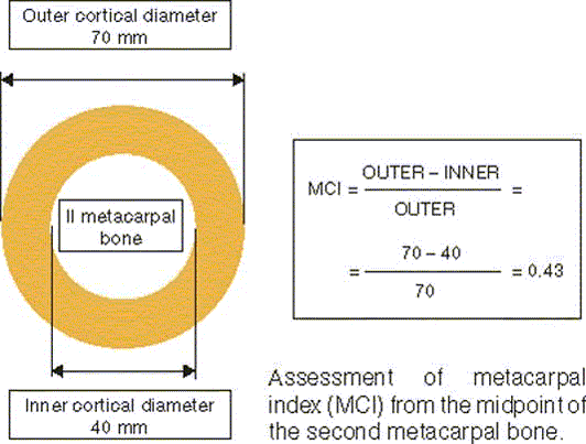
Posteroanterior radiographs of both hands were taken from 3,595 individuals who met at least 1 of the screening criteria for musculoskeletal diseases, or belonged to the random 20% sample. Hand radiographs were read mainly to diagnose osteoarthritis (Kärkkäinen Citation1985). A radiologist read the radiographs without any information on the clinical findings or on metacarpal measurements, which were assessed by another radiologist. The measurements for MCI were made to the nearest 0.1 mm with a digital caliper, at the midpoint of the second metacarpal bone of the right hand (n = 3,561) (Figure) and without any information on the clinical findings. The coefficients of intraobserver reliability were 0.91 for both the inner and outer cortical diameter (Kärkkäinen Citation1985).
Morbidity and mortality in the cohort have been continuously recorded since the baseline examination. Record linkage to the National Hospital Discharge Register identified 117 subjects who had been hospitalized for primary treatment of hip fracture by the end of 1994. Hip fractures as primary or secondary diagnoses were defined according to the eighth edition of the ICD, codes 820.00 to 820.99.
Statistics
We used the Statistical Analysis System (SAS) software (SAS Institute, Gary, NC) for statistical analysis. Cross-sectional associations between MCI and risk factors of hip fractures were based on the general linear models. Cox's life-table regression model was used to estimate the associations between MCI and hip fractures. Potential confounding and effect-modifying factors were also entered into the models. Significance of interaction was tested by entering interaction terms into the models. The 95% confidence intervals of the relative risk estimates and the likelihood ratio statistics (chi-squared values expressed as p-values) were based on the models.
Results
At baseline, high age, low body mass index, tall stature and smoking were significantly associated, independently of each other, with low MCI ().
Table 1. Metacarpal index (MCI) by age, sex, body mass index, body height, educational level, smoking status, alcohol intake, physical activity at leisure, and selfrated general health, adjusted independently of each other (n=3,561)
When adjusted for all the potential confounding factors, low MCI strongly predicted hip fracture ( and ). The prediction of hip fractures by MCI was not related to the type of hip fracture (data not shown).
Table 2. Adjusted relative risk (RR) and its 95% confidence interval (CI) of hip fracture by metacarpal index, age, sex, body mass index, height, level of education, smoking, alcohol intake, physical activity at leisure and self reported general health, adjusted independently of each other (n=3,561)
Table 3. Relative risk of hip fracture and its 95% confidence interval (CI) per decrement of metacarpal index by 1 standard deviation (0.1)
Discussion
Our results suggest that MCI, which can easily be determined at low cost, can be used to assess bone mineral status and future hip fracture risk. Thus, MCI may be useful for both epidemiological research and primary care, to find patients with osteoporosis.
One strength of our study is that it was based on a large, nationally representative health examination survey of people aged 30 years or over. Moreover, the follow-up time was more than 15 years, thus greatly exceeding the follow-up times in previous studies (Wishart et al. Citation1993, Dey et al. Citation2000, Bouxsein et al. Citation2002). Our average MCI values were similar to those found in recent studies (Black et al. Citation2001, Hyldstrup and Nielsen Citation2001).
The Finnish National Hospital Discharge Register, in operation since 1967, contains information on hospital admissions and discharges from every hospital in the country. This information includes the primary and secondary diagnoses according to the ICD, along with a personal identification code. Using these data, we were able to identify those study subjects who had sustained a hip fracture and who had been admitted to hospital for acute care since the baseline examination. In severe injuries leading to hospitalization, 92% to 97% of the diagnoses had been correctly registered at the three-digit level in the National Hospital Discharge Register, as compared with those in the original hospital documents (Honkanen Citation1990, Luthje et al. Citation1995). In view of the above, we feel justified in making generalizations from our results.
We found that low MCI strongly predicted hip fractures in a representative population sample. Similar results have been reported previously (Cooper et al. Citation1991, Wishart et al. Citation1993). In addition, a clear association has been shown recently between low bone mineral density measured by digital radiogrammetry, and hip fractures. This technique uses automated image analysis of standard hand radiographs to estimate bone mineral density (Bouxsein et al. Citation2002). Such an association, however, was not found between the metacarpal cortical area and hip fractures (Kiel et al. Citation2001), probably due to methodological differences between the measurements of the cortical area by Kiel et al. and our measurements of MCI.
Low BMD measured by DXA is known to be predictive of hip fracture in post-menopausal women (Cummings et al. Citation1990). Since MCI was associated with several risk factors of osteoporosis, such as high age, tall stature, low body mass index and smoking (Kanis Citation2002, Kröger et al Citation1994), it can also be expected to be associated with osteoporosis.
In DXA measurements, BMD depends to a great extent on bone size, whereas MCI is not affected by this (Hyldstrup and Nielsen Citation2001). Also, MCI is not dependent on calibration and measurement scales between instruments. Thus, MCI might prove preferable to DXA for comparisons between different populations (Dequeker Citation1976). Furthermore, MCI can be measured from any appropriate hand radiograph, thus enabling estimations of cortical bone loss from sets of successive earlier radiographs (Nielsen Citation2001). MCI may be a practical method for assessment of bone mineral status and fracture risk in developing countries, because of limited availability of DXA. WHO has estimated that in a few decades, most hip fractures will occur in developing countries.
The reduction in cortical bone mass with age and time since menopause in women results from both progressive intracortical porosity and reduction in cortical thickness (Iwamoto et al. Citation1998). However, increased bone loss after menopause is also associated with increased periosteal apposition, which partially preserves bone strength (Ahlborg et al. Citation2003). In women, bone loss occurs when a greater volume of bone is removed than is replaced in cortical layers of bone, because estrogen deficiency inhibits the periosteal apposition (Seeman Citation2003). MCI is therefore subject to changes resulting from bone resorption and remodeling.
MCI appears to have potential for use both in epidemiological studies and in clinical practice. We recommend that MCI should be determined in a patient if osteoporosis is suspected based on other risk factors. However, further studies are needed to establish cut-off values of MCI, in order to enable the clinician to decide whether a given patient should undergo further evaluation or not.
This study was supported in part by the EVO funding of Kuopio University Hospital and Norther-Savo Cultural Foundation. The authors thank Marianna Sunnari of the Uni-versity of Turku, Finland, for checking the language in this article.
No competing interests declared.
Author contributions
M Haara reviewed literature, planned and decided contents of the article and wrote the first draft of the paper. M Heliövaara participated in developing ideas of the study and carried out the statistical analysis. HK participated in planning and structuring the study and manuscript. JA participated in interpretating the data and revising critically for important content of the article. PM design, commented and revised the text. AK planned the standard reading and subsequently read all the hand radiographs. PK managed the databases and was respondible for the statistical methods. OI and AR contributed to the planning and execution of the Mini-Finland Health Survey and revised the manuscript in depth. AA was the project leader and main designer of the Mini-Finland Health Survey. All the authors took part in the interpretation of the results and prepared the final version of the paper. All authors have full access to data in the study and final responsibility for the decision to submit for publication.
- Ahlborg H G, Johnell O, Turner C H, Rannevik G, Karlsson M K. Bone loss and bone size after menopause. N Engl J Med 2003; 349: 327–34
- Aromaa A, Heliövaara M, Impivaara O, Knekt P, Maatela J, et al. Health, functional limitations and need for care in Finland. Basic results from the Mini-Finland Health Survey. Publications of the Social Insurance Institution, Helsinki and Turku 1989; AL32, (in Finnish with English Summary)
- Barnett E, Nordin B E. C. The radiological diagnosis of osteoporosis: a new approach. Clin Radiol 1960; 11: 166–74
- Black D M, Palermo L, Sorensen T, Jorgensen J T, Lewis C, et al. A normative reference database study for Pronosco X-posure System. J Clin Densitom 2001; 4: 5–12
- Bouxsein M, Palermo L, Yeung C, Black D M. Digital X-ray radiogrammetry predicts hip, wrist and vertebral fracture risk in elderly women: a prospective analysis from the study of osteoporotic fractures. Osteoporosis Int 2002; 13: 358–65
- Cooper C, Wickham C, Walsh K. Appendicular skeletal status and hip fracture in the elderly: 14-year prospective data. Bone 1991; 12: 361–4
- Cummings S R, Melton L J, III. Epidemiology and outcomes of osteoporotic fractures. Lancet 2002; 359: 1761–7
- Cummings S R, Black D M, Nevitt M C, Browner W S, Cauley J A, et al. Appendicular bone density and age predict hip fracture in women. The Study of Osteoporotic Fractures Research Group. JAMA 1990; 263: 665–8
- Dequeker J. Quantitative radiology: radiogrammetry of cortical bone. Br J Radiol 1976; 49: 912–20
- Dey A, McCloskey E V, Taube T, Cox R, Pande K C, et al. Metacarpal morphometry using a semi-automated technique in the assessment of osteoporosis and vertebral fracture risk. Osteoporosis Int 2000; 11: 953–8
- Honkanen R. Hospitalization due to injuries in Finland in 1980. Statistics and reviews 1/1990. University of Kuopio, KuopioFinland 1990
- Hyldstrup L, Nielsen S P. Metacarpal index by digital X-ray radiogrammetry: normative reference values and compar-ison with dual X-ray absorptiometry. J Clin Densit 2001; 4: 299–306
- Iwamoto J, Takeda T, Otani T, Yabe Y. Age-related changes in cortical bone in women: metacarpal bone mass measurement study. J Orthop Sci 1998; 3: 90–4
- Kanis J A. Diagnosis of osteoporosis and assessment of fracture risk. Review. Lancet 2002; 359: 1929–36
- Kärkkäinen A. Osteoarthrosis of the hand in the Finnish population aged 30 years and over. Publications of the Social Insurance Institution, Turku 1985, (in Finnish with English summary)
- Kiel D P, Hannan M T, Broe K E, Felson D T, Cupples L A. Can metacarpal cortical area predict the occurrence of hip fracture in women and men over 3 decades of follow-up? Results from the Framingham Osteoporosis Study. J Bone Miner Res 2001; 16: 2260–6
- Kröger H, Tuppurainen M, Honkanen R, Alhava E, Saarikoski S. Bone mineral density and risk factors for osteoporosis–a population-based study of 1600 perimenopausal women. Calcif Tissue Int 1994; 55: 1–7
- Luthje P, Nurmi I, Kataja M, Heliovaara M, Santavirta S. Incidence of pelvic fractures in Finland in 1988. Acta Orthop Scand 1995; 66: 245–8
- Marshall D, Johnell O, Wedel H. Meta-analysis of how well measures of bone mineral density predicts occurrence of osteoporotic fractures. BMJ 1996; 312: 1254–9
- Martikainen P, Aromaa A, Heliövaara M, Klaukka T, Knekt P, Maatela J, Lahelma E. Reliability of perceived health by sex age. Soc Sci Med 1999; 48: 1117–22
- Nielsen S P. The metacarpal index revisited: a brief overview. J Clin Densit 2001; 4: 199–207
- Seeman E. Periosteal bone formationa neglected determinant of bone strength. N Engl J Med 2003; 349: 320–3
- Wishart J M, Horowitz M, Bochner M, Need A G, Nordin B E. Relationships between metacarpal morphometry, forearm and vertebral bone density and fractures in post-menopausal women. Br J Radiol 1993; 66: 435–40