Abstract
Background Revision total hip arthroplasty (THA) in patients with juvenile chronic arthritis (JCA) is complicated by the young age of the patient, poor bone stock and small physical proportions. We report the complications and outcome of a prospective series of 17 revision THAs in Charnley class C JCA patients.
Methods 15 acetabular components and 10 femoral components were revised. 13 cementless cups, 2 reconstruction/roof rings and cemented cups, and 4 cemented and 6 cementless femoral stems were implanted. 2 proximal femoral allografts and 1 strut allograft were used. Age at revision was 32 (21–53) years. Follow-up averaged 7 (4–12) years.
Results 2 patients with cemented femoral stems developed loosening, osteolysis and fracture. Both were successfully revised to long-stem cementless implants with strut/proximal femoral allografts. 1 loose, worn cementless cup with osteolysis was revised. 1 patient with a peri-operative infection and late acetabular fracture had a loose, non-revised cementless cup. 1 case of sciatic nerve palsy occurred after revision using a reconstruction ring. 1 late infection necessitated resection arthroplasty. Harris hip scores improved from 53 (34–85) to 76 (47–96).
Interpretation Revision THA in JCA has a substantial complication rate, even in experienced hands. The problem of obtaining long-term stable fixation, osteolysis, and replenishment of lost bone stock are major difficulties.
Total hip arthroplasty (THA) in juvenile chronic arthritis (JCA) provides significant relief of pain, and improved range of motion and function (Scott et al. Citation1984, Lachiewicz et al. Citation1986, Ruddlesdin et al. Citation1986, Witt et al. Citation1991, Maric and Haynes Citation1993, Torchia et al. Citation1996, Chmell et al. Citation1997, Lehtimaki et al. Citation1997, Haber and Goodman Citation1998, Kumar and Swann Citation1998). The procedure is, however, complicated by the patient’s young age, small stature, insufficient bone stock and abnormal bony anatomy, which are the result of a chronic inflammatory process during growth (Rahimtoola et al. Citation2000). These factors limit the longevity of THA in JCA and may compromise subsequent revision surgery. Revision operations in JCA are particularly difficult because the original acetabulum is frequently small and dysplastic; the revised acetabulum is larger, with thin walls and poor bone stock. Furthermore, whereas the original femoral medullary canal is often extremely narrow with thin cortices, the femur undergoing revision is thinwalled and more capacious, making the use of standard or custom long-stem implants hazardous due to the risk of fracture and prosthetic failure.
To date, there have been no study series reporting the outcome of revision THA in patients with JCA. We report the clinical and radiographic out-come of a series of 17 THA revisions in 11 patients followed prospectively for 4–12 years.
Material and methods
All patients with juvenile chronic arthritis under-going revision total hip arthroplasty by the senior author (SBG) were followed prospectively over a 20-year period. Of the 22 revision cases in 15 patients, 5 hips in 4 cases were excluded from this review. 2 patients (2 hips) refused further clinical and radiographic follow-up because of their over-all disability and long traveling distance, and 2 patients (3 hips) had more recent surgery with less than 2 years of follow-up. This resulted in a series of 17 revision arthroplasties (5 bilateral, 6 unilateral, 1 hip revised twice) performed in 11 patients (7 women) with an average of 7 (4–12) years of follow-up (). The average age at time of surgery was 32 (21–53) years. The average height was 161 (142–175) cm and average weight was 54 (38–84) kg.
9 of 11 patients (12 hips) were diagnosed with polyarticular juvenile rheumatoid arthritis, and the 2 remaining patients (3 hip operations; one hip revised twice) were diagnosed with juvenile ankylosing spondylitis. All patients were on non-steroidal anti-inflammatory medication prior to surgery. The medication was discontinued for 10 days before surgery and up to 6 weeks afterwards if possible, because of the risk of intraoperative bleeding from anti-platelet effects and possible interference with bone graft incorporation due to these drugs. No patient was taking corticosteroid medication at the time of surgery. All patients were rated as Charnley class C (both hips arthritic as well as one or more major weight-bearing joint), and had major difficulties in performing their daily activities. The indication for surgery was aseptic loosening with severe functional impairment and disabling pain.
Surgical technique and components
All of the revision arthroplasties were preformed using a posterior approach. Trochanteric osteotomy and soft tissue release were used as necessary, to gain wide exposure for reconstruction. 8 of the hips had revision of both the acetabular and femoral components, while 7 hips had revision of the cup alone and 2 hips had revision of the stem alone. Thus, 15 hips had acetabular reconstruction. In 2 hips, combined segmental and cavitary acetabular deficiencies necessitated the use of a roof or reconstruction ring, cemented cup and cancellous allograft bone. In the remaining 13 hips, a cementless cup with screws was used in addition to allograft cancellous bone. 10 hips in 7 patients underwent revision of the stem. In 4 of the 10 hips, a cemented stem was implanted with a proximal femoral allograft (2 hips) or a strut allograft (1 hip). In the remaining 6 hips, a cementless stem was used ().
Figure 1. Case 5. Bilateral THR in a patient with bilateral failed resurfacing arthroplasties. Figure 1A. This patient has juvenile rheumatoid arthritis and underwent bilateral staged resurfacing arthroplasty several years previously.
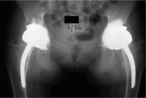
Figure 1B. 5 years later, the left hip was painful and the limb had shortened. Radiographs showed that the left femoral component had subsided and migrated into varus. The right hip was not painful, despite the extensive acetabular periprosthetic osteolysis.
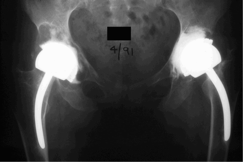
Figure 1C. The left hip was revised using a cementless porous-coated acetabular cup with screws and impacted cancellous allograft bone chips, and a cementless extensively porous-coated femoral component. A sliding trochanteric osteotomy was used for enhanced exposure.
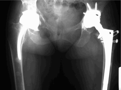
Figure 1D. The right hip was reconstructed in a similar fashion to the left hip at a later time.
All patients received broad-spectrum cephalo-sporins, low-molecular-weight heparin when available, thromboembolic stockings and sequential compression devices. No specific prophylaxis was used to prevent heterotopic ossification. In general, protected weight bearing with walker or crutches was recommended for 6–12 weeks postoperatively because of general weakness and unsteadiness after surgery.
Analysis of clinical outcome
A self-administered questionnaire encompassing the questions for the Harris hip score (Harris Citation1969) was answered by the patient, and clinical and radio-graphic evaluation by the surgeon and the same physiotherapist was undertaken preoperatively and at follow-up visits at 6 weeks, 3 months, 6 months, 12 months, and annually thereafter. The pre-and postoperative range of motion, strength and gait were assessed and documented by the same physiotherapist throughout the study. The Harris hip scores were calculated at the preoperative visit and at the most recent clinical visit.
Analysis of radiographs
Anteroposterior, cross-table and frog lateral radio-graphs were taken before surgery and at 6 weeks, 3 months, 6 months, and 1 year after the operation, and annually thereafter. They were examined for loosening, migration, and other outcome variables outlined below according to established grading systems.
Radiolucent lines around the femoral component were classified into 7 Gruen zones. The change in distance from the center of the head to the lesser trochanter was used to determine the axial subsidence of the stem. In hips with a cemented femoral component, definite loosening of cemented stems was defined as subsidence of more than 5 mm or continuous demarcation around the stem, and probable loosening as subsidence of 2–5 mm or a radiolucent line surrounding 50% of the stem or more, according to Barrack et al. (Citation1992). Non-progressive incomplete radiolucent lines less than 1 mm in width in the bone-cement interface were ignored. Analysis of stability in cementless femoral components was performed using the criteria of Engh and Bobyn (Citation1986) and Engh et al. (Citation1987): bone ingrowth, stable fibrous ingrowth and unstable implants. The femoral component was considered unstable if serial radio-graphs showed a progressive change in the position of the femoral component and/or progressive axial subsidence of more than 5 mm. Heterotopic ossification was graded using the system of Brooker et al. (Citation1973).
Juvenile chronic arthritis patients undergoing revision total hip replacement
The location of radiolucent lines around the acetabular component was classified into 3 Charnley zones (DeLee and Charnley Citation1976). The stability of the acetabular component was assessed by determination of vertical and horizontal migration. The components that had tilted more than 5 degrees or had migration of 2 mm or more, or a continuous radiolucent line more than 2 mm in width, were considered unstable.
In order to avoid interobserver variation, all radio-graphic measurements were performed independently by one observer (K-J O), an experienced orthopedic surgeon who was not involved in the surgical procedures. Ace-tabular and femoral defects were classified according to the grading system of the American Academy of Orthopaedic Surgeons (D'Antonio et al. Citation1989, Haddad et al. Citation2000). The radiographic classification of each case by the observer was confirmed with the surgeon postoperatively after review of the surgical details, the medical record and the radiographs.
Statistics
The clinical and radiographic data were analyzed statistically using SPSS version 10.0 for Windows. For continuous variables, a two-tailed t-test was used for comparison of pre- and postoperative measurements.
Results
Clinical outcome
In JCA patients, the Harris hip score is markedly affected by the systemic nature of the patient’s disease and the multiple joint involvement. Nevertheless, the scores improved from an average of 53 (34–85) points preoperatively to 76 (47–96) points postoperatively after revision surgery (p < 0.001). The changes in range of motion were not statistically significant.
Radiographic results
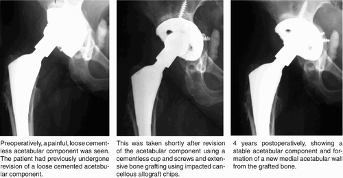
On the acetabular side, 1 cementless cup was radio-graphically loose and had migrated medially. This patient had a perioperative infection which was successfully treated with antibiotics, and a late fall in which an undisplaced periprosthetic acetabular fracture was sustained (see details under complications). The cup was not revised, according to the patient’s wishes. 1 other cementless cup was revised because of loosening associated with substantial polyethylene wear and periprosthetic osteolysis. This cup articulated with a well-fixed non-modular cementless stem with a 32-mm femoral head. All other acetabular reconstructions have been stable ().
Figure 2. Case 9. Revision of a loose acetabular component in a patient with juvenile rheumatoid arthritis.
The 2 fully cemented femoral stems (without allografts) have both become loose, subsided and have been associated with femoral fractures (see details below). Both stems have been revised. The remaining femoral stems are stable.
Heterotopic ossification was noted in 6 hips (grade I in 4 hips, grade II in 1, and grade III in 1).
Complications
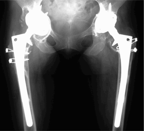
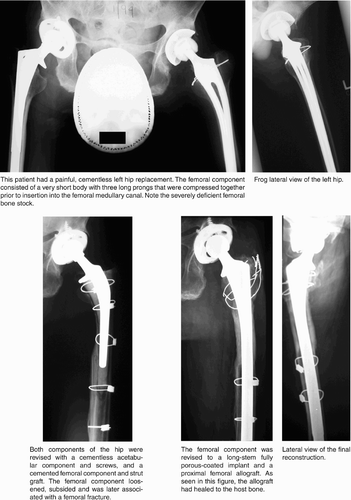
2 hips with revision cemented femoral stems loosened, subsided and were associated with extensive osteolysis. These cases subsequently sustained peri-prosthetic femoral fractures (1 minor nondisplaced cortical crack and 1 major displaced fracture). The first case (case 3 in the Table) was revised successfully using a cementless long-stem prosthesis with strut graft. The second case (1) was revised using a proximal femoral allograft and cementless long-stem prosthesis, cemented into the allograft portion only (). Both re-revisions are doing well (1 and 5 years postoperatively).
Figure 3. Case 1. Painful total hip replacement in a patient with ankylosing spondylitis.
1 patient (no. 8) underwent complex revision of the acetabulum with a reconstruction ring and bone graft. The sciatic nerve abutted the posterior aspect of the ring because of a large uncontained defect of the posterior column. A neurolysis was performed intraoperatively and the nerve was mobilized; however, a peroneal nerve palsy was diagnosed postoperatively, and has not improved.
1 patient (2A) with acetabular revision with cancellous autograft and allograft and a cementless cup had a deep infection with Pseudomonas aeruginosa postoperatively. The hip was debrided after 1 month and the patient was given systemic antibiotics. 8 years later, the patient fell and sustained a nondisplaced periprosthetic fracture of the medial wall of the acetabulum. This was treated nonoperatively with protected weight bearing for 3 months. The acetabular component has migrated several mm medially and has a complete circumferential radiolucent line, but the hip is stable and the patient does not want further surgery.
1 patient (10A) had acetabular loosening due to severe polyethylene wear and periprosthetic osteolysis, and has recently undergone revision with a structural bone graft, roof ring and cemented cup. It is noteworthy that his primary hip had a non-modular implant with a 32-mm femoral head.
1 patient (6) with 2 well-functioning revision THAs died many years after surgery, due to progression of her JRA to a lupus variant with multi-system organ failure. 1 hip became infected with several organisms and was excised 4.5 years post-operatively.
There were no dislocations or deep-vein thromboses.
Discussion
In young patients, THA will not usually last a lifetime. Although JCA patients are frequently lighter in weight and less active than those with osteoarthritis, initial prosthetic fixation is difficult because of abnormal anatomical development of the hip, small-proportioned more fragile bones with narrow medullary canals and thinner cortices, and limited sizes of implants (Scott et al. Citation1984, Lachiewicz et al. Citation1986, Witt et al. Citation1991, Maric and Haynes Citation1993, Torchia et al. Citation1996, Chmell et al. Citation1997, Lehtimaki et al. Citation1997, Haber and Goodman Citation1998, Kumar and Swann Citation1998). Revision surgeries in younger patients with chronic arthritis are even more complicated because of the additional loss of bone stock. As a result of continued prosthetic wear, osteolysis, and implant failure, increasing numbers of young patients with JCA will require complicated revision surgeries.
To date, there have been no studies which examine the outcome of revision total hip replacement in young patients with chronic arthritis. In this regard, our current series of 17 revisions in 11 patients with an average of seven years of follow-up, though small in number, is unique.
Observations from our series have resulted in a change in our surgical technique for future reconstructions. Firstly, as for primary total hip replacement in patients with chronic arthritis and small anatomic proportions, we no longer cement revision femoral stems. In both primary and revision cases, it is difficult to centralize the prosthesis and obtain an adequate cement mantle circumferentially; subsequent failure of the cemented femoral stem is accompanied by cement fracture, subsidence and osteolysis, further jeopardizing bone stock. Although cementless primary and revision stems are technically challenging, gradual reaming with smallersized reamers, the use of longer modular cementless stems, the taking of intraoperative radiographs to con rm adequate prosthetic fit, and the application of strut grafts in recent cases have provided a sound reconstruction and helped restore lost bone stock.
Secondly, on the acetabular side, excessive ante-version, hypoplasia and severe loss of bone stock often require structural and morselized bone grafts for bone defects. We prefer to use cementless cups in primary and revision cases of younger patients with chronic arthritis. However, if one cannot easily place the cup in the correct orientation on at least 70% firm host bone and obtain a satisfactory peripheral rim fit and adequate screw fixation, we use a roof ring or reconstruction cage. These devices protect the healing bone graft, come in numerous small sizes with more screw holes than modular acetabular shells, and allow subsequent cementing of a polyethylene liner in the desired degree of anteversion. Because of the patient’s young age and small pelvis, the use of a roof ring rather than reconstruction ring is usually preferred, because the ischium is often small and adequate purchase of the inferior flange is difficult to obtain.
Thirdly, because of small pelvic proportions, excessive acetabular anteversion and bone loss, the sciatic nerve is vulnerable to injury—especially in cases in which a structural graft or ring is used posteriorly. We identify the location of the nerve by palpation during exposure and reconstruction; in addition, retractors are positioned to avoid impingement of the sciatic nerve.
Finally, despite preoperative planning, changes in the surgical plan are often made during the reconstructions. The surgeon must have access to a variety of small-sized reamers, implants and bone graft options to obtain a stable articulation.
Author contributions
All authors participated in gathering and analyzing the data. In addition, the radiographs were asssessed by K-JO. All range of motion measurements were done by SI.
- Barrack R L, Mulroy R D., Jr, Harris W H. Improved cementing techniques and femoral component loosening in young patients with hip arthroplasty. A 12-year radio-graphic review. J Bone Joint Surg (Br) 1992; 74: 385–9
- Brooker A F, Bowerman J W, Robinson R A, Riley L H., Jr. Ectopic ossification following total hip replacement. Incidence and a method of classification. J Bone Joint Surg (Am) 1973; 55: 1629–32
- Chmell M J, Scott R D, Thomas W H, Sledge C B. Total hip arthroplasty with cement for juvenile rheumatoid arthritis. Results at a minimum of ten years in patients less than thirty years old. J Bone Joint Surg (Am) 1997; 79: 44–52
- D'Antonio J A, Capello W N, Borden L S, Bargar W L, Bierbaum B F, Boettcher W G, Steinberg M E, Stulberg S D, Wedge J H. Classification and management of acetabular abnormalities in total hip arthroplasty. Clin Orthop 1989, 243: 126–37
- DeLee J G, Charnley J. Radiological demarcation of cemented sockets in total hip replacement. Clin Orthop 1976, 121: 20–32
- Engh C A, Bobyn J D. Principles, techniques, results, and complications with a porous-coated sintered metal system. Instr Course Lect 1986; 35: 169–83
- Engh C A, Bobyn J D, Glassman A H. Porous-coated hip replacement. The factors governing bone ingrowth, stress shielding, and clinical results. J Bone Joint Surg (Br) 1987; 6945–55
- Haber D, Goodman S B. Total hip arthroplasty in juvenile chronic arthritis: a consecutive series. J Arthroplasty 1998; 13: 259–65
- Haddad F S, Masri B A, Garbuz D S, Duncan C P. Femoral bone loss in total hip arthroplasty: classification and pre-operative planning. Instr Course Lect. 2000; 49: 83–96
- Harris W H. Traumatic arthritis of the hip after dislocation and acetabular fractures: treatment by mold arthroplasty. An end-result study using a new method of result evaluation. J Bone Joint Surg (Am) 1969; 51: 737–55
- Kumar M N, Swann M. Uncemented total hip arthroplasty in young patients with juvenile chronic arthritis. Ann R Coll Surg Engl 1998; 80: 203–9
- Lachiewicz P F, McCaskill B, Inglis A, Ranawat C S, Rosenstein B D. Total hip arthroplasty in juvenile rheumatoid arthritis. Two to eleven-year results. J Bone Joint Surg (Am) 1986; 68: 502–8
- Lehtimaki M Y, Lehto M U, Kautiainen H, Savolainen H A, Hamalainen M M. Survivorship of the Charnley total hip arthroplasty in juvenile chronic arthritis. A follow-up of 186 cases for 22 years. J Bone Joint Surg (Br) 1997; 79: 792–5
- Maric Z, Haynes R J. Total hip arthroplasty in juvenile rheumatoid arthritis. Clin Orthop 1993, 290 197–9
- Rahimtoola Z O, Finger S, Imrie S, Goodman S B. Outcome of total hip arthroplasty in small-proportioned patients. J Arthroplasty 2000; 15: 27–34
- Ruddlesdin C, Ansell B M, Arden G P, Swann M. Total hip replacement in children with juvenile chronic arthritis. J Bone Joint Surg (Br) 1986; 68: 218–22
- Scott R D, Sarokhan A J, Dalziel R. Total hip and total knee arthroplasty in juvenile rheumatoid arthritis. Clin Orthop 1984, 182: 90–8
- Torchia M E, Klassen R A, Bianco A J. Total hip arthroplasty with cement in patients less than twenty years old Long term results. J Bone Joint Surg (Am) 1996; 78: 995–1003
- Witt J D, Swann M, Ansell B M. Total hip replacement for juvenile chronic arthritis. J Bone Joint Surg (Br) 1991; 73: 770–3