Abstract
Background There are many modalities of treatment for complex lower extremity deformity in hypophosphatemic rickets. We evaluated the outcomes of deformity correction using an external fixation and/or intramedullary nailing in hypophosphatemic rickets
Patients and methods 55 segmental deformities (20 femora, 35 tibiae) from 20 patients were examined retrospectively. There were 9 children and 11 adults. Distraction osteogenesis was used in 28 segments and acute deformity correction in 27. External fixation was applied in 24 segments, intramedullary nailing in 6, and external fixation and intramedullary nailing in 25.
Results There were 18 major and 13 minor complications in 26 of 28 segments with distraction osteogenesis, and 13 major and 10 minor complications in 19 of 27 segments with acute correction. Recurrent deformity or refracture occurred in 10 of 21 segments with distraction osteogenesis by external fixation only, 4 of 6 with acute correction by intramedullary nailing, and 1 of 25 with distraction osteogenesis or acute correction by external fixation and intramedullary nailing. Nail-related complications occurred in 3 of 6 with intramedullary nailing and 2 of 25 with external fixation and intramedullary nailing.
Interpretation External fixation and intramedullary nailing can be recommended to prevent complications during or after deformity correction in hypophosphatemic rickets.
Hypophosphatemic rickets involves complex deformities of the lower extremities, including coxa vara, genu varum or valgum, bowing of the femur or tibia, and torsional deformity of the tibia. External fixation or intramedullary nailing has been used for deformity correction in rickets. We compared the results and complications between external fixation only, intramedullary nailing only, and a combination of intramedullary nailing with external fixation for the treatment of deformities in rickets.
Patients and methods
Between 1991 and 2001, 20 patients (10 women) underwent deformity correction for hypophosphatemic rickets (20 femora, 35 tibiae) using an external fixation and/or intramedullary nailing. The average age at surgery was 20 (8–41) years. There were 18 segments in 9 children and 37 in 11 adults. 4 patients (2 femora, 6 tibiae) were referred from other hospitals for correction of recurrent deformity after prior treatment. 6 patients had a family history of rickets. Preoperatively, 19 patients had lower serum phosphate levels with a mean of 2.0 (1.4–2.8 mg/dL; normal range 4.0–7.0 mg/dL). Only 1 patient had a normal serum phosphate level (4.9 mg/dL) because of good compliance to medical treatment previously.
The deformities included 11 cases of genu varum, 7 cases of genu valgum, and 2 cases of windswept deformity. 5 patients had a unilateral leg deformity or a deformity in one femur only. The axial deformities of the bone segments were defined by the CORA (center of rotation of angulation) method and malalignment test (Paley et al. Citation1994). The maximum angulation in the oblique plane deformities was calculated using the magnitude of angulation in the anteroposterior and lateral views. The rotational deformities of the tibia and femur were assessed using a computed tomography (CT) scan (Murphy et al. Citation1987, Eckhoff and Johnson Citation1994).
Corrective osteotomies were performed at the single, double, or triple CORA levels. The Ilizarov ring fixator (U & I, Korea) was used for gradual or acute correction in all tibial deformities. A hybrid fixator with a distal ring and proximal arch was used for gradual deformity correction of the femur, and a lateral monofixator (Medixalign, Korea) was used for acute correction of the femoral deformity. Gradual correction was performed mainly in the skeletally immature patients, which was aimed at increasing the bone length, and in patients with severe deformities. This procedure was performed to avoid the resection of large amounts of bone that would have been necessary for deformity correction.
Among 55 segments, distraction osteogenesis was used in 28 segments and acute deformity correction in 27 (). External fixation was applied in 24 segments, intramedullary nailing in 6, and external fixation and intramedullary nailing in 25.
Table 1. Data concerning the 20 patients who underwent deformity correction of 55 segments
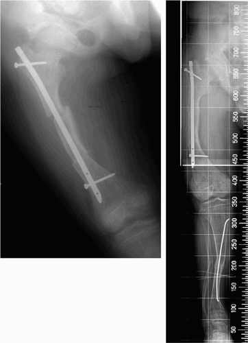
The distraction group was subdivided into 3 methods including only external fixation (method 1), external fixation and secondary late nailing (method 2), and external fixation over an intramedullary nail (method 3). The acute deformity correction group was further divided into 3 methods including only external fixation (method 4), external fixation and intramedullary nailing (method 5), and intramedullary nailing only (method 6). 18 segments in children underwent method 1 (n = 12), method 5 (n = 2), and method 6 (n = 4). 37 segments in adults underwent method 1 (n = 9), method 2 (n = 6), method 3 (n = 1), method 4 (n = 3), method 5 (n = 16), and method 6 (n = 2). Flexible intramedullary nailing was used in 26 segments and rigid intramedullary nailing was used in 5 (). Rigid interlocking nailings were used in 2 femora of method 6 and 1 tibia of method 3. Rigid nailing without interlocking was used in 1 femur and 1 tibia of method 5. Immobilization by cast or brace was used in patients with flexible or rigid, without interlocking, nailing.
Figure 1. Acute deformity correction after double-level osteotomy of the femur with an intramedullary nail (left).There was no mechanical axis deviation of the whole extremity, but the varus deformity was starting at the metaphysis of the distal femur (right).
The tibial frame in methods 1, 2, 3, and 5 consisted of 3 full rings at the proximal, middle, and distal tibia. In methods 1 and 2, the proximal ring was fixed with 2 wires and 1 half-pin and the middle ring with 1 wire and 1 half-pin. At the distal ring, 1 wire and 1 half-pin were placed for a single-level osteotomy at the proximal tibia whereas 2 wires and a 1 half-pin were placed for a double-level osteotomy at the proximal and distal tibia (). In method 5 with a flexible intramedullary nail, the proximal and distal rings were fixed with 2 wire after double-level osteotomy (). The hybrid femoral frame in methods 1, 2, and 4 was composed of 2 rings and 1 arch for a single-level osteotomy at the distal femur (). 3 half-pins were placed at the femoral arch and the proximal ring and 2 half-pins with 1 wire at the distal ring. In method 5, 2 or 3 half-pins were fixed at the most proximal and distal femoral fragments after a double- or triple-level osteotomy, and the middle segments were not fixed by pin or wire. In method 3, with a rigid intramedullary nail, the proximal and distal rings were fixed with 3 wires and the middle ring was not fixed with wire or pin (). The proximal and distal tibiofibulae were trans-fixed with 1 wire in all methods.
Figure 2. Gradual correction of varus deformity after double-level osteotomy of the tibia with an external fixator (left). The varus deformity was corrected successfully (right).
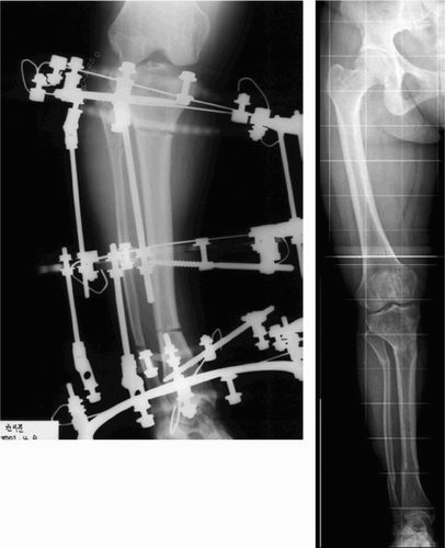
Figure 3. Acute deformity correction after triple-level osteotomies of the femur and double-level osteotomies of the tibia with an external fixator and intramedullary nail (left). After removal of external fixator, showing normal alignment of the mechanical axis of the leg (right).
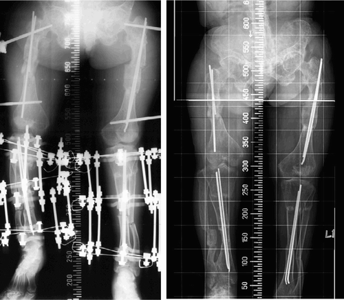
Figure 4. Gradual deformity correction after a single osteotomy at the distal femur and the proximal tibia with external fixator (left). Secondary late intramedullary nailing after a deformity correction (right).
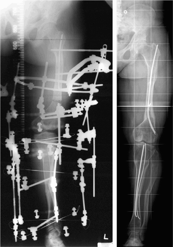
Figure 5. Distraction osteogenesis with an external fixator over an intramedullary nail (right). After removal of external fixator and intramedullary nail, showing normal alignment of the mechanical axis of the leg (right).
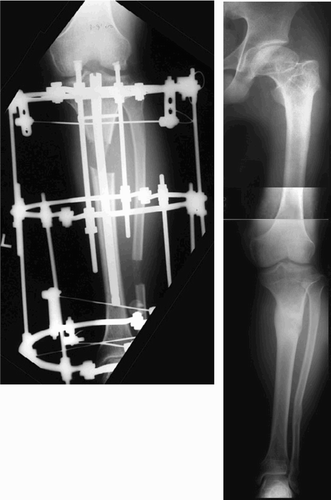
The distraction was started 7 days and 14 days after the osteotomy in children and adults, respectively. The distraction rates were 1 mm/day in children and 0.75 mm/day in adults with a single-level osteotomy at the distal femur or the proximal tibia. After the double-level osteotomy of the tibia, the distraction rates of the proximal tibia and the distal tibia were 1 mm/day and 0.5 mm/day in children and 0.5 mm/day and 0.5 mm/day in adults. The distraction rate was controlled according to the degree of callus formation. The healing time (months with external fixator) was used in methods without lengthening, and lengthening index (months with external fixator/cm lengthening) was used in methods with distraction osteogenesis. The external fixator was removed when there was callus formation at the 3 cortices of the distraction site in the gradual correction, or at the osteotomy site in the acute correction on radiographs in 2 planes (Kristiansen and Steen Citation1999).
Results
All osteotomies healed. The means of the preoperative angular deformity, the lateral tibial torsion, and the anteversion of the femoral neck were 22°, −12° and 7°, respectively, in the external fixation (methods 1 and 4), 33°, −10°, and 0° in the intramedullary nailing (method 6), and 35°, −10° and 9° in the external fixation and intramedullary nailing (methods 2, 3 and 5). The mean values for the corrected angulation and rotation were 20° and 20°, respectively, in the external fixation, 25° and 17° in the intramedullary nailing, and 30° and 20° in the external fixation with intramedullary nailing.
Of the 20 patients, 19 experienced an improvement in their gait after correction of the deformity. Of these 19 patients, 14 independent ambulators had improved walking with minimal or no pain compared to preoperative status. 4 crutch-dependent ambulators became independent ambulators, and 1 wheelchair user became a crutch walker. 1 child showed a deterioration in ambulation from an independent ambulator to a crutch walker, due to recurrent deformity of both tibiae. This patient died from complications during the treatment of pulmonary tuberculosis before the recurrent deformity could be corrected. The preoperative pain-free walking distances in independent ambulators were less than 0.1 km in 4 patients, 0.5 km in 3, and 1 km in 7. During the follow-up after surgery, 7 patients could walk long distances without pain. 4 patients could walk 1–2 km without pain and 3 patients had a pain-free walking distance of 0.5 km. 4 crutch walkers had a pain-free walking distance of 0.5 km without crutches after surgery. 1 wheelchair user could walk 1–2 km with one crutch after surgery.
There were 18 major complications and 13 minor complications in 26 of 28 segments with distraction osteogenesis, and 13 major and 10 minor complications in 19 of 27 segments with acute correction (). Refracture or recurrent deformity (n = 15) and nail-related problems (n = 5) were common major complications. Recurrent deformity or refracture occurred in 10 of 21 segments with method 1, in 4 of 6 segments with method 6, and in 1 of 18 segments with method 5. All the recurrent deformities occurred in the children. 10 of 12 segments with method 1 in the children had a recurrent deformity at the metaphysis and diaphysis, but 5 of 6 segments with method 5 and 6 had a recurrence only at the metaphysis because the nailing specifically prevents a recurrent deformity at the diaphysis. Nail-related problems occurred in 3 of 6 segments with method 6 and in 2 of 25 segments with methods 2, 3, and 5. There was migration of the nail in 3 segments, loss of stability due to loosening of interlocking screws in 1, and deep intramedullary infection in 1. A deep intramedullary infection was the most serious complication in the nailing group. 1 patient suffered this complication during tibial lengthening over the nail using the Ilizarov fixator. The distraction was discontinued after 3.5 cm of lengthening, the nail was removed, and the Ilizarov fixator was retained. Radical debridment and insertion of cement beads mixed with antibiotics was performed. Fortunately, callus formation had taken place at the posterior cortex of the lengthening site prior to onset of this complication. After control of the infection, the cement beads were removed and a cancellous bone grafting was done. Bone union was eventually obtained.
Table 2. Complications and treatments
Flexion of the knee was limited after correction of a severe valgus deformity in 3 segments (of 2 patients) and a quadricepsplasty had to be performed. This treatment included the release of the rectus femoris from the vastus lateralis and medialis, the release of the medial and lateral retinaticulum from the patella, and a reattachment of the vastus medialis to the lateral border of the patella. These segments had lateral subluxation of the patella prior to surgery, and the patellae were laterally dislocated after the correction due to tightness of the patellar retinaticulum, vastus lateralis, and iliotibial band.
1 patient had a persistant residual leg length discrepancy of 5 cm after a closing wedge osteotomy had been performed on both femora. Preoperatively, she had a severe flexion deformity of 100° on the right hip, with severe anterior bowing of the femur and tibia on the same side. This patient required a significant bone resection of the femur and tibia for acute correction and in order to avoid stretching of the femoral nerve. Her daughter also had rickets with a severe valgus deformity of both knees, but was treated with a gradual correction by external fixation and secondary late nailing. Like her mother, this patient also underwent a quadricepsplasty for the lateral dislocation of both patellae.
Discussion
The use of intramedullary nailing after an osteotomy for correction of a deformity has been advocated in order to prevent a progressive recurrent deformity and a refracture in the osteogenesis imperfecta (Sofield and Millar Citation1959), and in limb lengthening (Lin et al. Citation1996, Paley et al. Citation1997). There have been very few reports on the results of intramedullary nailing in rickets (Evans et al. Citation1980, Ferris et al. Citation1991, Eyres et al. Citation1993). However, these reports have emphasized that it is more beneficial to perform a metaphyseal osteotomy near maturity—in order to prevent the recurrence that often accompanies growth. Our study has also demonstrated that the intramedullary nailing method can prevent a progressive recurrent deformity at the diaphysis, but it cannot control the progression of the deformity at the metaphysis during growth in children due to a lack of metaphyseal fixation. The use of telescopic rods can be a treatment option because the drilling of the physis with a small-diameter drill bit may not disturb the growth of the physis (Bailey and Dubow Citation1981), and the placement of telescopic rods at the epiphysis and the metaphysis can prevent a recurrent deformity at the metaphysis and the physis. This method makes it difficult to maintain the bone length, and has limited deformity correction when there is a severe complex deformity because it requires resection of a large amount of bone. The use of intramedullary nailing only after a multiple osteotomy cannot allow early ambulation or joint motion because of the limited stability. Our study has also demonstrated that there is a high risk of complications associated with instability when using this method.
The use of external fixation for correction of a deformity has several advantages over other methods (Kanel and Price Citation1995, Choi et al. Citation2002). This method allows early ambulation and possible correction of multiplanar complex deformities with less neurovascular injuries. There are disadvantages, however, including delayed consolidation and a higher risk of recurrent deformity or a refracture at the lengthening site after removal of the external fixator. In our study, distraction osteogenesis only by external fixation in children had a high risk of recurrent deformity or refracture compared to other methods. Thus, intramedullary nailing during or after distraction osteogenesis is recommended in children in order to avoid complications.
External fixation combined with the nailing method can overcome the disadvantages of the treatments using external fixation or intramedullary nailing only. This method allows the early removal of the external fixation immediately upon callus formation, which provides rotational stability. In our study, increased stability with this method could mean less nail-related problems often associated with loss of stability, such as migration of the flexible nail and loosening of interlocking screws of the rigid nail, compared to other methods. Furthermore, the shortened external fixation time might reduce the incidence of a pin-tract infection and prevent joint stiffness. In our study, some patients had knee or ankle stiffness with this method; however, the knee stiffness was not related to the method because the patella subluxation associated with the severe genu valgus was already present before surgery, and the knee stiffness was inevitable after the correction of genu valgus. The postoperative equinus contracture in our study was related to distraction osteogenesis for correction of the severe anterior bowing of the tibia, which was expected before surgery. The use of a foot frame can avoid an equinus contracture, and no Achilles tendon lengthening is necessary.
Previous studies (Kristiansen and Steen Citation1999, Song et al. Citation2005) and our study have demonstrated that the most serious complication of lengthening over an intramedullary nail is deep intramedullary infection requiring the removal of the intramedullary nail and additional surgery—such as a radical debridment or bone grafting. A deep intramedullary infection can be avoided by preserving the space between the nail and the pin-tract (Herzenberg and Paley Citation1997, Paley et al. Citation1997). In addition, aggressive treatment of a pin-tract infection (which might contribute to a deep intramedullary infection) is necessary. Thus, before any of the methods described above are performed, all the patients should be informed of the possibility of developing a deep intramedullary infection as well as the possibility of requiring additional surgery to correct the complications associated with these procedures.
No competing interests declared.
Author contributions
HRS guarantor of integrity of the study, study concepts and design. HRS, SR, SK literature research, SHL data analysis. SWS, JRK, JSH manuscript preparation and editing.
- Bailey R W, Dubow H I. Evolution of the concept of an extensible nail accommodating to normal longitudinal bone growth: Clinical considerations and implications. Clin Orthop 1981, 159: 157–70
- Choi I H, Kim J K, Chung C Y, Cho T J, Lee S H, Suh S W, Whang K S, Park H W, Song K S. Deformity correction of knee and leg lengthening by Ilizarov method in hypophosphatemic rickets: Outcomes and significance of serum phosphate level. J Pediatr Orthop 2002; 22: 626–31
- Eckhoff D G, Johnson K K. Three-dimensional computed tomography reconstruction of tibial torsion. Clin Orthop 1994, 302: 42–6
- Evans G A, Arulanantham K, Gage J R. Primary hypophos-phatemic rickets. J Bone Joint Surg (Am) 1980; 62: 1130–8
- Eyres K S, Brown J, Douglas D L. Osteotomy and intramedullary nailing for the correction of progressive deformity in vitamin D-resistant hypophosphatemic rickets. J R Coll Surg Edinb 1993; 38: 50–4
- Ferris B, Walker C, Jackson A, Kirwan E. The orthopaedic management of hypophosphataemic rickets. J Pediatr Orthop 1991; 11: 367–73
- Herzenberg J E, Paley D. Tibial lengthening over nail(LON). Techniques in Orthopaedics 1997; 12: 250–9
- Kanel J S, Price C T. Unilateral external fixation for corrective osteotomies in patients with hypophosphatemic rickets. J Pediatr Orthop 1995; 15: 232–5
- Kristiansen L P, Steen H. Lengthening of the tibia over an intramedullary nail, using the Ilizarov external fixator. Major complications and slow consolidation in 9 lengthenings. Acta Orthop Scand 1999; 70: 271–4
- Lin C C, Huang S C, Liu T K, Chapman M W. Limb lengthening over an intramedullary nail. An animal study and clinical report. Clin Orthop 1996, 330: 208–16
- Murphy S B, Simon S R, Kijewski P K, Wilkinson R H, Griscom N T. Femoral anteversion. J Bone Joint Surg (Am) 1987; 69: 1169–76
- Paley D, Herzenberg J E, Tetsworth K, McKie J, Bhave A. Deformity planning for frontal and sagittal plane corrective osteotomies. Orthop Clin North Am 1994; 25: 425–65
- Paley D, Herzenberg J E, Paremain G, Bhave A. Femoral lengthening over an intramedullary nail. J Bone Joint Surg (Am) 1997; 79: 1464–80
- Sofield H A, Millar G A. Fragmentation, realignment and inramedullary rod fixation of deformities of long bones in children. J Bone Joint Surg (Am) 1959; 41: 1371–91
- Song H-R, Oh C-W, Mattoo R, Park B-C, Kim S-J, Park I-H, Jeon I-H, Ihn J C. Femoral lengthening over an intramedullary nail using the external fixator. Risk of infection and knee problems in 22 patients with a follow-up of 2 years or more. Acta Orthop 2005; 76: 245–52