Abstract
Background and purpose Increased intradiscal pressure and relative segmental hypermobility are in vitro observations supporting the idea of increased postoperative load being a reason for progressive degeneration of the free mobile segment adjacent to a lumbar fusion. These mechanisms have been difficult to confirm in clinical studies, and an alternative theory claims instead that the adjacent segment degeneration follows a natural degenerative course in patients who are predisposed. We examined 9 patients 5 years after lumbar fusion, to assess whether relative hypermobility of the segment adjacent to fusion could be correlated to progressive degeneration of the same segment.
Patients and methods The 9 patients, all of whom had been treated with a lumbar fusion after a preoperative intervertebral mobility assessment by spinal RSA, were re-examined 5 years after surgery. The intervertebral translations of the vertebra proximal to the fusion were determined by RSA and compared to the mobility of the same lumbar segment before fusion. The disc height and any progressive reduction at the two levels proximal to the one fused were measured on conventional radiographs.
Results Adjacent segment mobility 5 years after fusion—expressed as mean transverse, vertical, and sagittal translation of the vertebra proximal to fusion— was not significantly changed compared to the mobility measured before surgery. Increased mobility of the segment seen in 5 individual patients was not associated with progressive degeneration of the same segment or to a poor clinical outcome.
Interpretation Hypermobility of the segment adjacent to fusion is not a general finding. Increased mobility that can be seen in certain individuals does not impair the 5-year result. The significance of mechanical alterations in adjacent segment degeneration is uncertain, and it is possibly overestimated.
Progressive degeneration of the free mobile segment next to a spinal fusion is referred to as adjacent segment disease and includes disc degeneration, facet joint hypertrophy, spinal stenosis, and even aquired spondylolysis (Unander-Scharin Citation1951, Lee Citation1988). For mechanical reasons, this development is often considered a late complication (or sequelae) of the spinal fusion. In vitro investigations have suggested that fusion increases the intradiscal pressure of the adjacent segment (Weinhoffer et al. Citation1995) and also that a situation of relative hypermobility is induced (Lee and Langrana Citation1984). Both observations would mean increased load on the segment. The same observations are often referred to by spinal surgeons when discussing the relative merits of disc arthroplasty and fusion when treating patients with degenerative low back pain (Andersson and Rouleau Citation2004, Freeman and Davenport Citation2006). Maintaining mobility of the segment to treat would theoretically unload the adjacent parts of the spine, if the in vitro observations are applicable to the in vivo situation. The biomechanical arguments have, however, been difficult to confirm in clinical studies, and there is another theory that claims that degeneration of the adjacent segment rather reflects the natural degenerative course for the individual patient (Penta et al. Citation1995, Wai et al. Citation2006).
Intervertebral mobility (in mm) and grade of degeneration at the segment adjacent to fusion both before and 5 years after surgery
Radiostereometric analysis (RSA) (Selvik Citation1989) has previously been used to study intervertebral mobility over time in the early postoperative period after lumbosacral fusion (Axelsson et al. Citation1997). A transformation of mobility from the level fused to the adjacent L4-L5 level was verified in 2 patients, but this was not a general finding in the 6 patients examined. The aim of the current radiostereometric study was to assess the degree of hypermobility of the adjacent level at long-term follow-up and whether progressive disc degeneration or poor clinical outcome can be correlated to such kinematics.
Patients and methods
Patients
9 patients, all of whom had been treated with a lumbar fusion after a preoperative spinal RSA, were identified for radiostereometric, radiographic, and clinical reassessment 5 years after surgery. In order to study the effect(s) of fusion on the adjacent segment, only patients who were considered to have a solid lumbar fusion from radiographs taken 1 year after surgery were included. The group included 6 women and 3 men with a mean age of 45 (35–59) years at surgery. The diagnosis was painful degenerative disc disease at level L4-L5 and/or L5-S1. Before fusion, all patients had had lumbar pain without sciatica with a mean duration of 4 (1–15) years. No patients had undergone spinal surgery before the fusion procedure, but all had tried nonoperative treatment without success.
Surgery
A preoperative external pedicular fixation test leading to pain relief, supporting treatment by fusion, had been performed in all patients (Magerl Citation1984, Olerud et al. Citation1986, Elmans et al. Citation2005). Fusion was achieved by 3 techniques . 3 patients had a posterolateral fusion without instrumentation. The same surgical procedure but with a supplementary stabilization using posterior instrumentation was performed in 3 patients. The remaining 3 patients had an anterior lumbar interbody fusion with 2 threaded, cylindrical cages as a stand-alone procedure via a retroperitoneal approach.
Radiostereometry
A spinal RSA (Axelsson et al. Citation2006) was performed in all patients 2 months after finishing the external fixation test, but prior to the definitive fusion procedure. Tantalum indicators used for the RSA had been implanted by percutaneous technique in connection with the application of the pedicle screws for test fixation. The 0.8-mm indicators were placed into the bases of the tranverse processes and into the tip of the spinous process of the 3 most distal lumbar vertebrae. The lateral masses and the central crest were used in the sacrum. The implantation of tantalum indicators has been approved by the ethics committee of the Medical Faculty, Lund University.
The RSA procedures before fusion and at the 5-year follow-up were identical. Two 40-degree angulated roentgen tubes were used to provide exposures on 2 separate films. The lumbosacral spine and a combined reference plate and calibration device with tantalum indicators at known positions in front of the film plane were exposed simultaneously (Axelsson et al. Citation2006). Using the Kinema program for computed data processing according to Selvik (Citation1989), the intervertebral translations along the transverse (x-), vertical (y-) and sagittal (z-) axes were calculated. Each patient was examined in 2 standardized positions, supine and sitting, in order to avoid active movements of the spine—thus reducing the confounding effects caused by varying pain level and muscular spasm (Axelsson and Karlsson Citation2005). The sitting position in this context means semiflexed hips to allow Xrays to pass above the femoral shafts to the lumbosacral spine. The translatory movements induced by the patient changing from supine to this standardized sitting position were calculated for each vertebra relative to the adjacent, distal vertebra as a measure of the intervertebral mobility.
According to previous studies with double examinations of healed fusions (Johnsson et al. Citation2002), the minimum significant measurement with this RSA set-up is 0.5, 0.5, and 0.7 mm for the 3 axes: transverse, vertical, and sagittal. Translation values below these accuracy levels were considered insignificant.
Radiography
All patients were examined by conventional radiography (anteroposterior and lateral views) before surgery. The 5-year follow-up included an identical radiographic examination. Disc status preoperatively and 5 years after surgery was assessed for the 2 spinal levels adjacent and proximal to the fusion, using a classification in 4 groups with the following characteristics: normal disc height (I), disc height decreased by less than 50% (II), disc height decreased by at least 50% (III), and disc height obliterated (IV) (Saraste et al. Citation1985).
Clinical evaluation
At the 5-year follow-up, the overall clinical outcome was assessed by the patients and graded into one of three categories: good (minor or no residual pain), fair (some pain relief but residual pain), or poor (unchanged or worse compared to the situation preoperatively).
Statistics
For the statistical analysis, the Wilcoxon signedrank test was used to compare the intervertebral translations before fusion and at 5-year follow-up. The Chi-squared test was used to study the mobility effects on adjacent segment postoperatively, related to the clinical outcome (expressed as improvement or no improvement).
Results
Fusion healing with stabilization at the levels intended was confirmed in all patients by the disappearance of the intervertebral translations at radiostereometry 5 years after surgery.
The mean transverse, vertical, and sagittal translation 5 years after surgery at the proximal segment, adjacent to fusion, was 0.6 mm, 1.4 mm, and 2.3 mm. The corresponding values for the same segment before surgery were 0.4 mm, 1.7 mm, and 1.8 mm. There was no significant difference between pre- and postoperative translation along any of the 3 axes (Wilcoxon signed-rank test). Relative hypermobility was seen in individual cases at the segment adjacent to fusion, with no correlation to the type of surgical procedure used .
Preoperatively, 7 patients had no reduction of disc height at the level proximal and adjacent to the level to be fused . 2 patients were eclassified as category II, with the disc height being reduced by less than 50%. 5 years after fusion, their disc status was unchanged with no sign of progressive degeneration according to plain radiography.
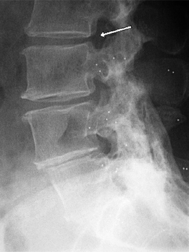
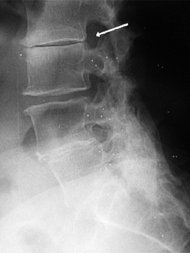
For the disc level adjacent to the adjacent segment, no reduction of disc height was seen in preoperative radiographs. 5 years after fusion, progressive disc degeneration was verified in 1 patient (case 1; see ) with the disc height almost obliterated . For the remaining 8 patients, the disc status at this level remained unchanged.
The Case 1, 3 years after fusion of the L4–S1 level. The patient had relapsing symptoms, but at that time no degeneration of the L2–L3 segment proximal to the segment adjacent to the fusion. Tantalum indicators can be seen placed in L3 and distally.

The same patient 5 years after fusion, with progressive degeneration of the L2–L3 level, where the disc is almost obliterated.

Clinical outcome was good in 5 patients, fair in 3 patients, and poor in 1 patient. Increased postoperative translations of the adjacent segment seen in 5 patients () could not be correlated to the clinical outcome (expressed as improvement or no improvement) (Chi-squared test).
Discussion
Adjacent segment disease is considered to be a potential long-term complication of spinal fusion. The condition includes disc degeneration, facet joint hypertrophy, and spinal stenosis with or without olisthetic deformity for the free mobile segment adjacent to fusion (Unander-Scharin Citation1951, Lee Citation1988). The reported incidence of symptomatic adjacent segment disease has ranged from 5 to 20% in studies with varying follow-up time and with different techniques to achieve fusion (Park et al. Citation2004). Surgical treatment includes decompression and extended fusion, but the results are modest and the treatment must be given with correspondingly moderate patient expectations (Whitecloud et al. Citation1994, Phillips et al. Citation2000, Chen et al. Citation2001).
The etiology of the degeneration adjacent to fusion has not been clarified. Two major mechanisms are ascribed varying importance (Park et al. Citation2004). One theory favors a mechanical explanation, which has been supported by the results of in vitro studies indicating that there is increased stress to the adjacent segment after fusion. The intradiscal pressure increases (Weinhoffer et al. Citation1995) and a situation of relative hypermobility is induced by shifting the center of rotation in flexion to a more proximal level (Lee and Langrana Citation1984). These arguments are often used to justify disc replacement instead of fusion, with the intention of preserving mobility of the segment to treat in order to unload the adjacent part of the spine. The second theory, on the other hand, claims that the findings for the adjacent segment instead reflect the natural progressive degenerative course of the ageing disc—with no correlation to the mechanics (Penta et al. Citation1995, Wai et al. Citation2006).
Radiostereometry allows a more refined possibility to measure spinal kinematics in vivo. The method has been used to study the adjacent segment during the early postoperative course after fusion at the L5-S1 level (Axelsson et al. Citation1997). The result from cadaveric studies, with relative hypermobility of the adjacent segment, has thus been confirmed with the transfer of lumbosacral mobility to the L4- L5 level. This result was, however, found in only 2 of 6 patients examined. According to the current study, this conclusion seems to be relevant even 5 years postoperatively. Thus, the mean mobility of the adjacent segments did not change significantly over time and hypermobility seems to be a possible but infrequent postoperative mobility pattern. 5 individuals had some increase in mobility of the adjacent segment , but the relative hypermobility did not lead to progressive degeneration or to a poor clinical outcome.
It is notable that, in our patients, progressive degeneration was seen in only 1 patient and affected the level adjacent to the adjacent segment (). This patient (case 1; see ) described an initial pain-free postoperative interval of 3 years before having relapsing symptoms, and had a poor outcome at the 5-year follow-up. Other authors have reported this type of finding to be as frequent as degeneration immediately next to the fusion (Schlegel et al. Citation1996). Unfortunately, our radiostereometric assessment did not include this level proximally, but for the adjacent level that was measured there was no sign of altered mobility that explained the progressive degenerative course in this patient.
In conclusion, hypermobility of the segment adjacent to fusion is not a general finding and cannot be demonstrated as an increased mean mobility 5 years after surgery. Increased mobility seen in some of our patients did not cause progressive degeneration. Although the number of patients available for this type of radiostereometric followup was limited, our findings imply that mechanical alterations have been overestimated as a long-term problem after fusion of the lumbar spine. The progressive degeneration seen at the segment adjacent to fusion is probably an expression of constitutional factors in the individual patient with ageing discs rather than being a consequence of altered kinematics or hypermobility.
This study was supported by grants from the Medical Faculty of Lund University, Stiftelsen för bistånd åt vanföra i Skåne, Stiftelsen Tornspiran, the Swedish Society of Medicine (the Trygger Fund), and the Swedish Medical Research Council (No. 17X-09509).
Contributions of authors
PA: involved in all aspects of the article including radiostereometry and writing of the manuscript. RJ: study design and surgery. BS: study design, surgery, and revision of the manuscript.
- Andersson P A, Rouleau J P. Intervertebral disc arthroplasty. Spine 2004; 29: 2779–86
- Axelsson P, Karlsson B S. Standardized provocation of lumbar spine mobility. Three methods compared by radiostereometric analysis. Spine 2005; 30: 792–7
- Axelsson P, Johnsson R, Strömqvist B. The spondylolytic vertebra and its adjacent segment. Mobility measured before and after posterolateral fusion. Spine 1997; 22: 414–7
- Axelsson P, Johnsson R, Strömqvist B. Radiostereometry in lumbar spine research. Acta Orthop 2006; 77(Suppl 323)1–42
- Chen W J, Lai PL, Niu C C, et al. Surgical treatment of adjacent instability after lumbar spine fusion. Spine 2001; 26: E519–24
- Elmans L, Willems P C, Anderson P G, van Limbeek J, de Kleuver M, van der Schaaf D. Temporary external transpedicular fixation of the lumbosacral spine. A prospective, longitudinal study in 330 patients. Spine 2005; 30: 2813–16
- Freeman B J C, Davenport J. Total disc replacement in the lumbar spine: a systematic review of the literature. Eur Spine J 2006; 15(Suppl 3)S439–47
- Johnsson R, Strömqvist B, Aspenberg P. Randomized radiostereometric study comparing osteogenic protein-1 (BMP-7) and autograft bone in human noninstrumented posterolateral lumbar fusion. Spine 2002; 27: 2654–61
- Lee C K. Accelerated degeneration of the segment adjacent to a lumbar fusion. Spine 1988; 13: 375–7
- Lee C K, Langrana N A. Lumbosacral spinal fusion. A biomechanical study. Spine 1984; 9: 574–81
- Magerl F P. Stabilization of the lower thoracic and lumbar spine with external skeletal fixation. Clin Orthop 1984, 189: 125–41
- Olerud S, Sjöström L, Karlström G, Hamberg M. Spontaneous effect of increased stability of the lower lumbar spine in cases of severe chronic back pain. The answer of an external transpeduncular fixation test. Clin Orthop 1986, 203: 67–74
- Park P, Garton H J, Gala V C, Hoff J T, McGillicuddy JE. Adjacent segment disease after lumbar or lumbosacral fusion. Review of the literature. Spine 2004; 29: 1938–44
- Penta M, Sandhu A, Fraser R D. Magnetic resonance imaging assessment of disc degeneration 10 years after anterior lumbar interbody fusion. Spine 1995; 20: 743–7
- Phillips F M, Carlson G D, Bohlman H H, Hughes S S. Results of surgery for spinal stenosis adjacent to previous lumbar fusion. J Spinal Disord 2000; 13: 432–7
- Saraste H, Broström L Å, Aparisi T, Axdorph G. Radiographic measurement of the lumbar spine. A clinical and experimental study in man. Spine 1985; 10: 236–41
- Schlegel J D, Smith J A, Schleusener R L. Lumbar motion segment pathology adjacent to thoracolumbar, lumbar, and lumbosacral fusions. Spine 1996; 21: 970–81
- Selvik G. Roentgen stereophotogrammetry. A method for the study of the kinematics of the skeletal system. Acta Orthop Scand 1989; 60(Suppl 232)1–51
- Unander-Scharin L. Spinal fusion in low back pain. Acta Orthop Scand 1951; 20: 335–41
- Wai E K, Santos E R G, Morcom R A, Fraser R D. Magnetic resonance imaging 20 years after anterior lumbar interbody fusion. Spine 2006; 31: 1952–6
- Weinhoffer S L, Guyer R D, Herbert M, Griffith S L. Intradiscal pressure measurements above an instrumented fusion. A cadaveric study. Spine 1995; 20: 526–31
- Whitecloud T S, III, Davis J M, Olive P M. Operative treatment of the degenerated segment adjacent to a lumbar fusion. Spine 1994; 19: 531–6