Abstract
Background and purpose Medial displacement of the femoral head reduces the force transmitted across the hip joint. Since 2005, we have performed a modified Ganz's osteotomy with curved periacetabular osteotomy (CPO) to obtain medialization of the femoral head. The modification involves cutting of the pubis at 30 degrees to the horizontal line. Here, we examined whether this modified CPO procedure medialized the femoral head more than the conventional CPO procedure.
Patients and methods 69 patients (mean age 37 years, 72 hips) treated with the modified CPO procedure (the M group) were compared with 68 patients (mean age 38 years, 72 hips) previously treated with conventional CPO (the C group). All patients were operated because of dysplastic hips. We used radiographic measurements from anteroposterior radiographs. The magnitude of the resultant hip force normalized with respect to the body weight (R/WB) and hip contact joint stress (Pmax/ WB) was calculated in all cases.
Results The average lateral center‐edge (CE) angle, acetabular roof obliquity (ARO), and acetabulum‐head index (AHI) improved in both groups. The CE angle, ARO, and AHI were similar in the 2 groups before and after surgery. Medialization of the femoral head was larger in the M group than in the C group (p < 0.001). The average value of the resultant hip force decreased from 3.2 to 2.9 in the M group and remained unchanged, at 3.1, in the C group. In addition, the average value of the peak contact stress decreased more in the M group (from 9.4 kPa/N to 3.4 kPa/N) than in the C group (from 9.1 kPa/N to 4.3 kPa/N).
Interpretation In dysplastic hips, the modified CPO reduces the contact hip stress more than the conventional CPO because of better medialization of the femoral head.
In dysplastic hips, the femoral head tends to shift laterally and superiorly as osteoarthritis (OA) advances. This shift can increase the stress in the hip joint, due to reduced femoral head coverage, which may increase the risk of development of OA (Pauwels Citation1976, Maxian et al. Citation1995, Michaeli et al. Citation1997). Factors that contribute to the higher peak stress in dysplastic hips include the smaller lateral coverage of the femoral head, larger inter‐hip distance, wider pelvis, and medial position of the greater trochanter (Mavcic et al. Citation2002). Hip dysplasia is an indication for surgical procedures to improve joint mechanics and reduce the risk of OA (Hadley et al.Citation1990, Brand et al. Citation2001, Vengust etal. Citation2001).
The shift of the center of rotation of the hip joint in the medial direction as a consequence of periacetabular osteotomy reduces joint stress considerably. The Bernese periacetabular osteotomy reduces the mean value of the peak contact stress from 5.2 kPa/N preoperatively to 3.0 kPa/N post‐operatively (Kralj et al.Citation2005).
On the contrary, lateral displacement of the center of the hip joint increases stress on the articular surface (Iglic et al. Citation1993). Thus, in peri‐acetabular osteotomy, medialization of the femoral head and improvement of acetabular coverage of the femoral head will help to obtain normal joint mechanics (Ganz et al. Citation1988, Brand Citation1997).
Since 1994, we have performed curved periacetabular osteotomy (CPO) (Naito et al. Citation2005), a modified Ganz's procedure, for the treatment of symptomatic acetabular dysplasia in adolescents and adults. 56 of 258 hips (22%) actually showed a postoperative lateralization of the femoral head. We have therefore used a modified pubic osteotomy procedure since 2005 in order to medialize the femoral head. Here, we examined whether this modification medialized the femoral head more than conventional pubic osteotomy in CPO.
Patients and methods
Study design and patients
We retrospectively reviewed the radiographs of 69 patients (mean age 37 years, 67 females, 72 hips) with acetabular dysplasia who had been treated with modified pubic osteotomy between April 2005 and December 2006 using a 30‐degree inclination to the horizontal line during CPO (the M group). We also reviewed the radiographs of 68 patients (mean age 38 years, 66 females, 72 hips) who had been treated before 2005 with conventional pubic osteotomy performed 90 degrees to the horizontal line (the C group) (). Indications for CPO included acetabular dysplasia with symptoms for more than 5 months, lateral center‐edge (CE) angle (Wiberg Citation1939) of less than 16 degrees on anteroposterior (AP) radiographs, and improvement of joint congruency on an AP radiograph in the abducted position (Murphy and Deshmukh Citation2002, Naito et al. Citation2005). This type of osteotomy is not recommended for patients with aggravation of joint congruency in the abducted position. Patients previously operated on were excluded from the study. All surgical procedures were performed by the senior author (MN). The patients were matched according to age and sex, using the Tönnis classification system (Tönnis Citation1987).
Figure 1. Cutting angle of pubic osteotomy (black line). The angle of the pubic osteotomy (right hip) is modified, with a 30‐degree inclination to the horizontal line (M group). The conventional pubic osteotomy (left hip) has a 90‐degree inclination to the horizontal line (C group).
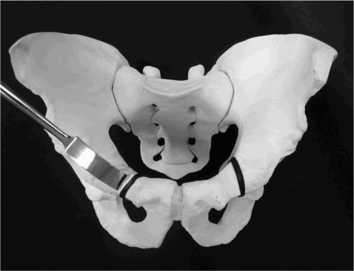
Preoperatively, the appearance of the hip following osteotomy was simulated. An AP radiograph of the pelvis with an abducted hip and a false‐profile view of the affected flexed hip in the supine position were taken. In addition, we observed the improvement of joint congruency and widening of the cartilage space in the abducted position. These functional radiographs documented that each hip showed a reduction in the subluxated joint or concentric motion.
Surgical technique
We performed CPO using a direct anterior approach in the supine position, as described previously (Naito et al. Citation2005). The surgical exposure was similar to those in Murphy and Millis (Citation1999) and Trousdale and Cabanela (Citation2003), and little damage was caused to the hip abductor strength because the gluteal muscles were not stripped from the bone (Murphy and Millis Citation1999, Ezoe et al. Citation2006). The iliac muscle of the supra‐acetabu‐lar portion was detached and the inner table of the pelvis was sharply stripped. A C‐shaped osteotomy line was marked using an airtome from the anteroinferior iliac spine to the distal part of the quadrilateral surface. The C‐shaped osteotomy line on the quadrilateral surface should be in front of the sciatic notch, but not more than 1.5 cm anterior to it (Shiramizu et al. Citation2004). The pubic osteotomy line is shown in . Pubic osteotomy using this technique was performed vertically to the horizontal line. However, in order to obtain medialization of the femoral head, we modified the pubic osteotomy procedures in 2005. In order to cut the pubis at 30 degrees to the horizontal line, a K‐wire was placed on the trans‐teardrop line under an image intensifier and used as the horizontal‐line landmark. The angle made by the K‐wire and the chisel was checked under the image intensifier. The C‐shaped osteotomy enabled acetabular re‐orientation because the osteotomy surfaces had the same curvatures. The image intensifier was used to ensure that the desired goals were achieved, i.e. that the femoral head was adequately covered by the re‐oriented acetabular fragment and the hip was medialized.
Active motion exercises were begun on postoperative day 2, and partial weight bearing with crutches was allowed on postoperative day 3. Full weight bearing was allowed after 8 weeks.
Radiographic evaluation
The severity of the secondary OA was graded using the Tönnis classification system (Tönnis Citation1987). In the M group, 28 hips were classified as grade 1, 22 hips as grade 2, 19 hips as grade 3, and 3 hips as grade 4. In the C group, 28 hips were classified as grade 1, 22 hips as grade 2, 19 hips as grade 3 and 3 hips as grade 4. Radiographic measurements included the CE angle, acetabular roof obliquity (ARO) (Massie and Howorth Citation1950), acetabulum head index (AHI) (Heyman and Herndon Citation1950), acetabular head lateralization index (HLI) (Yasunaga et al. 2003), inter‐hip distance (l), pelvic height (H), lateral pelvic width from the center of the femoral head (C), coordinates of the insertion point of the abductors on the greater trochanter in the frontal plane (point T), and radius of the femoral head (r). The coordinates of point T (Tx and Tz) were measured with respect to the center of the femoral head (Mavcic et al. Citation2002) obtained from the AP radiographs taken pre‐ and postoperatively (). The ratio of the lateralization of the femoral head was calculated using the HLI and modified Ninomiya measurements (Ninomiya Citation1989) (). The distance between the two Kohler ilioischial lines (t) was designated to the inter‐hip distance. The HLI was calculated using a / t / 2, and the ratio of the lateralization of the femoral head was calculated as postoperative HLI divided by preoperative HLI. Based on the value of the ratio of the lateralization of the femoral head, the hips were divided into 3 groups postoperatively: (1) a medialization group, when the ratio of the lateralization was less than 0.975 (), () a non‐displacement group, when the ratio of the lateralization was between 0.975 and 1.025, and (3) a lateralization group, when the ratio of the lateralization was more than 1.025 (). We also calculated the inclination of the hip resultant force (θR), magnitude of the resultant hip force normalized with respect to body weight (R/WB), and hip contact joint stress (Pmax/WB in units of kPa/N) in all cases in each group (medialization, no displacement, and lateralization) using the method described by Mavcic et al. (Citation2002). We confirmed the measurements using the nomograms described by Daniel et al. (Citation2001). Cases with extreme negative CE angle were excluded from the mechanical study because Pmax/WB was high using this method.
Figure 2. Pelvic radiographic parameters: interhip distance (l), pelvic width (C), pelvic height (H), radius of the femoral head (r), center‐edge angle of Wiberg (θCE), and the frontal coordinates of point T (the midpoint of the straight line connecting the most lateral point (point 1) with the highest point (point 2) on the greater trochanter) were used as input data for computation of the resultant hip force R.
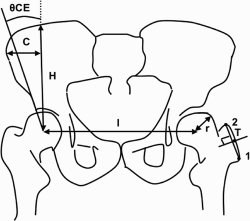
Figure 3. Radiographic indices measured from AP radiographs: Kohler's ilioischial line (b‐c), distance between the bilateral Kohler ilioischial lines (t), and distance from each Kohler ilioischial line to the center of the femoral head (a). Head laterailzation index (HLI) was calculated using the following formula: HLI = a /t / 2.
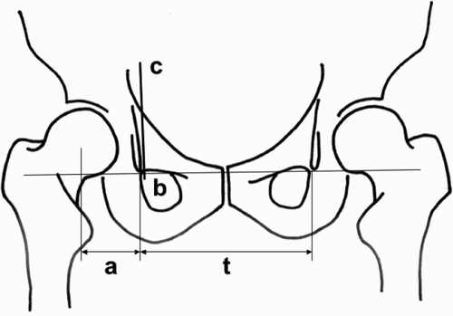
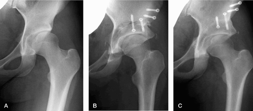
Figure 4. Medialization type. (A) A 24‐year‐old female with Tönnis classification grade 1 OA before CPO in 2006. The CE angle and ARO are 5° and 22°, respectively. (B) Immediately after surgery, showing a modified pubic osteotomy with a 30° inclination from the horizontal line in CPO. (C) Bony union at the osteotomy sites is observed at 4 months after CPO. The CE angle and ARO are 44° and ‐7°, respectively. Since the ratio of lateralization of the femoral head is 0.73, this hip is classified as the medialization type.
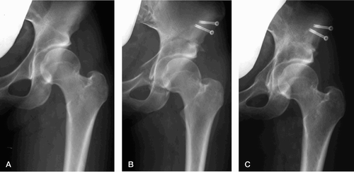
Figure 5. Lateralization type. (A) An 18‐year‐old female with Tönnis classification grade 1 OA before CPO in 2004. The CE angle and ARO are 7° and 14°, respectively. (B) Immediately after surgery, showing a conventional pubic osteotomy performed vertically to the horizontal line in CPO. (C) The CE angle and ARO have improved to 30° and ‐2°, respectively. Since the ratio of lateralization of the femoral head is 1.16, this hip is classified as the lateralization type.
AP radiographs of the pelvis were taken with the patient in the supine position. The tube‐to‐film distance was 120 cm, and the tube was perpendicular to the table. The center beam was directed toward the midpoint between the upper border of the symphysis and a horizontal line connecting both anterior and superior iliac spines. To judge the extent of the pelvic inclination, we used the method described by Siebenrock et al. (Citation2003). No corrections were made for radiographic magnification.
We made the radiographic measurements using a digital caliper (Mitutoyo, Tokyo, Japan) with an accuracy of ± 0.02 mm. Measurements of the involved hips were carried out by two authors (TT, YN) after repeated training sessions. In addition, the radiographs were reviewed 3 times on different days by the same observers, and the average values were calculated to assess intraobserver reliability. We assessed intraobserver and interobserver reliabilities for HLI measurements using the intraclass correlation coefficient. Intraobserver reliabilities of HLI measurements were 0.91 preoperatively and 0.89 postoperatively in the M group, and 0.92 preoperatively and 0.90 postoperatively in the C group. Interobserver reliabilities of HLI measurements were 0.83 preoperatively and 0.80 postoperatively in the M group, and 0.82 preoperatively and 0.81 postoperatively in the C group.
Statistics
We used the Mann‐Whitney U test to compare the corresponding radiographic parameters between the groups. A Wilcoxon signed ranks test was used to compare changes in radiographic parameters within the same group. Statistical significance was assumed at p‐values of < 0.05. The power of the analysis was 0.85.
All investigations were conducted in conformity with ethical principles of research and the study was approved by the ethical review board of Fukuoka University.
Results
There were no statistically significant differences in age, sex, and Tönnis classification of the OA grades between the two groups. In addition, there were no statistically significant differences in the CE angle, ARO, and AHI between the two groups on pre‐ and postoperative radiographic evaluation. The modified pubic osteotomy procedure (M group) had a positive influence on the medialization of the femoral head compared to conventional pubic osteotomy (C group). The results for all radiographic measurements are given in The ratio of lateralization of the femoral head differed between the two groups (p < 0.001), being 0.93 in the M group and 1.03 in the C group. In the M group, 58 hips were classified as the medialization type, 8 hips as the non‐displacement type, and 6 hips as the lateralization type (). Preoperatively, 3 of the 6 lateralization‐type hips were grade‐3 OA according to the Tönnis classification, and 2 of the 3 remaining hips were grade‐4 OA. The remaining lateralization‐type hip was classified as grade‐2 OA. In the C group, 35 hips were classified as the medialization type, 24 hips as the non‐displacement type, and 13 hips as the lateralization type. Preoperatively, 7 of the 13 lateralization‐type hips were advanced OA of grade 3 or 4 according to the Tönnis classification. The measurements of radiographic and mechanical parameters for the 3 displacement types are shown in . The ratio of lateralization of the femoral head in all cases of each type was 0.93 for the medialization type, 0.99 for the non‐displacement type, and 1.04 for the lateralization type. There were no statistically significant differences in the θCE angle, or the values of l, C, H, Tx, Tz, and r among the three types. The average inter‐hip distance (l) decreased by 13 mm in the medialization type and by 2 mm in the non‐displacement type, but it increased by 12 mm in the lateralization type. The resultant hip forces and hip contact joint stress were calculated using the method described by Mavcic et al. (Citation2002). The average value of the resultant hip force normalized to the body weight, R/WB, decreased from 3.0 preoperatively to 2.8 postoperatively in the medialization type, remained unchanged at 2.9 in the non‐displacement type, and increased from 2.9 preoperatively to 3.3 postoperatively in the lateralization type. On the other hand, the average value of the peak contact stress normalized to the body weight, Pmax/WB, was 8.4 kPa/N in the medialization type, 10.4 kPa/N in the non‐displacement type, and 9.6 kPa/N in the latelalization type preoperatively, and 3.2 kPa/N in the medialization type, 4.4 kPa/N in the non‐displacement type, and 4.7 kPa/N in the lateralization type postoperatively.
Table 1. Radiographic evaluations. Values are mean (SD)
Table 2. Distribution of postoperative femoral head centers
Table 3. Radiographic and mechanical parameters for the three displacement types. Values are mean (SD)
The average inter‐hip distance (l) decreased by 11 mm in the M group, but increased by 5 mm in the C group. Also, R/WB decreased from 3.2 preoperatively to 2.9 postoperatively in the M group and remained unchanged at 3.1 in the C group. On the other hand, Pmax/WB was 9.4 kPa/N in the M group and 9.1kPa/N in the C group preoperatively, and 3.4 kPa/N in the M group and 4.3 kPa/N in the C group postoperatively ().
Table 4. Radiographic and biomechanical parameters for the M and C groups. Values are mean (SD)
Discussion
The center of rotation of the hip joint shifted as a consequence of the periacetabular osteotomy. This change may have considerable effects on the resultant force of the hip joint and therefore also on the pressure on the femoral head, which could cause the development of OA (Srakar et al. Citation1992). Various attempts have been made to medialize the femoral head, including excision of the capital drop of the femoral head, partial excision of the transferred acetabulum, excision of the quadrilateral surface of the pelvis, osteotomy of the tear drop including the quadrilateral surface, and eccentric osteotomy in rotational acetabular osteotomy (Hasegawa et al. Citation2002).
In previously reported cases, the location of the center of adduction‐rotation during lateral displacement of the acetabular fragment appears to have been distal to the femoral head (Ganz et al. Citation1988). In the non‐displacement type, the acetabular fragment was rotated around the center of the femoral head. Proximal location of the center of rotation resulted in the medialization type. In our study, 35 of 72 hips in the C group and 58 of 72 hips in the M group were classified as the medialization type. We found that cutting the pubis at a 30‐degree inclination to the horizontal line and performing spherical osteotomies of the ilium and ischium were useful in order to obtain medialization of the femoral head. When the pubic osteotomy was performed vertically to the horizontal line, the distal pubis hindered rotation of the acetabular fragment. Furthermore, in cases requiring excessive rotation of the acetabulum in severe acetabular dysplasia of the hip, there was a smaller contact area of the pubic osteotomy site and the risk of dehiscence and nonunion of the pubic osteotomy increased. When the pubic osteotomy was performed using a 30‐degree inclination and the teardrop was lifted up, the bony obstruction for rotating the acetabular fragment at the osteotomy site was reduced and the contact area at the pubic osteotomy was better preserved. In addition, performing spherical osteotomies of the ilium and ischium enabled sufficient medialization of the femoral head.
Due to the asphericity of the osteotomy surfaces of the original Ganz osteotomy procedure, anterior displacement of the hip or anterior overcorrection may occur in patients who require extensive acetabular re‐orientation. Since a gap is created at the site of the osteotomy, a bone graft and relatively rigid fixation with hardware are needed to secure the rotated acetabular fragment, and resultant nonunion of the ilium has been reported (Hussell et al. Citation1999). CPO produces osteotomy surfaces with the same curvatures. As a result, greater contact between the bony surfaces and solid union of the iliac osteotomy can be accomplished with a low complication rate (Naito et al. Citation2005). We could also control the medialization of the femoral head safely due to direct visualization of the medial side of the pubis (Murphy and Millis Citation1999). However, in our study 5 of 6 hips with postoperative lateralization of the femoral head in the 30‐degree group showed advanced OA preoperatively. In cases with a non‐spherical femoral head, medialization of the femoral head seemed to be difficult despite the modified pubic osteotomy procedure. We found that two major causes of lateralization of the hip in advanced OA were osteophyte formation of the acetabular fossa and deformation of the femoral head. Thus, we could not easily rotate the acetabular fragment smoothly, or easily acquire medialization of the femoral head.
In cases requiring excessive rotation of the acetabulum in severe acetabular dysplasia of the hip, the risk of dehiscence and of nonunion of the pubic osteotomy site increases. In the one‐legged stance, the empirical relationship between the moment arms and the inter‐hip distance was described by McLeish and Charnley (Citation1970). A change in the parameter of the inter‐hip distance causes a change in the moment arms and the resultant hip force. In our study, the average value of the peak contact stress normalized to the body weight, and Pmax/WB was improved in all 3 types. However, the average value of the resultant hip force normalized to the body weight, R/WB, decreased in the medialization type, remained unchanged in the non‐displacement type, and increased in the lateralization type. Also, R/WB was more reduced and Pmax/WB was more improved in the M group than in the C group. Medialization of the femoral head reduces the peak contact stress due to the change in magnitude/direction of the resultant hip force, even if the CE angle is the same (Kralj et al. Citation2005). Thus, although it is a small change, the medialization of the femoral head in periacetabular osteotomy appears to decrease the peak contact stress and the resultant hip force, which may prevent progression of OA.
In this study, we did not investigate the relationship between the medialization of the femoral head and clinical outcome due to the short follow‐up period for many patients. In the future, indications for medialization of the femoral head should be established using long‐term clinical outcome.
Contributions of authors
TT: data collection, data proceeding, statistical analysis, and writing of manuscript. MN: initiator of project and supervisor. KS: data collection and writing of manuscript. YN: data collection and data proceeding. SM: data anlysis.
No competing interests declared
- Brand R A. Hip osteotomies: A biomechanical consideration. J Am Acad Orthop Surg 1997; 5: 282–91
- Brand R A, Iglic A, Kralj-Iglic V. Contact stress in the human hip: implications for disease and treatment. Hip Int 2001; 11: 117–26
- Daniel M, Antolic V, Iglic A, Kralj‐Iglic V. Determination of contact hip stress from nomograms based on mathematical model. Med Eng Phys 2001; 23: 347–57
- Ezoe M, Naito M, Asayama I. Muscle strength improves after abductor-sparing periacetabular osteotomy. Clin Orthop 2006, 444: 161–8
- Ganz R, Klaue K, Vinh T S, Mast J W. A new periacetabular osteotomy for the treatment of hip dysplasias. Technique and preliminary results. Clin Orthop 1988, 232: 26–36
- Hadley N A, Brown T D, Weinstein S L. The effects of the contact pressure elevations and aseptic necrosis on the long-term clinical outcome of congenital hip dislocation. J Orthop Res 1990; 8: 504–13
- Hasegawa Y, Iwase T, Kitamura S, Yamauchi K, Sakano S, Iwata H. Eccentric rotational acetabular osteotomy for acetabular dysplasia. J Bone Joint Surg (Am) 2002; 84: 404–10
- Heyman C H, Herndon C H. Legg‐Perthes disease: a method for the measurement of the roentgenographic results. J Bone Joint Surg (Am) 1950; 32: 767–78
- Hussell JG, Rodriguez JA, Ganz R. Technical complications of the Bernese periacetabular osteotomy. Clin Orthop 1999, 363: 81–92
- Iglic A, Kralj-Iglic V, Antolic V, Srakar F, Stanic U. Effect of the periacetabular osteotomy on the stress on the human hip joint articular surface. IEEE Trans Rehab Eng 1993; 1: 207–12
- Kralj M, Mavcic B, Antolic V, Iglic A, Kralj‐Iglic V. The Bernese periacetabular osteotomy: clinical, radiographic and mechanical 7–15‐year follow‐up of 26 hips. Acta Orthop Scand 2005; 76: 833–40
- Massie W K, Howorth M B. Congenital dislocation of the hip. J Bone Joint Surg (Am) 1950; 32: 519–31
- Mavcic B, Pompe B, Antolic V, Daniel M, Iglic A, Kralj-Iglic V. Mathematical estimation of stress distribution in normal and dysplastic human hips. J Orthop Res 2002; 20: 1025–30
- Maxian T A, Brown T D, Weinstein S L. Chronic stress tolerance levels for human articular cartilage: two nonuniform contact models applied to long term follow up of CDH. J Biomech 1995; 28: 159–66
- McLeish R D, Chanley J. Abduction forces in the one‐legged stance. J Biomech 1970; 3: 191–209
- Michaeli D A, Murphy S B, Hipp J A. Comparison of predicted and measured contact pressures in normal and dysplastic hips. Med Eng Phys 1997; 19: 180–6
- Murphy S B, Millis M B. Periacetabular osteotomy without abductor dissection using direct anterior exposure. Clin Orthop 1999, 364: 92–8
- Murphy S, Deshmukh R. Periacetabular osteotomy. Preop-erative radiographic predictors of outcome. Clin Orthop 2002, 405: 168–74
- Naito M, Shiramizu K, Akiyoahi Y, Ezoe M, Nakamura Y. Curved periacetabular osteotomy for treatment of dysplastic hip. Clin Orthop 2005, 433: 129–35
- Ninomiya S. Rotational acetabular osteotomy for the severely dysplastic hip in the adolescent and adult. Clin Orthop 1989, 247: 127–37
- Pauwels F. Biomechanics of the normal and diseased hip. Springer‐Verlag, Berlin, Heidelberg, New York 1976; 129–35
- Shiramizu K, Naito M, Asayama I, Yatsunami M. A quantitative anatomic characterization of the quadrilateral space for periacetabular osteotomy. Clin Orthop 2004, 418: 157–61
- Siebenrock K A, Schoeniger R, Ganz A. Anterior femoro-acetabular impingement due to acetabular retroversion. J Bone Joint Surg (Am) 2003; 85: 278–86
- Srakar F, Iglic A, Antolic V, Herman S. Computer simulation of the periacetabular osteotomy. Acta Orthop Scand 1992; 63: 411–2
- Tönnis D. Congenital dysplasia and dislocation of the hip in children and adults. Springer‐Verlag, New York 1987; 167–71
- Trousdale R T, Cabanela M E. Lessons learned after more than 250 periacetabular osteotomies. Acta Orthop Scand 2003; 74: 119–26
- Vengust R, Daniel M, Antolic V, Zupanc O, Iglic A, Kralj‐Iglic V. Biomechanical evaluation of the hip joint after innominate osteotomy: a long‐term follow‐up study. Arch Orthop Trauma Surg 2001; 121: 511–6
- Wiberg G. Studies on dysplastic acetabula and congenital subluxation of the hip joint, with special reference to the complication of osteoarthritis. Acta Chir Scand (Suppl 58) 1939; 83: 1–35
- Yasunaga Y, Takahashi K, Ochi M, Ikuta Y, Hisatome T, Nakashiro J, Yamamoto S. Rotational acetabular osteotomy in patients forty‐six years of age or older: Comparison with younger patients. J Bone Joint Surg (Am) 2003; 85: 266–72