Abstract
A case is presented of iliac artery perforation during percutaneous coronary intervention. This resulted in retroperitoneal bleeding complicated by the development of the abdominal compartment syndrome and multi‐organ failure, which was successfully treated by abdominal decompression.
Introduction
As the number of patients undergoing invasive cardiological procedures increases, complications related to the procedure are becoming more readily recognized. These include dissection of the cannulated artery and bleeding, which is usually from the vascular access site. However, bleeding may also occur following rupture of a vessel at a more distal portion of the angiography catheter. We present a previously unreported consequence of such a complication.
Case report
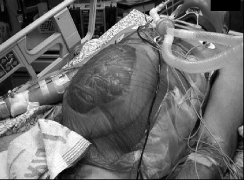
A 67‐year‐old man presented to the hospital within 2 h of onset of chest discomfort. An electrocardiogram revealed ST segment elevation in the precordial leads. A transthoracic echocardiogram demonstrated apical akinesia and mild to moderate left ventricular dysfunction. He was treated with aspirin 300 mg, intravenous unfractionated heparin 5000 units, clopidogrel 300 mg, and propranolol 20 mg and he underwent emergent primary percutaneous coronary intervention (PCI). At catheterization, it was noted that the iliac artery was very tortuous, and difficulty was encountered in advancing the guide wire of the catheter. Following repeated attempts, an abrupt decrease in blood pressure was noted. Perforation of the vessel was confirmed by the finding of extravasation of contrast dye from the iliac artery (). The patient underwent tracheal intubation and mechanical ventilation was initiated. Fluid resuscitation with the rapid infusion of crystalloids, blood, and fresh frozen plasma was given. However, hypotension persisted and vasopressor support with noradrenalin was commenced. A covered stent graft was implanted in the area of the perforation and a repeat contrast injection revealed no further extravasation. No further attempts at coronary angiography and PCI were made and the patient was transferred to the cardiac intensive care unit (ICU). Intra‐arterial blood pressure remained low (85/50 mm Hg) and the central venous pressure was found to be 12 cm H2O. A repeat echocardiogram revealed mild left ventricular dysfunction with a hypovolemic chamber and the patient received further fluid infusions. Ten hours after the acute event, the patient was anuric, and the serum creatinine had increased to 3.5 gm%. Blood gas analysis revealed a severe metabolic acidosis (pH 7.21, base excess—10 mmol/L, serum bicarbonate 16 mEq/L) with a markedly elevated serum lactate (29 mg/dl, normal <20 mg/dl)). In addition, the patient required an inspired oxygen content (FiO2) of 1.0 to maintain blood oxygen saturation >90%, and static lung compliance was markedly decreased (20ml/cm H2O, normal >50ml/cm H2O). Physical examination revealed that the abdomen was tense and distended, tympanic to percussion, but not tender to palpation. Measurement of intra‐abdominal pressure (IAP) via the intravescical route revealed a pressure of 32 mm Hg. An ultrasound examination revealed dilated loops of small bowel with a moderate amount of ascites. Two hours later the IAP had increased to 36 mm Hg. A diagnosis of abdominal compartment syndrome was made and the patient was immediately transferred to the operating room. At laparotomy, the small bowel was markedly edematous. There was no evidence of bowel ischemia and exploration of the rest of the abdomen was unremarkable. Temporary closure of the abdomen was achieved with a Bogota bag ().
Figure 1 Extravasation of contrast dye from ileac artery.
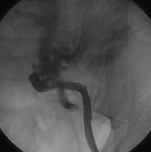
Figure 2 Temporary abdominal closure with Bogota bag.
The patient was transferred to the general ICU, and within hours of admission, began to pass urine, vasopressor therapy could be discontinued, and there was significant improvement in pulmonary function. The IAP had decreased to 19 mm Hg. A CT scan of the abdomen performed the following day revealed an extensive retroperitoneal hematoma. By the fourth day the serum creatinine had returned to normal. The patient’s subsequent course was characterized by repeated episodes of pulmonary infection, prolonged mechanical ventilation, and eventually tracheotomy. The abdomen was definitively closed three weeks after the initial operation. The patient was eventually weaned from the ventilator and discharged to a rehabilitation hospital.
Discussion
This is the first reported case of the abdominal compartment syndrome occurring in a patient following primary PCI for acute myocardial infarction. IAP, normally ±5 mm Hg, increases when the volume of the retroperitoneal or abdominal contents increases Citation[1]. In our case, this was the result of retroperitoneal bleeding together with massive fluid resuscitation (typically >5 l in a 24‐h period), but may also follow emergency surgery for abdominal trauma, intra‐abdominal bleeding, or be the consequence of tense ascites, ileus or intestinal obstruction Citation[1].
IAP should be assessed via the intravescical route, since clinical examination is unreliable Citation[2]. Fifty milliliters of fluid is inserted into the bladder via an indwelling urinary catheter, allowing it to act as a passive reservoir, so that changes in intravescical pressure reflect changes in IAP. A plastic cannula or needle inserted into the specimen collecting port is connected to a transducer with arterial pressure tubing and the IAP, expressed in mm Hg, can then be determined. The transducer should be zeroed at the level of the mid‐axillary line and the measurement made at the end of expiration.
As occurred in our case, sustained increases in IAP⩾20 mm Hg (recorded during three measurements performed over 1–6 h apart) may result in the abdominal compartment syndrome, a condition characterized by significant organ dysfunction that was not previously present Citation[3]. This is due to an increase in both intra‐abdominal and resultant intra‐thoracic pressures. Significant hemodynamic compromise is common, an effect which is aggravated by hypovolemia. The decrease in venous return may result in peripheral venous pooling, predisposing to venous thrombosis. The effects on the respiratory system, which may be even more significant for patients receiving mechanical ventilation, include reduced thoracic volume and compliance and airway pressures usually need to be increased to maintain adequate ventilation. Compressive atelectasis results in ventilation‐perfusion mismatch, hypoxemia, hypercarbia and respiratory acidosis. An IAP>15 mm Hg causes impairment of renal blood flow and glomerular filtration rate, resulting in oliguria which may progress to anuria when the IAP>30 mm Hg. Mesenteric and ileal blood flow decreases further when the IAP exceeds 20 mm Hg. Hepatic blood flow is also impaired and severe mucosal acidosis, indicative of ischemia, becomes apparent when the IAP exceeds 40 mm Hg. Although not relevant in our case, cerebral venous outflow may be impaired and may aggravate raised intracranial pressure in patients with cerebral trauma.
Early recognition and treatment of the syndrome is essential. While some patients may benefit from fluid resuscitation, especially in the early stages of oliguria, over‐aggressive fluid replacement may actually aggravate the IAP. Due to the raised intrathoracic pressure, assessment of central venous pressure and pulmonary artery occlusion pressure may give inaccurately elevated results when the IAP is markedly elevated (⩾30 mm Hg), despite severe intravascular volume depletion Citation[4]. Volumetric methods, such as right ventricular end‐diastolic volume and echocardiography, provide more accurate assessments Citation[4].
Our patient underwent early surgical decompression, which is the definitive treatment, and this was associated with significant improvement of organ dysfunction, as occurs in 80% of cases Citation[5]. However, the mean survival rate is only 53%. Following decompression, the abdomen is typically left open and covered with a Bogota bag (a cystoscopy irrigation bag, which is sutured to the wound edges) or a mesh.
This case demonstrates a complication of PCI, as yet unreported, namely the abdominal compartment syndrome, the result of retroperitoneal bleeding and massive fluid resuscitation. As the caseload of interventional cardiologists becomes more challenging with an increasing proportion of older and sicker patients, this complication may be more common. Thus, the interventional cardiologist should be acquainted with this ominous complication.
References
- Sugrue M. Abdominal compartment syndrome. Curr Opin Crit Care 2005; 11: 333–8
- Fusco M. A., Martins R. S., Chang M. C. Estimation of intra‐abdominal pressure by bladder pressure measurement: validity and methodology. J Trauma 2001; 50: 297–302
- Malbrain M., Deeren D., De Potter T. J. Intra‐abdominal pressure in the critically ill: it is time to pay attention. Curr Opin Crit Care 2005; 11: 156–71
- Ridings P. C., Bloomfield G. L., Blocher C. R., Sugerman H. J. Cardiopulmonary effects of raised intra‐abdominal pressure before and after intravascular volume expansion. J Trauma 1995; 39: 1071–5
- Moore A. F. K., Hargest R., Martin M., Delicata R. J. Intra‐abdominal hypertension and the abdominal compartment syndrome. Br J Surg 2004; 91: 1102–10