Abstract
Percutaneous intervention of the vertebral artery stenosis is associated with the risk of distal embolization and restenosis, which can manifest with stroke. We describe a case of vertebral artery percutaneous intervention utilizing the embolic protection FilterWire and a drug‐eluting Taxus stent, to overcome these issues of distal embolization and restenosis respectively.
Introduction
Vertebral artery stenosis has been successfully treated using percutaneous intervention with stenting. We describe successful percutaneous treatment of symptomatic patient with bilateral vertebral artery stenosis using a distal embolic protection device and a drug‐eluting stent. This combination of an embolic protection device and a drug eluting stent may enhance the safety and long‐term success of extra‐cranial vertebral interventions.
Case report
A 64‐year‐old male with diabetes mellitus type 2, chronic renal failure on maintenance hemodialysis, previous coronary artery bypass graft surgery had been experiencing episodic vertigo and postural dizziness. Carotid ultrasound followed by aortic arch, carotid, coronary and bypass angiography revealed significant left internal carotid artery and bilateral ostial vertebral artery stenoses in the range of 90–95%, and a dominant right vertebral artery. The patient underwent successful stenting of the left internal carotid artery stenosis using Guidant RX ACCULINK stent with a Guidant RX ACCUNET embolic protection system.
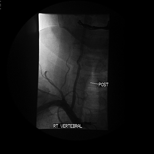
Vertebral percutaneous intervention was performed through transfemoral approach using a 7 F IMA guiding catheter to selectively cannulate the right vertebral artery (). Heparin was administered intravenously to achieve a therapeutic ACT. A Boston Scientific FilterWire EZ (Boston Scientific Corporation, Natick, MA) was used to cross the right vertebral artery and the filter was deployed under cinefluoroscopic guidance. A drug‐eluting Taxus 3.5×12 mm stent (Boston Scientific Corporation, Natick, MA) was positioned () and deployed without pre‐dilatation at 12 atmospheres, followed by additional high‐pressure post dilatation of the stent to achieve a luminal diameter of 4 mm to match the reference caliber of the right vertebral artery. Check angiograms showed an excellent angiographic result () without any dissection or slow flow. The filter wire was retrieved. There were no neurological complications. The patient was discharged next day on clopidogrel and aspirin.
Figure 1 Selective‘roadmap’ angiogram before the percutaneous intervention showing a critical ostial lesion in the dominant right vertebral artery.
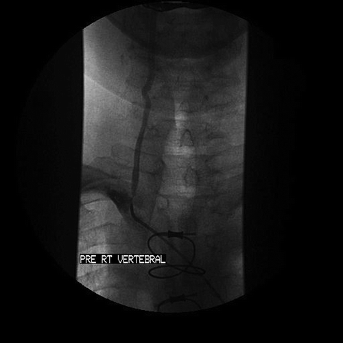
Figure 2 Vertebral angiogram showing the Boston Scientific FilterWire deployed and the Taxus stent being positioned before deployment.
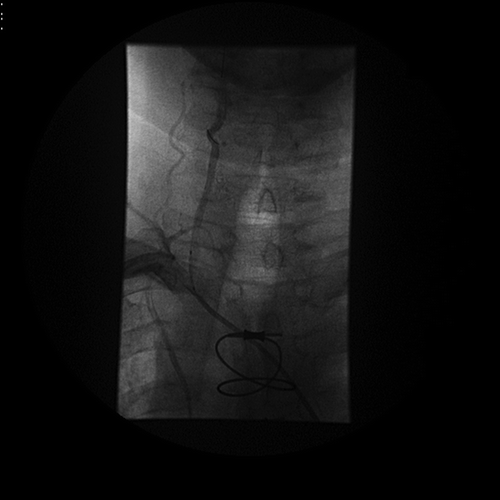
Figure 3 Vertebral angiogram immediately after stenting of the ostial lesion and retrieval of the FilterWire showing the final angiographic result.
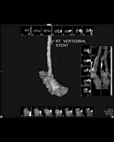
Our patient has been maintained on aspirin and clopidogrel therapy and he continues to be asymptomatic without any cerebrovascular events. A computed tomographic (Toshiba Aquilion 64 multislice CT scanner) angiogram of the carotids and vertebral arteries performed six months after the vertebral percutaneous intervention, showed patency of the stented arteries without any evidence of restenosis ().
Figure 4 Computed tomographic angiogram with three‐dimensional reconstruction image of the right vertebral artery performed six months after the percutaneous intervention showing patency of the right vertebral stent.
Discussion
Vertebral artery stenosis is often asymptomatic because of good collateral supply from the contra‐lateral vertebral artery Citation[1]. However, 50% of patients present initially with a stroke and 26% with a transient ischemic attack (TIA) rapidly followed by a stroke Citation[2]. Vertebrobasilar stroke is associated with a mortality of 20–30% Citation[1], Citation[3]. Atherosclerotic disease most commonly involves the origin of the vertebral artery Citation[4]. The treatment options for extracranial atherosclerotic disease of the vertebral artery include medical, surgical or endovascular options. Surgical vertebral endarterectomy or bypass operations have reasonable success but are associated with a high incidence of non‐stroke complications (Horner's syndrome, lymphatic injury, vertebral artery thrombosis, cranial nerve injury, etc.) Citation[5]. Percutaneous intervention (angioplasty and stenting) offers a less invasive approach with a fewer risk of complications.
The indications of treatment of vertebral artery stenosis include symptomatic disease causing vertebrobasilar ischemia or asymptomatic high grade (>70%) stenosis in a dominant or singular vertebral artery with evidence of resting posterior‐fossa hypoperfusion Citation[1]. Angioplasty and stenting of the vertebral artery has been accomplished using the standard coronary intervention equipment Citation[6–9]. The size of the vertebral artery is usually 3–5 mm, and thus balloon‐mounted coronary stents are favored given the high radial strength, limited foreshortening, low crossing profile and appropriate diameters. However, embolic complications were described in these series, as distal protection devices were not utilized. There has been a single previous case report of distal protection embolic filter used in vertebral artery stenting with a bare metal stent with good results Citation[10]. Embolic distal protection devices are routinely employed in carotid artery stenting. We believe that since the vertebro‐basilar circulation is as ‘unforgiving’ regarding distal embolization (distal embolism is likely to present with manifest symptoms as commonly as in the carotid circulation), distal embolic protection using a filter should be undertaken on a routine basis. Since the caliber of the vertebral artery matches the coronary arteries (3–5 mm), our choice of utilizing the Boston Scientific FilterWire EZ, which is approved for saphenous vein graft intervention, was an obvious choice. The 110‐micron pore filter allows continual perfusion reducing concerns about potential ischemia. The filter accommodates contrast injections for good lesion visibility and accurate stent placement. The suspended Nitinol wire loop provides 360° apposition, reducing particle drift between filter loop and vessel wall. It allows the filter to be placed in both straight and curved vessels, helping maintain vessel wall apposition even with changing wire bias, in vessels varying 3.5 to 5.5 mm in size and of varying shape. The loop mouth closes during retrieval for effective particle retention.
Since vertebral artery stenting has been associated with a high incidence of restenosis in some series, we elected to use a drug‐eluting Taxus stent for prevention of the risk for restenosis. Angiographic restenosis (>50% diameter reduction on follow‐up angiography) has been reported in 3–43% of cases Citation[6–8].
Restenosis occurs in vertebral arteries more frequently than in the carotid circulation; however, vertebral restenosis is largely asymptomatic. Vascular factors may account for the higher incidence of restenosis in the vertebral circulation. Since the diameter of the vertebral artery ranges from 3–5 mm, proximal vertebral lesions are likely to behave like the coronary arteries ‐with high elastic recoil and with any significant late‐loss due to intimal proliferation likely to manifest as restenosis. The ostial location of most vertebral lesions is also likely to subject these to high shear stress, accounting for a higher incidence of restenosis, as is observed in ostial renal artery lesions. Drug eluting stents are associated with minimal rates of restenosis due to the anti‐proliferative action of the drugs (paclitaxel, sirolimus) on the smooth muscle cells. As such, these stents should be employed in the vertebral artery interventions on a routine basis, given their proven benefit in terms of preventing restenosis. Computed tomographic (CT) angiography showed no evidence of restenosis in the vertebral stent six months after the percutaneous intervention in our patient.
The risk of late stent thrombosis is a major concern in drug‐eluting stents given delayed endolthelialization. It would be prudent continuing combination anti‐platelet therapy with aspirin and clopidogrel on a long term, possibly indefinite basis to prevent the risk of delayed stent thrombosis.
We believe that percutaneous intervention with filter wire and a drug‐eluting stent confers the dual benefit in terms of preventing the risk of distal embolization and late restenosis.
Acknowledgements
The authors should like to thank Girish Patel, MD and Janice Adams, RT for their assistance in providing computed tomographic imaging services and reconstruction of the 3‐D images.
References
- Wehman J. C., Hanel R. A., Guidot C. A., et al. Atherosclerotic occlusive extra cranial vertebral artery disease: indications for intervention, endovascular techniques, short term and long term results. J Interv Cardiol 2004; 17: 219–32
- Wityk R. J., Chang H. M., Rosengart A., Han W. C., DeWitt L. D., Pessin M. S., et al. Proximal extracranial vertebral artery disease in the New England Medical Center Posterior Circulation Registry. Arch Neurol 1998; 55: 470–8
- Patrick B. K., Ramirez‐Lassepas M., Synder B. D. Temporal profile of vertebrobasilar territory infarction. Prognostic implications.Stroke 1980; 11: 643–8
- Schwartz C. J., Mitchell J. R. Atheroma of the carotid and vertebral arterial systems. Br Med J 1961; 5259: 1057–63
- Buerger R. Long term results of vertebral artery reconstruction. Long term results in Vascular Surgery, J Yao, W Pearce. Appleton & Lange, Norwalk 1993; 69–79
- Chastain H. D 2nd., Campbell M. S., Iyer S., et al. Extracranial vertebral artery stent placement:In‐hospital and follow‐up results. J Neurosurg 1999; 91: 547–52
- Albuquerque F. C., Fiorella D., Han P., et al. A reappraisal of angioplasty and stenting for the treatment of vertebral origin stenosis. Neurosurgery 2003; 53: 607–16
- Lustep H. L., Barnwell S. L., Mawad M., et al. Stenting of Symptomatic Atherosclerotic Lesions in the Vertebral or Intracranial Arteries (SSYLVIA): Study results (abstract P83A). Stroke 2003; 34: 253
- Jenkins J. S., White C. J., Ramee S. R., et al. Vertebral artery stenting. Catheter Cardiovasc Interv 2001; 54: 1–5
- Mintz E. P., Gruberg L., Kouperberg E., Beyar R. Vertebral artery stenting using distal emboli protection and transcranial Doppler. Catheter Cardiovasc Interv 2004; 61: 12–5