

ABSTRACT
Background
In recent times, stem cell therapy has recently gained popularity, and is used for regenerating oral and maxillofacial structures. This study aimed to review stem cell applications in the reconstructive treatment of cleft lip and palate and other craniofacial anomalies.
Methods
A search on the PubMed database, Dental and Oral Science and CINAHL was conducted to identify relevant studies on stem cells and their implications in the reconstructive treatment of cleft lip and palate.
Results
Studies included in this review reported stem cell usage in the reconstruction of defects in cleft lip and palate cases, hemifacial microsomia, Treacher-Collins syndrome, Parry-Romberg syndrome and craniosynostosis. Stem cell knowledge is being utilized for several different tissues and organs. Preclinical data has displayed the potential of stem cells for craniofacial surgery.
Conclusions
Fortunately, stem cell-based oral and maxillofacial regeneration has shown promising results. Understanding stem cell practice will open a new era of research and proposed advancement in treating various diseases.
Introduction
In the human body, stem cells are unspecialized cells that can develop into specialized cells, each with new specialized cell functions. A stem cell, in essence, remains uncommitted until it receives a signal to differentiate into a specialized cell.Citation1 The bone marrow stem cell is the best example of a stem cell as it is unspecialized and can specialize into blood cells with pre-assigned functions, such as white blood cells and red blood cells. As a result, one cell type develops from another, giving rise to the term “stem cell.”Citation1
Under certain conditions, these undifferentiated stem cells can act as pluripotent (able to give rise to cells from all three germ layers, namely ectoderm, mesoderm, and endoderm) or multipotent (ability to give rise to a limited number of other specialized cell types).Citation2 These cells can be classified into three main categories: adult stem cells (ASCs), embryonic stem cells (ESCs) and induced pluripotent stem cells. ASCs have been detected and successfully extracted from adult tissues such as bone marrow, umbilical cord, amniotic fluid, brain tissue, liver, adipose tissue, and dental pulp.Citation2 The therapeutic use of ASCs encompasses a wide range of pathologies in which tissue and organ replacement and repair are required to restore form and function. As a result, stem cells, growth factors, and scaffolds are considered necessary when applying this technology to humans or animals.Citation3
Currently, stem cell knowledge is being utilized for a number of different tissues and organs. The multilineage differentiation capacity, qualified cell collection accessibility, noninvasive separation technique, and rapid in vitro extension of stem cells make them excellent sources for therapeutic purposes.Citation4 Rehabilitation and reconstruction of tissues and organs are two possible applications for stem cells.Citation5 Replacement of an oromaxillofacial structure is challenging because functions such as facial expression, articulation, mastication and swallowing are delicate processes involving complex anatomical structures composed of soft and hard tissues. Stem cells, biomimetic materials, and growth factors are required to form these three-dimensional structures. Stem cell therapy, which has recently gained popularity, can regenerate oromaxillofacial structures.Citation5
Lip and palate clefts are congenital craniofacial malformations that affect the lip, hard or soft palate, or all three structures simultaneously.Citation6 Cleft lip and palate treatment is multidisciplinary and must be guided by a distinct philosophy.Citation7 The pursuit of ideal surgical techniques is an ongoing endeavor. Finding the best method for correcting anatomical tissue repositioning is a significant challenge.Citation7 The use of stem cells and regenerative medicine opens new avenues for improving outcomes in various pathways. The use of regenerative medicine through tissue engineering with mesenchymal stem cells has been studied for at least 10 years. Many studies have shown that bone marrow, dental pulp, umbilical cord blood, and adipose tissue can be a source of osteoblastic, adipogenic, and chondrogenic cell lines.Citation8–10
Mesenchymal stem cells (MSCs) are now considered “research trends” in biology and medicine, and their use in regenerative medicine is expanding.Citation11 Some techniques involve directly implanting MSCs into the defect site, while others employ appropriate scaffolds to support the cells. MSCs are drifted by an osteoconductive scaffold and differentiated into osteogenic cells using osteoinductive growth factors in bone tissue engineering. Growth factors and scaffoldings of various types are then used to regenerate maxillofacial bone defects.Citation11 This study aimed to review stem cell applications in the reconstructive treatment of cleft lip and palate and other craniofacial anomalies.
Cleft Lip and Palate
One of the most common congenital anomalies, cleft lip and palate (CLP), is caused by the failure of the nasal process and the oropalatal shelves to fuse. This malformation affects 0.36–0.83 out of every 1,000 live-born infants.Citation12 Around 10,000 cases of cleft lip and palate were identified in Pakistan in the year 2015. The data provided by Smile Train show that from 2008 to 2015, around 50,000 cleft lip and palate patients were treated surgically in 25 different centers in Pakistan.Citation13 The prevalence of cleft lip and palate in various geographical regions has been mentioned in .Citation14–19
Table 1. Prevalence of cleft lip and palate in various geographical regions.Citation14–19
The severity of cleft varies from occult to overt; the etiology of this anomaly ranges from genetic to environmental to idiopathic. The management is often complex, ranging from simple surgical closure to multidisciplinary surgical and cleft team care.Citation20 CLP patients may exhibit alveolar bone defects, maxillary deficiency, facial deformity, hypodontia and, swallowing and speech difficulties ().Citation21 Malformed alveolar bone repair is essential for the closure of the oronasal fistula, the eruption of teeth, and alar base support.Citation22,Citation23 The gold standard for alveolar reconstruction in CLP patients is autogenous cancellous bone grafts.Citation24 The most common site for obtaining autogenous bone for grafting is the anterior iliac crest.Citation25 In terms of bone resorption, iliac crest bone grafting to the alveolar cleft has an 88% success rate.Citation26 Tissue engineering techniques have provided substitutes for conventional iliac crest bone grafting techniques. The formation of new bone occurs in areas requiring repair when MSCs are transplanted.Citation27 In a study conducted by Behnia et al., approximately 50% fill of the bone defect was seen three months postoperatively.Citation28 Whereas, Hibi et al. reported 79.1% bone regeneration in the cleft area.Citation29 A study conducted by Pradel et al. showed that when autologous osteoblasts were cultured on a demineralized bone matrix, the size of the defect demonstrated a reduction in comparison to the control group.Citation30 After 18 months postoperatively, biomaterial with autogenous osteogenic cells into the alveolar cleft resulted in the spontaneous eruption of the canine in its proper position.Citation31 Osteogenically differentiated fat-derived stem cells and poly-L-lactic acid demonstrated significant bone regeneration in palatal defects.Citation32
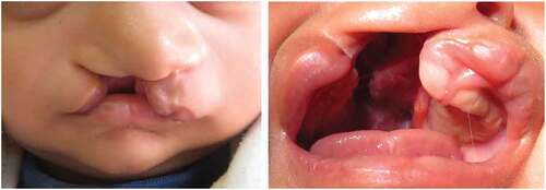
Figure 1. Unilateral right-sided cleft lip and palate.
A study by Garcia et al.Citation33 to assess the effectiveness of umbilical cord blood stem cells in gingivoperiosteoplasty procedures reported satisfactory results after 18 months of follow-up. In their study, Mazzetti et al.Citation34 proclaimed the use of umbilical cord blood and placenta blood stem cells for the first surgery of cleft palate repair to promote the healing of both soft and hard tissues. The results on soft tissue were substantial; however, there was no bone formation, only a reduction in scar formation and an inflammatory response in the soft tissue of the lip. After the second surgery (hard palate surgery), there was less fibrous tissue formation, and no palatal fistula was observed.Citation34 Bajestan et al.Citation35 conducted a study to determine the safety and effectiveness of cell therapy with ex vivo expanded stem cell populations in the regeneration of extensive alveolar defects in patients. They found that while stem cells are safe to treat broad alveolar defects, their ability to complete the reconstitution of enormous alveolar defects is limited. This approach requires further modifications to achieve the outcomes observed with current methods for treating broad defects, particularly those caused by cleft palate.Citation35
Tanikawa et al.Citation36 conducted a study in 2020 to assess the use of deciduous dental pulp stem cells (DDPSC) associated with a hydroxyapatite-collagen sponge (Bio-Oss Collagen® 250 mg, Geistlich) for repairing alveolar defects during the dental eruption. They isolated the DDPSC from each patient and then associated those cells with the biomaterial. They used this bone tissue-engineered set to fill the alveolar defect and evaluated the outcome with cone beam computed tomography at a six-month and 12-month follow-up. The researchers observed progressive alveolar bone union in all patients. DDPSC therapy resulted in adequate healing of alveolar defects with excellent feasibility and safety, and the researchers concluded that stem cell therapy resulted in satisfactory bone regeneration with dental eruption and reduced morbidity.Citation36
Literature suggests that stem cell use in alveolar defect repair can reduce defect size through bone formation and that stem cells have the potential for bone regeneration in the oral and maxillofacial region, have less postoperative morbidity than autogenous bone grafting and enable teeth in the defect area to erupt in their proper position.Citation28,Citation31,Citation32,Citation37 Furthermore, stem cells in cleft lip and palate surgery reduce inflammation and produce fewer scars than the classic series.Citation34
Other Craniofacial Anomalies
After cleft lip and palate, hemifacial microsomia (HFM) is the second most common type of congenital facial malformation.Citation37 HFM is caused by unilateral abnormal morphogenesis of the first and second pharyngeal archesCitation38 and is distinguished by unilateral hypoplasia of the craniofacial skeleton and its overlying soft tissue.Citation38 Autologous fat grafting is used to reconstruct soft tissue defects in the treatment of congenital and post-traumatic malformations.Citation1 Numerous initiatives and improvements in surgical techniques have been reported to overcome problems associated with fat grafting, such as unpredictable clinical outcomes and a low rate of graft survival. Recently, attention has been drawn to the use of adipose-derived stromal cells (ASCs) for tissue regeneration.Citation39 ASC supplementation has demonstrated 88% fat volume survival after six months in patients with HFM.Citation39,Citation40 For soft tissue reconstruction, ASCs have shown improvement in angiogenesis and graft survival.Citation39,Citation40 Studies suggest that isolation and supplementation of ASCs is an efficient, secure and superior method for facial recontouring in patients with craniofacial microsomia.Citation39,Citation40
Treacher-Collins syndrome (TCS) is a congenital disorder caused by a genetic mutation that results in the partial or complete absence of the lateral and inferolateral orbital rims, and zygomatic arches along with the deficiency in overlying soft tissues.Citation41 ASCs have also shown tremendous results in the repair of defects related to TCS.Citation41,Citation42
Parry-Romberg syndrome (PRS), also known as Romberg disease, is characterized by progressive hemifacial atrophy of the skin, dermis, subcutaneous fat, muscle, cartilage, and bone. EulenbergCitation38 classified it as an acquired disease in 1871, naming it “progressive facial hemiatrophy,” which affects one side of the face and has aesthetic, functional, and psychological effects on affected patients.Citation43,Citation44 In such patients, facial soft tissue defects can be repaired by placing microfat grafts in the affected area. With the advent of stem cells, these defects can be fixed in a convenient and efficient way. In these patients, microfats with simultaneous injection of ACSs have shown better prognosis and provided the patients with marvelous results.Citation45
Craniosynostosis, a premature fusion of cranial sutures, is a congenital craniofacial disorder that affects one in 2,500 live births in the United States. It can occur either as an isolated pathology or as part of a recognized genetic syndrome. Infants with syndromic craniosynostosis may present with other symptoms or deformities that include neurological and respiratory problems.Citation46 There are multiple treatment options to correct this sutural deformity, including pharmacological, surgical and stem cell-based treatment. Later mentioned treatment methodology has gained popularity in the recent era. The removal of fused sutures followed by the implantation of MSCs has shown tremendous results in rats.Citation46 These results highlight a potential paradigm shift in the future treatment of craniosynostosis, away from extensive surgery that incurs significant blood loss to a less invasive stem cell-based biological solution.Citation42,Citation46
In general, PRS treatment has centered on the reconstruction of facial soft tissue defects. In the past, free dermal-fat flaps, free silicone injections, autologous fat grafts, free omental transfer or free groin, parascapular, serratus anterior muscle, and anterolateral thigh flaps were used with considerable success as well as challenges.Citation47–49 Autologous fat transplantation for the repair of soft tissue defects has recently become common in clinical practice, owing primarily to theoretical advantages such as the wide availability of autogenic and easy-to-harvest adipose tissue, fairly low-cost procedures, and evasion of potential adverse effects and risks when allogeneic fillers and implants were used.Citation45 Koh et al.Citation45 utilized ASCs to improve the survival of fat tissues grafted into the face, and the findings revealed that a microfat graft with concurrent ASCs injection could be used to treat PRS without the need for microvascular free flap transfer. In a similar study, a patient with a five-year history of progressive right facial hemiatrophy underwent facial volumetric restoration using cell-assisted lipotransfer (CAL), which consists of an autologous fat graft enriched with ASCs retrieved from the same patient. The graft’s permanence and stability in all injected areas demonstrated that autologous fat grafts infused with stem cells could be a promising technique for correcting defects caused by this syndrome.Citation45
Fat grafting aided by bone marrow-derived mesenchymal stem cells (BMSCs) was used to treat PRS patients in another study, and the outcome was compared to autologous fat graft treatment. The findings indicate that BMSC-assisted fat graft is safe and effective for soft tissue augmentation and that it may be superior to traditional lipoinjection.Citation50 In light of these results, combining stem cells with various scaffolds may be an effective method for the regeneration and reconstruction of facial and alveolar defects. Stem cells are a valuable and easily accessible source with potential use in regenerative dental therapy and orthodontics. ()
Table 2. Types of stem cells and their applications in craniofacial anomalies.
Conclusion
Autologous bone transplants are commonly used to treat patients suffering from maxillofacial deficiencies, but this therapy has a number of drawbacks, such as postoperative pain, donor site morbidity, inadequate bone regeneration, additional costs, and extended surgical time. On the other hand, cell-based therapies combined with scaffolds have proven to be a promising alternative to autogenous bone grafts. Preclinical data has highlighted stem cells’ potential for craniofacial surgery; fortunately, stem cell-based oral and maxillofacial regeneration has shown promising results. Understanding stem cell practice will open a new era of research and prospective advancement in treating various diseases.
Disclosure statement
No potential conflict of interest was reported by the author(s).
Additional information
Notes on contributors
Leelan Kanwal
Leelan Kanwal, BDS, Resident Orthodontics, Section of Dentistry, Department of Surgery, The Aga Khan University Hospital, Karachi, Pakistan.
Mariam Khawaja
Mariam Khawaja, BDS, Research Assistant, School of NursingThe Aga Khan University Hospital, Karachi, Pakistan.
Wafa Idrees
Wafa Idrees, BDS, Resident Orthodontics, Section of Dentistry, Department of Surgery,The Aga Khan University Hospital, Karachi, Pakistan.
Rashna Hoshang Sukhia
Rashna Hoshang Sukhia, BDS, MSc (Epidemiology and Biostatistics), FCPS, MOrth RCSEd, FFD Orth RCSI, FHEA Consultant Orthodontist/Assistant Professor, Program Director Orthodontics Residency Program, Section of Dentistry, Department of Surgery, The Aga Khan University Hospital, Karachi, Pakistan.
Mubassar Fida
Mubassar Fida, BDS, MCPS & FCPS (Orthodontics), MCPS (Periodontology) MCPS (Community Dentistry), PGD HIMS Consultant Orthodontist/ Professor, Associate Program Director Orthodontics Residency Program, Section of Dentistry, Department of Surgery, The Aga Khan University Hospital, Karachi, Pakistan.
References
- Pourlak T, Ghodrati M, Mortazavi A, Dolati S, Yousefi M. Usage of stem cells in oral and maxillofacial region. J Stomatol Oral Maxillofac Surg. 2021;122:441–5. doi:10.1016/j.jormas.2020.10.003.
- Zakrzewski W, Dobrzyński M, Szymonowicz M, Rybak Z. Stem cells: past, present, and future. Stem Cell Res Ther. 2019;10(1):1–22. doi:10.1186/s13287-019-1165-5.
- Clevers H, Watt FM. Defining adult stem cells by function, not by phenotype. Annu Rev Biochem. 2018;87:1015–1027. doi:10.1146/annurev-biochem-062917-012341.
- Barui A, Chowdhury F, Pandit A, Datta P. Rerouting mesenchymal stem cell trajectory towards epithelial lineage by engineering cellular niche. Biomaterials. 2018;156:28–44. doi:10.1016/j.biomaterials.2017.11.036.
- Shang F, Yu Y, Liu S, et al. Advancing application of mesenchymal stem cell-based bone tissue regeneration. Bioact Mater. 2021;6(3):666–683. doi:10.1016/j.bioactmat.2020.08.014.
- Martín-Del-Campo M, Rosales-Ibañez R, Rojo L. Biomaterials for cleft lip and palate regeneration. Int J Mol Sci. 2019;20(9):2176. doi:10.3390/ijms20092176.
- Luzzi V, Zumbo G, Guaragna M, et al. The role of the pediatric dentist in the multidisciplinary management of the cleft lip palate patient. Int J Environ Res Public Health. 2021;18(18):9487. doi:10.3390/ijerph18189487.
- Bobis S, Jarocha D, Majka M. Mesenchymal stem cells: characteristics and clinical applications. Folia Histochem Cytobiol. 2006;44:215–230.
- Bueno DF, Kerkis I, Costa AM, et al. New source of muscle-derived stem cells with potential for alveolar bone reconstruction in cleft lip and/or palate patients. Tissue Eng Part A. 2009;15:427–435. doi:10.1089/ten.tea.2007.0417.
- de Mendonça Costa A, Bueno DF, Martins MT, et al. Reconstruction of large cranial defects in nonimmunosuppressed experimental design with human dental pulp stem cells. J Craniofac Surg. 2008;19:204–210. doi:10.1097/scs.0b013e31815c8a54.
- Safari S, Mahdian A, Motamedian SR. Applications of stem cells in orthodontics and dentofacial orthopedics: current trends and future perspectives. World J Stem Cells. 2018;10:66–77. doi:10.4252/wjsc.v10.i6.66.
- Moos S, Marcolin F, Tornincasa S, et al. Cleft lip pathology diagnosis and foetal landmark extraction via 3D geometrical analysis. Int J Interact Des Manuf. 2017;11:1–8. doi:10.1007/s12008-014-0244-1.
- Sharif F, Mahmood F, Azhar MJ, et al. Incidence and management of cleft lip and palate in Pakistan. J Pak Med Associat. 2019;69:632–639.
- Alonso RR, Brigetty GP. Analysis of the prevalence and incidence of cleft lip and palate in Colombia. Cleft Palate Craniofac J. 2020;57:552–559. doi:10.1177/1055665619886455.
- Watkins SE, Meyer RE, Strauss RP, Aylsworth AS. Classification, epidemiology, and genetics of orofacial clefts. Clin Plast Surg. 2014;41(2):149–163. doi:10.1016/j.cps.2013.12.003.
- Gasca-Sanchez FM, Santos-Guzman J, Elizondo-Dueñaz R, et al. Spatial clusters of children with cleft lip and palate and their association with polluted zones in the monterrey metropolitan area. Int J Environ Res Public Health. 2019;16:2488–2510.
- Montanher RC, de Barros SP, Montanher PL, Ribas Filho D, Alonso N, Tonello C. Nutritional status of patients at the moment of primary cleft lip and palate surgery: a retrospective observational study. Int J Nutr. 2022;15:1–8. doi:10.54448/ijn22405.
- Chen G, Li MX, Wang HX, et al. Identification of key genes in cleft lip with or without cleft palate regulated by miR-199a-5p. Int J Pediatr Otorhinolaryngol. 2018;111:128–137. doi:10.1016/j.ijporl.2018.06.005.
- Fell M, Butterworth S, Humphries K, Sainsbury DC, Mehendale FV, Richard B. Image data sharing for patients born with a cleft lip: a call for action to the UK cleft community. J Plast Reconstr Aesthet Surg. 2022;75:2831–2870. doi:10.1016/j.bjps.2022.06.082.
- Candotto V, Oberti L, Gabrione F, et al. Current concepts on cleft lip and palate etiology. J Biol Regul Homeost Agents. 2019;33:145–151.
- Le BT, Woo I. Alveolar cleft repair in adults using guided bone regeneration with mineralized allograft for dental implant site development: a report of 2 cases. J Oral Maxillofac Surg. 2009;67(8):1716–1722. doi:10.1016/j.joms.2009.04.012.
- Brito LA, Paranaiba LM, Bassi CF, et al. Region 8q24 is a susceptibility locus for nonsyndromic oral clefting in Brazil. Birth Def Res Clin Mol Teratol. 2012;94:464–468. doi:10.1002/bdra.23011.
- Sultana N, Wang M. Fabrication of tissue engineering scaffolds using the emulsion freezing/freeze-drying technique and characteristics of the scaffolds. Integr Bio Mater Tissue Eng. Apr 2012;2012:63–89.
- Nwoku AL, Al Atel A, Al Shlash S, Oluyadi BA, Ismail S. Retrospective analysis of secondary alveolar cleft grafts using iliac of chin bone. J Craniofac Surg. 2005;16:864–868. doi:10.1097/01.scs.0000179742.45424.0a.
- Eufinger H, Leppänen H. Iliac crest donor site morbidity following open and closed methods of bone harvest for alveolar cleft osteoplasty. J Craniofac Surg. 2000;28:31–38. doi:10.1054/jcms.2000.0105.
- Schultze-Mosgau S, Nkenke E, Schlegel AK, Hirschfelder U, Wiltfang J. Analysis of bone resorption after secondary alveolar cleft bone grafts before and after canine eruption in connection with orthodontic gap closure or prosthodontic treatment. J Oral Maxillofac Surg. 2003;61(11):1245–1248. doi:10.1016/S0278-2391(03)00722-5.
- Horswell BB, Henderson JM. Secondary osteoplasty of the alveolar cleft defect1. J Oral Maxillofac Surg. 2003;61(9):1082–1090. doi:10.1016/S0278-2391(03)00322-7.
- Behnia H, Khojasteh A, Soleimani M, Tehranchi A, Atashi A. Repair of alveolar cleft defect with mesenchymal stem cells and platelet derived growth factors: a preliminary report. J Cranio-Maxillofac Surg. 2012;40:2–7. doi:10.1016/j.jcms.2011.02.003.
- Hibi H, Yamada Y, Ueda M, Endo Y. Alveolar cleft osteoplasty using tissue-engineered osteogenic material. Int J Oral Maxillofac Surg. 2006;35:551–555. doi:10.1016/j.ijom.2005.12.007.
- Pradel W, Lauer G. Tissue-engineered bone grafts for osteoplasty in patients with cleft alveolus. Ann Anat. 2012;194:545–548. doi:10.1016/j.aanat.2012.06.002.
- Pradel W, Tausche E, Gollogly J, Lauer G. Spontaneous tooth eruption after alveolar cleft osteoplasty using tissue-engineered bone: a case report. Oral Surg Oral Med Oral Pathol Oral Radiol Endod. 2008;105(4):440–444. doi:10.1016/j.tripleo.2007.07.042.
- Conejero JA, Lee JA, Parrett BM, et al. Repair of palatal bone defects using osteogenically differentiated fat-derived stem cells. Plast Reconstr Surg. 2006;117(3):857–863. doi:10.1097/01.prs.0000204566.13979.c1.
- Garcia BA, Prada MR, Ávila-Portillo LM, Rojas HN, Gómez-Ortega V, Menze E. New technique for closure of alveolar cleft with umbilical cord stem cells. J Craniofac Surg. 2019;30:663–666. doi:10.1097/SCS.0000000000004967.
- Mazzetti MP, Alonso N, Brock RS, Ayoub A, Massumoto SM, Eça LP. Importance of stem cell transplantation in cleft lip and palate surgical treatment protocol. J Craniofac Surg. 2018;29:1445–1451. doi:10.1097/SCS.0000000000004766.
- Bajestan MN, Rajan A, Edwards SP, et al. Stem cell therapy for reconstruction of alveolar cleft and trauma defects in adults: a randomized controlled, clinical trial. Clin Implant Dent Relat Res. 2017;19:793–801. doi:10.1111/cid.12506.
- Tanikawa D, Pinheiro CC, Almeida MC, et al. Deciduous dental pulp stem cells for maxillary alveolar reconstruction in cleft lip and palate patients. Stem Cells Int. 2020;2020:1–9. doi:10.1155/2020/6234167.
- Cohen N, Cohen E, Gaiero A, et al. Maxillofacial features and systemic malformations in expanded spectrum hemifacial microsomia. Am J Med Genet A. 2017;173:1208–1218. doi:10.1002/ajmg.a.38151.
- Chen Q, Zhao Y, Shen G, Dai J. Etiology and pathogenesis of hemifacial microsomia. J Dent Res. 2018;97(12):1297–1305. doi:10.1177/0022034518795609.
- Tanikawa DY, Aguena M, Bueno DF, Passos-Bueno MR, Alonso N. Fat grafts supplemented with adipose-derived stromal cells in the rehabilitation of patients with craniofacial microsomia. Plast Reconstr Surg. 2013;132:141–152. doi:10.1097/PRS.0b013e3182910a82.
- Kølle SF, Fischer-Nielsen A, Mathiasen AB, et al. Enrichment of autologous fat grafts with ex-vivo expanded adipose tissue-derived stem cells for graft survival: a randomised placebo-controlled trial. Lancet. 2013;382:1113–1120. doi:10.1016/S0140-6736(13)61410-5.
- Sanchez E, Laplace-Builhé B, Mau-Them FT, et al. POLR1B and neural crest cell anomalies in Treacher Collins syndrome type 4. Genet Med. 2020;22(3):547–556. doi:10.1038/s41436-019-0669-9.
- Clauser L, Gardin C, Ferroni L, et al. New trends in Treacher Collins syndrome: bony reconstruction and regenerative therapy. Am J Surg Res Rev. 2021;4:22.
- Jianhui Z, Chenggang Y, Binglun L, et al. Autologous fat graft and bone marrow–derived mesenchymal stem cells assisted fat graft for treatment of Parry-Romberg syndrome. Ann Plast Surg. 2014;7399–103. doi:10.1097/SAP.0000000000000238.
- Schultz KP, Dong E, Truong TA, Maricevich RS. Parry Romberg syndrome. Clin Plast Surg. 2019;46(2):231–237. doi:10.1016/j.cps.2018.11.007.
- Koh KS, Oh TS, Kim H, et al. Clinical application of human adipose tissue–derived mesenchymal stem cells in progressive hemifacial atrophy (Parry-Romberg disease) with microfat grafting techniques using 3-dimensional computed tomography and 3-dimensional camera. Ann Plast Surg. 2012;69:331–337. doi:10.1097/SAP.0b013e31826239f0.
- Stanton E, Urata M, Chen JF, Chai Y. The clinical manifestations, molecular mechanisms and treatment of craniosynostosis. Dis Model Mech. 2022;15(4):1–18. doi:10.1242/dmm.049390.
- Xie Y, Li Q, Zheng D, Lei H, Pu LL. Correction of hemifacial atrophy with autologous fat transplantation. Ann Plast Surg. 2007;59:645–653. doi:10.1097/SAP.0b013e318038fcb7.
- Hunstad JP, Shifrin DA, Kortesis BG. Successful treatment of Parry-Romberg syndrome with autologous fat grafting: 14-year follow-up and review. Ann Plast Surg. 2011;67:423–425. doi:10.1097/SAP.0b013e31820b3aa8.
- Cheng J, Shen G, Tang Y, Zhang Z, Qiu W, Lu X. Facial reconstruction with vascularised serratus anterior muscle flap in patients with Parry–Romberg syndrome. Br J Oral Maxillofac Surg. 2010;48(4):261–266. doi:10.1016/j.bjoms.2009.06.021.
- Cui Y, Zhao J, Hu X, Fang B, Mao L. Combined surgical-orthodontic treatment of patients with severe Parry-Romberg syndrome. J Craniofac Surg. 2022;33:e564–9. doi:10.1097/SCS.0000000000008572.
- Ahmed A, Qabool H, Habib S, Fida M, Rukh Shah SG. Mesenchymal stem cells and its translation into clinical orthodontics: current trends and future perspectives. J Pak Med Assoc. 2023;73:S62–8. doi:10.47391/JPMA.AKUS-10.