

ABSTRACT
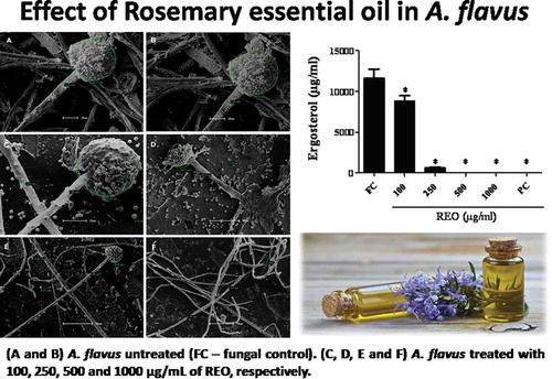
The increased risk to health by diverse pathologies, such as cancer, liver diseases, and endocrine alterations, caused by chemical residues in food, has led to the search for sustainable agricultural management alternatives, such as the use of essential oils for the development of natural and eco-friendly fungicides. The aim of this study was to evaluate the antifungal and antiaflatoxigenic activity of Rosmarinus officinalis L. essential oil (REO) against Aspergillus flavus Link. REO was obtained by hydrodistillation and its major components were identified as 1,8-cineole (eucalyptol, 52.2%), camphor (15.2%) and α-pinene (12.4%) by GC/MS and NMR. The minimum inhibitory concentration (MIC) and minimum fungicidal concentration (MFC) were both 500 µg/mL. REO reduced the mycelial growth of A. flavus at a concentration of 250 µg/mL (15.3%). The results obtained from scanning electron microscopy (SEM) demonstrated a reduction in the size of conidiophores and in the thickness of hyphae in A. flavus caused by treatment with REO (250 µg/mL). The production of ergosterol and the biomass of mycelium were both reduced as the REO treatment concentration increased. The production of aflatoxins B1 and B2 was inhibited after treatment with 250 µg/mL REO, a concentration below the MIC/MFC, indicating that the antiaflatoxigenic effect of REO is independent of its antifungal effect and is likely due to its direct action upon toxin biosynthesis. The data demonstrated that REO may be used as an alternative to synthetic fungicides.
Graphical abstract
Acknowledgments
This paper was part of the M.Sc. thesis work that was presented by the first author and funded by Coordenação de Aperfeiçoamento de Pessoal de Nível Superior – CAPES (Grant # 028054/2009), for the first and second author’s masters fellowships. We thank the Microscopy Central (CIM) of Complex of Central Research Support (COMCAP), Laboratory of Mycology, Laboratory of Histology and Laboratory of Inflammation at State University of Maringa for their cooperation in this study.
Disclosure statement
No potential conflict of interest was reported by the authors.