
Abstract
The aim was to investigate the long-term outcome after corrective osteotomy for malunion of distal radius fractures. Radiological findings, function, activity performance, pain, health-related quality of life and self-efficacy were studied. Evaluation of 37 patients 3–10 years after osteotomy fixated with a volar plate. Conventional radiographs were taken. Grip strength and range of motion were evaluated. Scores from the Patient Rated Wrist Evaluation (PRWE) were compared with normative values. The RAND-36 was used for evaluation of health-related quality of life and the General Self-Efficacy scale (S-GSE) for self-efficacy. Radial height, volar tilt, and ulnar variance improved postoperatively. In the long term, the corrections were maintained. Radiographs showed significantly more advanced osteoarthritis. Mean grip strength was 31 kg (SD 13) 89%, and range of motion varied between 80% and 95% compared to the uninjured side. The median PRWE was 12 points (0–99). The study group experienced higher levels of pain than reference values. There was a moderate correlation between the PRWE and volar tilt (rs = 0.453, p = .006) and grip strength (rs = 0.40, p = .014). At long-term follow-up functional outcome after a corrective osteotomy is generally good, but patients may experience some degree of pain. Corrective osteotomy might be considered for patients with a poor functional outcome after a distal radius fracture.
Introduction
Distal radius fracture is a common injury with an incidence of 26 per 10,000 person-years. The standard treatment consists of a reduction, in the event of displacement, which is reported to occur in up to 60% of patients, and cast immobilisation. Unstable fractures are usually treated surgically [Citation1]. One of the most common complications for initially displaced distal radius fractures is malunion. Malunion is reported to occur in approximately 35% of non-surgically treated fractures and up to 10% of surgically treated fractures [Citation2,Citation3].
Malunion may lead to difficulty performing activities of daily life due to pain or decreased range of motion and loss of grip strength. The degree of malunion can have an effect on the severity of the functional impairment [Citation4,Citation5].
Corrective osteotomy after a distal radius fracture is a procedure undertaken in the event of symptomatic malunion [Citation6]. There is no consensus about the exact radiographic indications when patients benefit from a corrective osteotomy.
The surgery aims to restore the anatomy of the distal radius and involves the controlled creation of a fracture near the original fracture, after which the malposition of the fragments of the distal radius is corrected. There are several ways to perform the surgery [Citation6].
In the short run, the results appear to be satisfactory both radiologically [Citation7] and in terms of range of motion and grip strength [Citation6]. Previous studies, using different patient-rated outcome measurements (PROM:s), have shown that patients also regain functional ability [Citation8]. However, studies indicate that the results may be hampered in the long term due to osteoarthritis of the wrist [Citation9]. Other factors, such as psychological aspects can also influence the outcome negatively [Citation10].
Self-efficacy is a central concept in the social psychological theory of Bandura, which is related to the strength of the individual’s belief in his/her own ability to handle different situations and achieve different goals [Citation11]. Studies have shown that self-efficacy has an impact on results after orthopaedic procedures in general [Citation10]. It is, therefore, of interest to study self-efficacy as a part of a long-term outcome.
The aim of this study was to investigate the long-term outcome, defined as more than 3 years, after corrective osteotomy for malunion of distal radius fractures, in terms of radiological findings, function, activity performance, pain, health-related quality of life and self-efficacy.
Materials and methods
Patients
The study was conducted at the Mölndal campus of the Sahlgrenska University Hospital, Gothenburg, Sweden. The inclusion criteria at the retrospective analysis were patients 16 years of age or more, who underwent an open wedge volar corrective osteotomy, after a symptomatic malunion of a distal radius fracture with a dorsal tilt of >25° or volar tilt of >20°, fixed with a volar plate between 2007 and 2014. In two cases, the indication was delayed primary osteosynthesis and as the fractures were already healed, osteotomy had to be carried out. Exclusion criteria were fixation with a dorsal plate or a concomitant ulna shortening.
The study was approved by the Ethics Committee in Gothenburg, Sweden (no. 153-17) and by the local committee for radiation protection (no SU 2017-01064 17-06).
Methods
The participants were evaluated once. The median time from osteotomy to follow up was 64 months (range 33–119).
Image acquisition and evaluation
All the patients underwent conventional radiographic investigation of the wrist. It was performed according to the standard clinical protocol with an anterior-posterior (AP) and a lateral exposure of the wrist, the latter with 10–15° distal inclination of the radiographic tube. Radiographs performed before and within the first postoperative week after the corrective osteotomy were identified retrospectively through the medical records. All the images were stored in the same radiological archive and measured using the hospital PACS software (Centricity Radiology RA 600 Clinical v.8.0, General Electric Healthcare, Barrington, IL). On the AP view, the radial height, radial inclination and ulnar variance were measured, while, on the lateral view, dorsal/palmar tilt was estimated. These measurements have previously been described by Cole et al. [Citation12]. See illustration –Citation4(b).
Figure 1. Initial CR of a 59 year old woman with a comminute intra-articular distal radial fracture and a fracture of the ulnar styloid process. (a) AP view. (b) Sagittal view showing dorsal tilting of the radial articular surface.
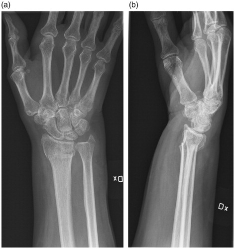
Figure 2. Preoperative CR of the same patient as in Figure 1. One year later, during conservative treatment, the distal radial fracture has healed in malunion and the patient is scheduled for corrective osteotomy. (a) AP view showing axial shortening. (b) Sagittal view showing severe volar tilt.
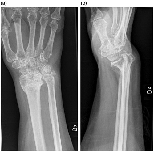
Figure 3. First postoperative control of the same patient as in Figures 1 and 2.The open-wedge corrective osteotomy is fixated with a volar plate. (a) AP view showing axial shortening and ulnar position is now restored. (b) Sagittal view showing volar tilt close to normal.
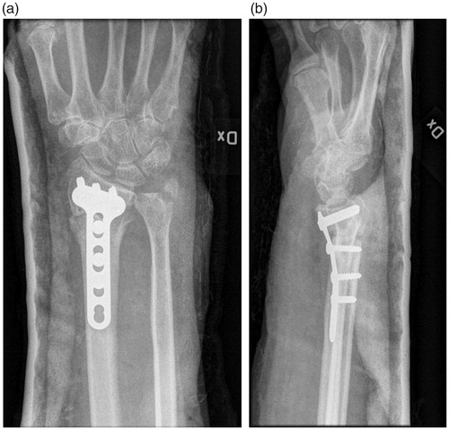
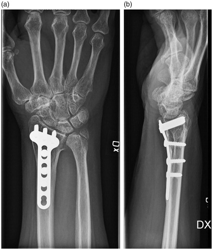
Figure 4. Long-term follow-up, 9 years post-operatively, of the same patient as in Figures 1–3. (a) AP view showing development of a slight axial shortening. No osteoarthritis is seen. (b) Sagittal view showing that the major improvement in joint positioning is maintained.
Radiographic osteoarthritis (OA) was scored according to Knirk and Jupiter [Citation13] as follows: grade 0 = none; 1 = slight joint space narrowing; 2 = marked joint space narrowing; 3 = bone on bone. Measurements were made by the same senior radiologist (Y. A.). The three sets of radiographs from each patient, preoperative-, postoperative- and long-term images, were evaluated twice with an interval of at least 1 week between evaluations.
Function: grip strength, range of motion
Grip strength and range of motion were evaluated comparing the injured side with the uninjured. The grip strength of both hands was measured using a Jamar Hand Dynamometer. The patient was in a seated position with the elbow in 90° of flexion. The mean value of three measurements was used [Citation14]. The range of motion of both wrists was measured using a goniometer in a standardised manner according to a national measurement manual [Citation15].
Subjective outcome
Activity performance and pain
Activity performance was evaluated by comparing the scores from the Patient Rated Wrist Evaluation (PRWE) [Citation16] and the shortened Disabilities of the Arm Shoulder and Hand questionnaire (Quick-DASH) [Citation17] with reference values. Pain was evaluated using the pain score of the PRWE [Citation16]. The PRWE quantifies patient-rated wrist pain and disability of daily living. The questionnaire consists of two subscales assessing pain (5 questions) and function (10 questions). Each question is scored on a categorical scale ranging from no pain/difficulty (0 points) to worst pain/unable to do (10 points). The pain score is the sum of the five items. The function score is the sum of the 10 items divided by two. The total score is the sum of the pain score and the function score. The PRWE thus ranges from 0 (a perfectly well-functioning wrist free of pain) to a total of 100 (a completely dysfunctional and painful wrist) [Citation16,Citation18]. The Swedish version of the PRWE has been tested for validity and reliability and sensitivity to change [Citation19].
The Disabilities of the Arm, Shoulder and Hand Questionnaire (DASH) evaluates symptoms and disability in the upper extremity and has been shown to be a reliable and valid tool to evaluate both proximal and distal function in the upper extremity [Citation20].
The Quick-DASH, used in this study, was derived from the DASH and consists of 11 items related to pain and function and ranges from 0 to 100 points, with lower scores indicating better function and less pain [Citation17].
Overall satisfaction with the surgical procedure was evaluated with a question constructed for this study: ‘How would you describe the way your wrist is functioning today?’ The answer was marked on a Likert scale using the following alternatives: very bad, bad, quite good, good and excellent. In an attempt to enrich the quantitative and qualitative data, the patients were also given the opportunity to comment on their wrist function in their own words.
Health-related quality of life and self-efficacy
The Swedish version of the RAND-36 was used to evaluate health-related quality of life. It consists of 36 items, measuring eight different domains of health. The score for each domain ranges from 0 to 100 points, with a higher score indicating better health [Citation21]. To evaluate self-efficacy the Swedish version of the General Self-Efficacy scale (S-GSE) was used. The S-GSE consists of 10 questions that are graded on a Likert scale with four steps, each step given 1–4 points. The score is the sum of the points and thus ranges from 10 to 40 points. The score can be presented as a total score or as a mean for the 10 items. There is no cut-off but higher scores indicate better self-efficacy [Citation22].
The functional assessments were made by the main author who is an occupational therapist experienced in hand therapy. If the patient needed an examination by a physician, based on clinical or radiological findings, the patient was referred to a specialist in orthopaedics (M. U.).
Statistical analysis
The mean, SD, median and range were used to describe demographics and outcome. The Shapiro–Wilks test was used for tests of distribution for range of motion and grip strength and radiographic parameters. Values for range of motion and grip strength were normally distributed. A paired-sample t-test was used to perform pair-wise comparisons between operated and non-operated hands in terms of range of motion and grip strength.
As the radiographic variables were not normally distributed, Wilcoxon’s signed-rank test for non-parametrically distributed variables was used for comparisons at group level.
The Mann–Whitney U test was used to compare activity performance between men and women and Wilcoxon’s signed-rank test was used between the studied group and age and gender specific reference values. All significance tests were two tailed and conducted at the 0.05 significance level.
Spearman’s rho coefficient (rs) was used to test the possible correlation between radiological parameters and activity performance, measured with the PRWE, and range of motion. Spearman’s rho was also used to test if there was a correlation between degree of osteoarthritis and age and between osteoarthritis and duration between surgery and follow-up. To describe the strength of the correlations, the following categories were used: 0–0.19 (very weak), 0.20–0.39 (weak), 0.40–0.59 (moderate), 0.60–0.79 (strong) and 0.80–1.0 (very strong) [Citation23]. Analyses were performed with SPSS for Windows (Version 20, SPSS, Inc., Chicago, IL).
Results
Sixty patients were identified through medical records. Eight patients were deceased. The remaining 52 were initially contacted by mail with information about the study. Two weeks later, the patients were contacted by telephone and given more detailed oral information and an invitation to participate. Five patients could not be reached by either phone or mail. Eight women, aged 49–78 years, declined participation; three were satisfied with their function and had no interest in participation, four because of time constraints and one was in hospital, for other reasons, at the time of the study. A total of 39 patients were enrolled in the study and gave their written, informed consent to participate. Two patients were incorrectly coded in the medical records and did not have volar plates and were, therefore, excluded. Finally, 20 women and 17 men were included. One patient declined radiological examination but took part in the functional assessment. Twenty-eight of the patients were of working age at the time they were injured. The dominant arm was affected in 18 patients and the non-dominant arm in 19. Twenty-five fractures were due to low-energy trauma, 12 to high-energy trauma, 16 fractures were extra-articular and 17 intra-articular, while data were missing in four cases. The primary treatment was a plaster following closed reduction in 16 cases, and no reduction in 10 cases and 11 fractures underwent surgery with volar plates with or without Euloc-pins (Euloc-pin®, Swemac AB, Linköping, Sweden). For detailed information on demographics, see .
Table 1. Demographics.
The surgery was performed by five different surgeons, of which one performed 27 of the osteotomies. None of them was engaged in the analysis of the results.
Surgical procedure
The osteotomy was carried out through a dorsal incision between the 2nd and the 4th extension compartment, after release of the EPL tendon. An oscillating saw was used and the volar cortex was fractured with a chisel. The osteotomy was widened with a distractor until the correct angle was achieved and temporarily fixed with two K-wires. Then a modified volar approach according to Henry was performed and the osteotomy fixed with a conventional volar titanium plate (DVR® Medica, Stryker Variax®) or a polyetheretherketon plate (PEEK-plate, DiphosR®, Lima Corporation, San Daniele del Friuli, Italy). The osteotomy gap was filled with autologous or synthetic graft (Hydroset®, Stryker Corp, Kalamazoo, MI) or left empty. The skin was closed using resorbable intra-cutaneous sutures (Monocryl 4-0). Then a plaster was applied for 2–4 weeks according to the surgeons’ discretion.
One patient underwent a median-nerve decompression and eight patients had the plate removed 1–4 years after the osteotomy. One patient underwent a tendon transfer due to an EPL-rupture 2 years after the osteotomy.
Radiological findings
The results of the radiological findings in terms of remaining malunion and osteoarthritis are presented in .
Table 2. Number of patients related to the level of osteoarthritis pre-operatively (N = 37) and at follow-up (N = 36), measured using the scale of Knirk and Jupiter.
There was an improvement, with respect to anatomical parameters, with an increase in radial height (p = .001) and volar tilt (p = .016) after surgery compared with before, and significantly less ulnar variance (p < .001). However, no significant difference was found in radial inclination preoperatively as compared with postoperatively.
The degree of osteoarthritis had increased significantly at the follow-up compared with postoperatively (p < .001). There was a weak correlation between age and osteoarthritis at follow up (rs= 0.37, p = .03). There was no difference in terms of osteoarthritis with respect to gender. There was no significant correlation between the degree of osteoarthritis and time from surgery to follow-up. No significant differences were found in terms of radial height, radial inclination, ulnar variance and volar tilt during this time. None of the patients had an implant failure at follow-up.
Function: grip strength, range of motion
In , the results in terms of grip strength and range of motion are presented. At long-term follow-up, the grip strength of the injured hand was 31 kg (SD 13), (89%) on average compared with the uninjured hand. For men, the grip strength was 38 kg (SD 14) (86%) on average compared with the uninjured hand, while it was 25 (SD 7) (94%) for women.
Table 3. Grip strength (KG) and range of motion (degrees).
The range of motion in the injured wrist was 80–95% on average in different movement directions compared with the uninjured side. The greatest restoration of range of motion was observed in pronation, dorsal extension followed by supination, while the least was seen in radial deviation. There was a significant difference between injured and uninjured hands in all movement directions (p < .05). There was no correlation between radiological findings and range of motion.
Activity performance and pain
The median total score for the PRWE was 12 points (IQR 23), (women 19.5 (IQR 50), men 5 (IQR 17)). The total mean score, calculated in relation to individual age and gender specific reference-values, was for the whole group for the PRWE 21.3 points (SD 26.3). This was significantly higher (p = .001) than the reference mean value of 7.7 points (SD 15.0) [Citation24]. When dividing the group according to gender, it was found that the women scored significantly higher than the reference values, 27.9 (SD 26.0) versus 8.6 (SD 15.9) (p = .001). The same was not seen among the men, 13.7 (SD 23.7) versus 6.5 (SD 13.6) (p = .075).
The median pain score was 9 points (IQR 23) in the whole group, women 14 (IQR 30) and men 4 (IQR 14). This value is significantly higher than the reference values (women 4.7 (SD 9.0) and men 3.8 (SD 8.1)) for the whole group (p = .002) and among the women (p = .003) [Citation24].
There was a moderate correlation between a lower PRWE-total score grip strength (rs= 0.40, p = .014) and between a lower PRWE-total score and more volar tilt (rs= 0.453, p = .006) and a very weak, albeit non-significant, correlation between a higher PRWE-pain score and increased osteoarthritis (rs=0.108, p = .53).
The median score for the Q-DASH was 11.4 points (IQR 19.8). The reported reference values are 10–15 points (SD 14.7) [Citation25].
Health-related quality of life and self-efficacy
The results for health-related quality of life and self-efficacy are presented in . The domains of the RAND-36 that had the lowest median scores were Bodily pain, Vitality/fatigue and General health. The domains with the highest median scores were Role physical, Social functioning and Role emotional.
Table 4. RAND-36 scores N = 37 median (min–max), mean (SD).
The results for the S-GSE showed a mean score of 3.2 points (SD 0.6), median 3.3 (IQR 0.8), which was significantly higher (p = .002) than the reference values; mean 2.9 (SD 4.6) [Citation22].
The ratings for a self-constructed question about overall satisfaction with the wrist were as follows: very bad (n = 1), bad (n = 5), quite good (n = 8), good (n = 14) and excellent (n = 8). One patient did not answer this question.
In answer to the question of whether they would have chosen surgery, if they had known the results, 33 patients answered yes and 3 no. One patient did not answer this question.
Of the patients who made comments in their own words (n = 16), five stated they were dissatisfied with the waiting time for surgery, five commented on being satisfied with the result of surgery, one was dissatisfied with the result of surgery because of restraints in fine dexterity, five had complaints related to communication with caregivers, one was satisfied with the communication with caregivers, one had a question about the implant and one commented on being free from pain since plate removal.
At the long-term follow-up, six patients were referred to a specialist in orthopaedics (MU) due to pain and/or crepitation.
Discussion
According to the results of the present study the surgical correction has resulted in a significant improvement in radial height, volar tilt and ulnar variance postoperatively. In the long term, the corrections were maintained. The level of osteoarthritis increased, however. The study also showed that the function was regained in terms of grip strength to 89% compared to the non-injured hand and the greatest restoration of range of motion was observed in pronation, dorsal extension followed by supination, while the least was seen in radial deviation. The activity performance measured by the PRWE was significantly lower than reference values [Citation24].
Activity performance showed a moderate correlation with volar tilt. Batra and Gupta [Citation26] found that radial length, followed by volar tilt, was the most important predictor of functional recovery 1 year after the treatment (both surgical and non-surgical) for distal radius fractures [Citation26]. Another recent study has shown that there is an association between ulnar variance as well as radial inclination and patient-reported outcomes [Citation27]. This indicates that the restoration of the wrist to a more anatomical position may have an influence on activity performance. However, the evidence supporting this in other studies is inconclusive and the results should be interpreted cautiously [Citation28].
Earlier research on functional outcome after the surgical treatment of distal radius fractures show the importance of recovery of grip strength in order to perform different tasks and activities [Citation29]. The present long-term study showed a moderate correlation between the PRWE-score and grip strength. It also showed that the grip strength of the injured hand was restored to 89% compared with the uninjured hand, which is a slightly higher level, compared with previous studies that have reported results of 71–85% of the grip strength of the injured hand [Citation8,Citation9,Citation30].
For different movement directions, the restoration of the range of motion arc varied from 0 (for two individuals the radial deviation was 0° in the operated wrist) to 100%. It can be presumed that, for the individual, it is the sum of range of motion in all directions that matters. In any case, earlier studies have shown that supination is of great importance for the ability to perform activities [Citation29]. In the present study, supination was restored to 89%, which can be regarded as an excellent result. However, only 61% of the patients rated their function as good or excellent compared with 72–76% in some previous studies [Citation30,Citation31]. This indicates that range of motion and the grip strength are not the only outcome measurements that should be used to determine the function of the wrist and hand.
The results of the PRWE, which is sensitive to wrist problems, showed more difficulties in carrying out activities in the study group, compared with reference values [Citation24].
The women experienced more pain than the men. Reference values for PRWE-pain are higher for women than for men, but also, in comparison with the reference values, the women in the study group experienced more pain [Citation24]. The study cohort is too limited to determine whether there is a difference between men and women with respect to pain in the cohort. In any case, the result might indicate that a certain amount of residual pain can be expected in the long term after a corrective osteotomy, something that it is important to communicate to patients waiting for surgery, in order for them to have realistic expectations. One patient had extremely poor outcome and had high scores on both the PRWE (99 p) and the Q-DASH (90.9 p) due to pain, low grip strength and a poor result in restoration of movement capacity in supination and volar flexion.
Pain possibly explains why the ratings for satisfaction with the wrist were not higher. Osteoarthritis is mentioned in an earlier study as a possible explanation of poorer outcome [Citation9] and may explain why the study group experienced higher levels of pain compared with the normal population. The degree of OA was significantly increased at the long-term follow-up, but there was no significant correlation between an increase in OA and an increase in the PRWE pain score. Further, there was no difference in the distribution of OA with respect to gender.
In terms of health-related quality of life, the two dimensions with the lowest scores were ‘Bodily pain’ and ‘Vitality/fatigue’. These domains reflect pain anywhere in the body and physical impairment experienced in a range of different activities such as climbing stairs or lifting heavy objects and the experience of fatigue. The scores reflect that a few individuals experienced bodily pain, not only in the wrist but also at other sites in the body, as well as fatigue. The study group had high scores in terms of ‘Social functioning’, ‘Role physical’ and ‘Role emotional’, which indicates that they experienced good quality of life in their social relationships and different life roles. These results are expected in this population.
Perceived function is a complex phenomenon in which different aspects, such as general self-efficacy, might have an influence. Self-efficacy has been shown to have an impact on results after orthopaedic procedures in general [Citation10] and also in relation to functional impairment and pain reported among persons suffering from chronic pain [Citation32]. Self-efficacy develops over time [Citation11] and measured 3–11 years after surgery it probably measures coping with the consequences of a long-standing pain problem rather than shows a predictive value of how it influences functional results of surgery. It is not clear if self-efficacy influences the ability to perform activities or contrary, if it is the ability to perform activities that influences self-efficacy, or rather, both. It might have been more appropriate to measure self-efficacy preoperatively to measure its predictive value on the long-term results.
Thirty-three (89%) of the 37 patients would have chosen surgery again, had they known the result. One possible explanation of this is that the patients experience less pain and greater function than before surgery and, even if the wrist is not fully restored, their ability to use their hand in everyday activities has improved.
Limitations
The relatively low number of eligible patients for this study may be a result of the choice to invite only patients operated on at the campus of Mölndal. Fifteen patients were operated on at the campus of Sahlgrenska during the period of study. Another explanation might be that patients left the waiting queue and were operated on at other hospitals. This has not been investigated, which is a limitation of the study. An additional reason may be the strict exclusion criterion where we chose to exclude patients where the osteotomy was stabilized using a dorsal plate. This exclusion criterion was chosen because patients more often experience problems with tendons after dorsal plating and were considered not being comparable to patients with volar plates.
Another limitation is the retrospective character of this study because no pre-operative measurements of range of motion, grip strength or scores from the different PROM:s were available for comparisons.
Conclusion
In the present study, the initial treatment of the patients varied and some had undergone different surgical procedures prior to the osteotomy. In spite of this, they felt that the osteotomy was worthwhile. This is consistent with the findings of another recent study [Citation8]. The present study contributes to the growing evidence relating to the long-term results of corrective osteotomy. This by showing that the correction is maintained over time and that function and ability to perform activities are restored to a high degree, even if function is not restored fully, compared to normal values, and patients may experience some residual pain. Corrective osteotomy might be considered for patients who experience a poor outcome after a distal radius fracture.
Disclosure statement
The authors declare no conflicts of interests.
Additional information
Funding
References
- Brogren E, Petranek M, Atroshi I. Incidence and characteristics of distal radius fractures in a southern Swedish region. BMC Musculoskelet Disord. 2007;8(1):48.
- Sharma H, Khare GN, Singh S, et al. Outcomes and complications of fractures of distal radius (AO type B and C): volar plating versus nonoperative treatment. J Orthop Sci. 2014;19(4):537–544.
- Wilcke MK, Hammarberg H, Adolphson PY. Epidemiology and changed surgical treatment methods for fractures of the distal radius: a registry analysis of 42,583 patients in Stockholm County, Sweden, 2004–2010. Acta Orthop. 2013;84(3):292–296.
- Gliatis JD, Plessas SJ, Davis TR. Outcome of distal radial fractures in young adults. J Hand Surg Br. 2000;25(6):535–543.
- Beumer A, McQueen MM. Fractures of the distal radius in low-demand elderly patients: closed reduction of no value in 53 of 60 wrists. Acta Orthop Scand. 2003;74(1):98–100.
- Prommersberger KJ, Pillukat T, Muhldorfer M, et al. Malunion of the distal radius. Arch Orthop Trauma Surg. 2012;132(5):693–702.
- Wada T, Tatebe M, Ozasa Y, et al. Clinical outcomes of corrective osteotomy for distal radial malunion: a review of opening and closing-wedge techniques. J Bone Joint Surg Am. 2011;93(17):1619–1626.
- Mulders MAM, d’Ailly PN, Cleffken BI, et al. Corrective osteotomy is an effective method of treating distal radius malunions with good long-term functional results. Injury. 2017;48(3):731–737.
- Lozano-Calderon SA, Brouwer KM, Doornberg JN, et al. Long-term outcomes of corrective osteotomy for the treatment of distal radius malunion. J Hand Surg Eur Vol. 2010;35(5):370–380.
- Flanigan DC, Everhart JS, Glassman AH. Psychological factors affecting rehabilitation and outcomes following elective orthopaedic surgery. J Am Acad Orthop Surg. 2015;23(9):563–570.
- Bandura A. Self-efficacy: toward a unifying theory of behavioral change. Psychol Rev. 1977;84(2):191–215.
- Cole RJ, Bindra RR, Evanoff BA, et al. Radiographic evaluation of osseous displacement following intra-articular fractures of the distal radius: reliability of plain radiography versus computed tomography. J Hand Surg Am. 1997;22(5):792–800.
- Knirk JL, Jupiter JB. Intra-articular fractures of the distal end of the radius in young adults. J Bone Joint Surg Am. 1986;68(5):647–659.
- Mathiowetz V, Weber K, Volland G, et al. Reliability and validity of grip and pinch strength evaluations. J Hand Surg Am. 1984;9(2):222–226.
- Nationell mätmanual. Manual for rorelse styrka version 1: Handkirurgiskt kvalitetsregister; 2011. [cited 2011 01112018]. Available from: https://hakir.se.
- MacDermid JC, Turgeon T, Richards RS, et al. Patient rating of wrist pain and disability: a reliable and valid measurement tool. J Orthop Trauma. 1998;12(8):577–586.
- Gummesson C, Ward MM, Atroshi I. The shortened disabilities of the arm, shoulder and hand questionnaire (QuickDASH): validity and reliability based on responses within the full-length DASH. BMC Musculoskelet Disord. 2006;7(1):44.
- MacDermid JC. The Patient-Rated Wrist Evaluation (PRWE) User Manual June 2011 [cited 2019 190904]. Available from: http://srs-mcmaster.ca/wp-content/uploads/2015/05/English-PRWE-User-Manual.pdf.
- Mellstrand Navarro C, Ponzer S, Tornkvist H, et al. Measuring outcome after wrist injury: translation and validation of the Swedish version of the patient-rated wrist evaluation (PRWE-Swe). BMC Musculoskelet Disord. 2011;12(1):171.
- Beaton DE, Katz JN, Fossel AH, et al. Measuring the whole or the parts? Validity, reliability, and responsiveness of the Disabilities of the Arm, Shoulder and Hand outcome measure in different regions of the upper extremity. J Hand Ther. 2001;14(2):128–146.
- Orwelius L, Nilsson M, Nilsson E, et al. The Swedish RAND-36 Health Survey – reliability and responsiveness assessed in patient populations using Svensson's method for paired ordinal data. J Patient Rep Outcomes. 2018;2(1):4.
- Schwarzer R. Everything you want to know about the General Self-Efficacy Scale 2014. [updated 2014 May 30; cited 2016 Mar 23]. Available from: http://userpage.fu-berlin.de/health/selfscal.htm.
- Statstutor. Spearman´s correlation. [cited 2018 Nov 01]. Available from: http://www.statstutor.ac.uk/resources/uploaded/spearmans.pdf
- Mulders MAM, Kleipool SC, Dingemans SA, et al. Normative data for the Patient-Rated Wrist Evaluation questionnaire. J Hand Ther. 2018;31(3):287–294.
- Aasheim T, Finsen V. The DASH and the QuickDASH instruments. Normative values in the general population in Norway. J Hand Surg Eur Vol. 2014;39(2):140–144.
- Batra S, Gupta A. The effect of fracture-related factors on the functional outcome at 1 year in distal radius fractures. Injury. 2002;33(6):499–502.
- Ali M, Brogren E, Wagner P, et al. Association between distal radial fracture malunion and patient-reported activity limitations: a long-term follow-up. J Bone Joint Surg Am. 2018;100(8):633–639.
- Plant CE, Parsons NR, Costa ML. Do radiological and functional outcomes correlate for fractures of the distal radius? Bone Joint J. 2017;99-B(3):376–382.
- Swart E, Nellans K, Rosenwasser M. The effects of pain, supination, and grip strength on patient-rated disability after operatively treated distal radius fractures. J Hand Surg Am. 2012;37(5):957–962.
- Flinkkila T, Raatikainen T, Kaarela O, et al. Corrective osteotomy for malunion of the distal radius. Arch Orthop Trauma Surg. 2000;120(1-2):23–26.
- Krukhaug Y, Hove LM. Corrective osteotomy for malunited extra-articular fractures of the distal radius: a follow-up study of 33 patients. Scand J Plast Reconstr Surg Hand Surg. 2007;41(6):303–309.
- Jackson T, Wang Y, Wang Y, et al. Self-efficacy and chronic pain outcomes: a meta-analytic review. J Pain. 2014;15(8):800–814.