
ABSTRACT
We are reporting a case of pneumonitis in an 81 year old-aged woman due to gemcitabine who was successfully managed with steroids. We also reviewed the literature and found previous case reports of gemcitabine induced pneumonitis.We reported this case to alert physicians to be aware of this infrequent and sometimes fatal complication from gemcitabine.
1. Introduction
Gemcitabine is one of the chemotherapy drugs used in the treatment of breast cancer. The most common adverse effect is myelosuppression; lung toxicity is rarely seen. Here, we present a case of an 81 year old female with metastatic breast cancer who presented with fever and dyspnea, two weeks after the second cycle of gemcitabine. Chest Xray and Computed Tomography (CT) scan chest revealed diffuse bilateral ground glass opacities. She then underwent bronchoscopy and bronchoalveolar lavage (BAL) which ruled out any infection or malignancy progression. Complete clinical and radiological response was achieved with steroid treatment.
2. Case description
Eighty one year old female with a past medical history of breast cancer with metastasis to the lung and pleura. She had radiation to the lung lesion 10 months ago and was started on gemcitabine every 2 weeks one month ago. She had finished 2 cycles of the chemotherapy and developed fever and shortness of breath, ten days after her second cycle. On presentation, she was hemodynamically stable but hypoxic with saturations at 88% on room air, which improved to 96% on two litres nasal cannula. Physical exam revealed decreased breath sounds bilaterally and moderate rales throughout, especially on the left side but no wheezing. Her initial work up included chest x ray which showed left upper lobe opacity. A CT scan chest was done which ruled out pulmonary embolism (PE) but revealed bilateral diffuse multifocal pneumonia, which was worse in the left upper lobe (). She was started on treatment with broad spectrum antibiotics and supplemental oxygen through a nasal cannula.
Figure 1. Bilateral ground glass opacities
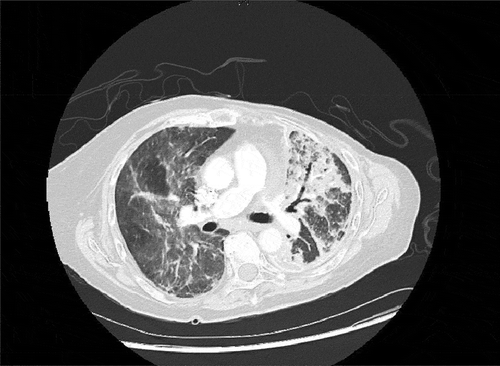
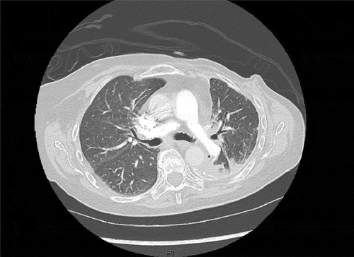
On Day 3 of her hospital admission, she became more hypoxic leading to increased oxygen requirements. A repeat chest x ray demonstrated bilateral airspace consolidations and interstitial markings which had worsened in comparison to the prior study. Blood cultures and sputum cultures still revealed no infection. Because of her worsening clinical condition, an alternative diagnosis such as gemcitabine induced pneumonitis was pursued and the patient was started on intravenous (IV) methylprednisolone 40 mg twice daily. She underwent flexible bronchoscopy with bronchoalveolar lavage (BAL) because of minimal improvement on steroids which was ultimately negative for infection and malignant cells. Despite her initially worsening picture, she gradually started to show improvement and was able to be off oxygen. Her IV steroids were switched to per oral (PO) prednisone 60 mg once daily with gradual tapering by 10 mg every 5 days. A repeat CT scan chest 2 months later showed complete resolution of pneumonitis ().
Figure 2. Resolution of pneumonitis
3. Discussion
Gemcitabine is a chemotherapy drug used in the treatment of multiple types of cancers including non-small cell lung cancer, pancreatic cancer, bladder cancer, breast cancer and esophageal cancer. It is a pyrimidine analogue which replaces cytidine during DNA replication thereby halting tumor growth by inducing apoptosis [Citation1].
Pulmonary toxicity from gemcitabine is relatively rare as evidenced by the low incidence of 0–5% grade III/IV toxicity in patients with various solid tumors. Increased risk of gemcitabine-induced pulmonary toxicity was found in patients with pre-existing lung disease, as well as in patients with previous thoracic irradiation and combination chemotherapy with drugs known to cause lung injury [Citation2].
The exact mechanism of this injury is unclear, but it has been speculated to be due to gemcitabine induced release of proinflammatory cytokines leading to dysregulation of tissue repair [Citation3]. Another speculated pathogenesis is from the fact that Cytarabine, a pyrimidine analogue which is structurally and metabolically similar to gemcitabine can cause damage to capillary endothelial cells leading to interstitial and intra alveolar proteinaceous edema resulting in acute respiratory distress syndrome (ARDS) [Citation4–6].
We performed a PubMed based comprehensive literature review. The search terms included ‘Gemcitabine’ and ‘lung toxicity’. We reviewed all results and found the following previous case reports as outlined below ().
Table 1. Cases of Gemcitabine induced pneumonitis
The timing of toxicity varies. It may occur immediately within a few hours of administration or within a few days. However, as evidenced by the date in , the majority of patients acquired it after their second cycle. The most common clinical finding is dyspnea in 70%, followed by fever and lung infiltrate in 35% and 21.9% respectively [Citation7]. Our patient presented ten days after her second cycle with fever and shortness of breath and was found to have lung infiltrates consistent with the most common presentation of gemcitabine induced lung toxicity.
Diagnosis is by exclusion and is usually made by a combination of a patient’s clinical picture, radiological evidence predominantly showing bilateral pulmonary or interstitial infiltrates and responsiveness to steroids. Bronchoalveolar lavage is usually also performed to rule out other causes of pulmonary toxicity. Lung biopsy is not an essential part of the work up but confirms the diagnosis [Citation2]. From the above, almost all patients had a CT scan but a few of them had BAL and lung biopsy. Our patient had a CT scan on admission which showed diffuse bilateral ground glass opacities. Given
her initial minimal improvement on steroids, she underwent BAL to rule out infection, malignancy, and other non-iatrogenic causes.
As gemcitabine induced lung injury is a diagnosis of exclusion and shares common clinical characteristics with other common lung pathologies like pneumonia, almost everyone receives antibiotics first. It’s the progression of symptoms on antibiotics that triggers alternate diagnosis.
Systemic steroids are widely used, and most patients show clinical improvement. As seen in , almost all patients received steroids. Fenocchio et al reported a case of a 69 year old male with pancreatic cancer who developed lung toxicity after 2 cycles of gemcitabine which ultimately responded to imatinib mesylate as steroids did not show improvement [Citation8]. Our patient was initially on antibiotics for suspected pneumonia and was later switched to steroids after which improvement was noted. Her IV methylprednisolone was switched to an equivalent dose of oral prednisone, which was gradually tapered.
In conclusion, lung toxicity due to gemcitabine is infrequent and usually presents with a clinical picture similar to pneumonia. Pre-existing lung disease, prior radiation exposure with concomitant chemotherapeutics known to cause lung injury will increase the risk of pulmonary toxicity. Diagnosis is by exclusion. CT and BAL are helpful in ruling out other causes, and lung biopsy helps in confirming t he diagnosis. Standard treatment is with systemic steroids, and most patients usually show a complete resolution. It is imperative that clinicians are aware of this adverse but treatable side effect of gemcitabine.
Disclosure statement
No potential conflict of interest was reported by the authors.
References
- Ullah K, O’Reilly A, Power DG, et al. A case series of patients on chemotherapy with dyspnoea and pulmonary infiltrates. BMJ Case Rep. 2013 Published 2013 Jun 5;2013:bcr2013009105.
- Barlési F, Villani P, Doddoli C, et al. Gemcitabine-induced severe pulmonary toxicity. Fundam Clin Pharmacol. 2004;18:85–91.
- Rübe CE, Wilfert F, Uthe D, et al. Increased expression of pro-inflammatory cytokines as a cause of lung toxicity after combined treatment with gemcitabine and thoracic irradiation. Radiother Oncol. 2004;72(2):231–241.
- Haupt HM, Hutchinson GM, Moore GW. Ara-C: non-cardiogenic pulmonary edema complicating cytosine arabinoside therapy in acute leukemia. Am J Med. 1981;70:256–261.
- Pavlakis N, Levi JA, Wheeler HR, Begbie SD, Bell DR: Novel pulmonary and neurological (GNS) toxicities with Gemcitabine. Proc ASCO 15: 181, 1996.
- Tham R, Peters WG, de Bruine FJ, et al. Pulmonary complications of cytosine arabinoside therapy: radiological findings. Am J Roentgenol. 1987;70:251–261.
- Comito F, Grassi E, Poerio A, Freier E, Calculli L, Zompatori M, Ricci C, Casadei R, Di Marco M. Organizing pneumonia after pancreatic cancer treatment with nab-paclitaxel and gemcitabine: a case report. BJR| case reports. 2018 Jun;4(2):20170086.
- Fenocchio E, Depetris I, Campanella D, et al. Successful treatment of gemcitabine-induced acute interstitial pneumonia with imatinib mesylate: a case report. BMC Cancer. 2016;16(1). DOI:10.1186/s12885-016-2833-9