
ABSTRACT
Rothia species are gram positive, round to rod-shaped bacteria that are normally oral and respiratory tract flora. They were first isolated in 1967 from dental caries. We present a 69-year-old male with no risk factors for aforementioned bacteria however was found to have thickened anterior leaflet of the mitral valve with a small isoechoic lesion consistent with vegetation on Transthoracic echocardiogram. Blood cultures grew pan sensitive Rothia dentocariosa. Patient was treated with long-term antibiotics. This case adds to the limited number of cases of Rothia dentocariosa Endocarditis.
1. Introduction
Infective endocarditis (IE) is a relatively uncommon disease as mentioned in the epidemiological study by Pant et.al. [Citation1] This aforementioned study published in 2015 estimated that the incidence of IE in the USA from 2000 to 2011 to be around 11 to 15 cases per 100,00 population [Citation1]. A reservoir of pathogens, most commonly floral in nature, is often involved; in addition to an access pathway into the patient’s bloodstream, and a suitable environment for the pathogen to proliferate and vegetate, i.e. an abnormal endocardium or a prosthetic heart valve. Some unmodifiable risk factors include age >60 years and male gender. On some rare occasions, IE might develop despite the lack of most identified risk factors. In this report, we discuss a case of IE caused by an unlikely pathogen, Rothia dentocariosa, in a patient who is lacking most of the aforementioned risk factors.
2. Case presentation
A 69-year-old male with a past medical history of diabetes mellitus, hypertension, hepatitis-C, and end-stage renal disease on hemodialysis initially presented to the hospital after he was found on the floor and brought to the emergency department by EMS. The patient reported that he missed his last dialysis session and complained of chronic upper back and neck pain. He otherwise denied any other complaints. He was afebrile and hemodynamically stable on admission. His physical exam was unremarkable except for the left upper arm AV graft in place. He had complete upper and lower dentures in place.
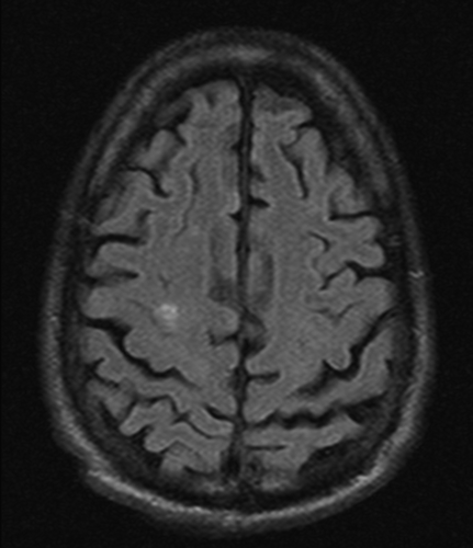
His blood work was remarkable for leukocytosis with WBCs 15.4 mg/dl with 86% neutrophils, normocytic normochromic anemia with hemoglobin 10.7 mg/dl, BUN 81 mg/dl, creatinine 15 mg/dl, potassium 6 mmol/l. CT head showed small foci of parenchymal hemorrhage at the bilateral frontal centrum semiovale.
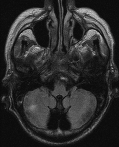
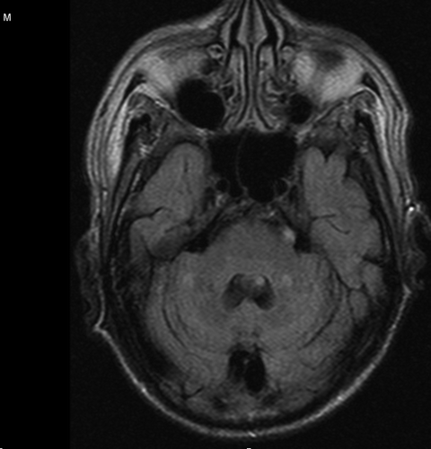
CT spine showed destructive disc and endplate changes at C6-C7 suggestive of diskitis and osteomyelitis, which was confirmed later by MRI with no evidence of cord compression. A cervical collar was placed, and the patient was placed on broad-spectrum antibiotics and transferred to the CCU for closer monitoring. MRI of the brain showed multiple embolic hemorrhagic strokes (Figures 1–3).
Transthoracic echocardiogram showed thickened anterior leaflet of the mitral valve with a small isoechoic lesion, 6 mm in length, attached to the atrial side of the mid-portion of the thickened anterior mitral valve leaflet with independent motion consistent with vegetation (video1).
Two blood culture bottles showed Rothia dentocariosa sensitivity for ceftriaxone and penicillin. PICC line was placed inpatient, and he was discharged to subacute rehab for long-term antibiotics.
3. Discussion
Rothia species are gram-positive, round- to rod-shaped bacteria that are part of the normal flora residing in the oral cavity and respiratory tract. It was first isolated from dental caries in 1967. Species include R. mucilaginosa, R. dentocariosa, R. aeria, R. nasimurium, and R.amarae [Citation2]. Rothia is a rare cause of endocarditis. However, endocarditis is the most common infection associated with Rothia. It has been reported to cause pneumonia, septic arthritis, peritonitis [Citation3], endophthalmitis, and bacteremia [Citation4]. Rothia dentocariosa is an aerobic coccoid to a rod-shaped, non-sporogenic, non-motile, catalase-positive gram-positive bacterium found in 30% of the oral cavity of healthy individuals. Risk factors associated with R. dentocariosa endocarditis include periodontal disease or manipulation, IV drug use [Citation5], pre-existing heart defects, prosthetic devices, especially prosthetic heart valves [Citation6], immunosuppression. Endocarditis is found to be more common in males than in females. The periodontal infection seems to be the initial source of bacteremia. Sometimes it could be an occult oral source with no overt dental disease as noted in 2 cases of R. dentocariosa with no apparent risk factors [Citation7,Citation8]. Surprisingly our patient is edentulous and has upper and lower dentures making periodontal disease an unlikely source. He also denied any recent IV drug use, which would be another source of infection. Identification of R. dentocariosa can be challenging. It could be mistaken by other organisms such as Corynebacterium, which may result in misidentifying as a contaminant and result in a delay in diagnosis [Citation5]. A transesophageal echocardiogram gives better diagnostic views of the heart valves and is more sensitive in detecting valve lesions and associated complications. However, due to the presence of cervical diskitis and osteomyelitis at C6-C7, the patient could not go for TEE. A literature review was done in 2003 by Boudewijns et al. It identified 20 cases of R. dentocariosa endocarditis [Citation10]. In a recent literature review published in 2020 by Franconieri et al., which included 51 cases of rothia induced endocarditis, 55% of them were due to Rothia dentocariosa [Citation11]. Of the cases of R. dentocariosa endocarditis, 64% had pre-existing valve disease, and 46% had Bucco dental diseases. The most commonly involved valve was the aortic, followed by the mitral valve. R. dentocariosa endocarditis has a low mortality rate of about 14%, based on the previous two reviews. However, there was a high rate of associated complications; Cardiac complications include paravalvular aortic abscess. Central nervous system complications include Septic embolic strokes [Citation3], Intracranial hemorrhagic strokes, and subarachnoid hemorrhages [Citation10]. Mycotic aneurysms, in particular, seem to be common, with a reported incidence in 25% of the cases [Citation9]. Other complications include vertebral osteomyelitis, brain abscesses, and abdominal aneurysms. No definite guidelines regarding treatment are present due to the rarity of Rothia endocarditis. The organism is usually susceptible to penicillin and cephalosporins. Treatment is by beta-lactam antibiotics for 4–6 weeks; options include penicillin with or without gentamicin or ceftriaxone [Citation11-12]. Our patient had mitral valve endocarditis, which was complicated by vertebral osteomyelitis and hemorrhagic embolic strokes believed to be septic emboli as a result of endocarditis. Our patient also had multiple comorbidities, which would make him a poor surgical candidate.
7. Conclusion
Infectious Endocarditis certainly follows common predisposing factors, but that is not always the case. As in this case report, our patient did not have the recognized propensity to develop IE. Rothia dentocariosa belongs to an uncommon group of bacteria implicated in IE and is generally regarded as one of the less severe agents. But that stratification of virulence is not absolute as rare microbes are still capable of producing major morbidity and mortality in the context of infective endocarditis.
Disclosure statement
No potential conflict of interest was reported by the authors.
References
- Pant S, Patel NJ, Deshmukh A, et al. Trends in infective endocarditis incidence, microbiology, and valve replacement in the USA from 2000 to 2011. J Am Coll Cardiol. 2015 May 19;65(19):2070–2076. PMID: 25975469.
- Tsuzukibashi O, Uchibori S, Kobayashi T, et al. Isolation and identification methods of Rothia species in oral cavities. J Microbiol Methods. 2017 Mar;134:21–26. Epub 2017 Jan 10. PMID: 28082174.
- Keng TC, Ng KP, Tan LP, et al. Rothia dentocariosa repeat and relapsing peritoneal dialysis-related peritonitis: a case report and literature review. Ren Fail. 2012;34(6):804–806. Epub 2012 Apr 17. PMID: 22506572
- Graevenitz AV. Rothia dentocariosa: taxonomy and differential diagnosis. Clin Microbiol Infect. 2004;10(5):399–402.
- Ramanan P, Barreto JN, Osmon DR, et al. Rothia bacteremia: a 10-year experience at Mayo Clinic, Rochester, Minnesota. J Clin Microbiol. 2014 Sep;52(9):3184–3189. Epub 2014 Jun 20. PMID: 24951810; PMCID: PMC4313135
- Chowdhary M, Farooqi B, Ponce-Terashima R. Rothia dentocariosa: A Rare Cause of Left-Sided Endocarditis in an Intravenous Drug User. Am J Med Sci. 2015 Sep;350(3):239–240. PMID: 26241376
- Bruminhent J, Tokarczyk MJ, Jungkind D, et al. Rothia mucilaginosa prosthetic device infections: a case of prosthetic valve endocarditis. J Clin Microbiol. 2013 May;51(5):1629–1632. Epub 2013 Mar 6. PMID: 23467598; PMCID: PMC3647895
- Willner S, Imam Z, Hader I. Rothia dentocariosa Endocarditis in an Unsuspecting Host: A Case Report and Literature Review. Case Rep Cardiol. 2019 Jan 23;2019:7464251. PMID: 30809399; PMCID: PMC6364109.
- Fridman D, Chaudhry A, Makaryus J, et al. Rothia dentocariosa Endocarditis: an Especially Rare Case in a Previously Healthy Man. Tex Heart Inst J. 2016 Jun 1;43(3):255–257. PMID: 27303245; PMCID: PMC4894708.
- Boudewijns M, Magerman K, Verhaegen J, et al. Rothia dentocariosa, endocarditis and mycotic aneurysms: case report and review of the literature. Clin Microbiol Infect. 2003;9(3):222-229.
- Franconieri F, Join-Lambert O, Creveuil C, et al., Rothia spp. infective endocarditis: A systematic literature review. Med Mal Infect. 2020 Oct 22:S0399-077X(20)30761–7. Epub ahead of print. PMID: 33164836. DOI:10.1016/j.medmal.2020.10.021
- Droz S, Zbinden R, Zbinden R Rothia dentocariosa. Available at: http://www.antimicrobe.org/b230.asp.