
Introduction
An epithelioid trophoblalstic tumour (ETT) is an extremely rare form of gestational trophoblastic disease (GTD).1 Usually, patients are of child-bearing age with a prior gestation.2 The uterus is the most common site for an ETT.Citation3 This tumour has the same clinical behaviour as that of a placental site trophblastic tumour, but the treatment options may differ. Histologically, an ETT is a distinct neoplasm whose cytological features and growth patterns mimic those of squamous cell carcinoma.3 An ETT does not appear to be as chemosensitive as other GTDs, making hysterectomy the treatment of choice in patients with disease confined to the uterus.3 This tumour may present a diagnostic challenge to the pathologist, while clinicians often face problems with treating this tumour because of its rarity. We describe two cases of an ETT occurring in two women, one aged 44 years and the other 42 years.
Case studies
Case study 1
A 42-year-old gravida 3, para 3, presented to a gynaecologist in February 2003 with offensive vaginal discharge, lower abdominal pain, post-coital bleeding and dyspareunia. She was not using contraception, and had no history of pregnancy complications.
A gynaecological examination revealed discharge from the cervical os, but was otherwise unremarkable. The laboratory results, including haemogram, coagulation panel and biochemical tests, did not reveal any data of interest. The serum beta-human chorionic gonadotropin (β-hcG) level was negative, cancer antigen 125 was 9.7 U/ml and carcinoembryonic antigen 2 ng/ml. The chest X-ray and abdominal ultrasound were normal. The Papanicolaou (Pap) smear was reported to be inadequate.
The patient underwent a total abdominal hysterectomy (TAH) and bilateral salpingo-oophorectomy (BSO). A patho-logical examination showed a uterine neoplasm, charac-terised as an epithelioid trophoblastic tumour (ETT) with a positive resection margin. She declined adjuvant therapy and was not seen again until six years later, when she pre-sented with deep vein thrombosis of the left leg. A comput-ed tomography (CT) scan of the abdomen and pelvis showed tumour recurrence in the pelvis, with encasement of the left external iliac vein.
She was treated with thrombectomy, iliac vein stenting and anticoagulation therapy. She received two cycles of three-day bleomycin, etoposide, cisplatin combination chemotherapy (bleomycin 15 IU for days 1-3, etoposide 120 mg/m2 for days 1-3 and cisplatin 35 mg/m2 for days 1-3). The patient tolerated the chemotherapy poorly, acquired neutropenic sepsis, and required hospital admission. There was no appreciable response on the CT scan to the chemotherapy. Therefore, she received external beam radiation to the pelvis of 45 Gy administered in 25 fractions. Unfortunately, she did not respond to the radiotherapy. Her condition continued to deteriorate, and she subsequently underwent ileal conduit urinary diversion for a malignant vesicovaginal fistula, as well as an above-knee amputation for increasing vascular compromise of the left leg due to tumour progression on the pelvic sidewall. She demised eight years after the initial diagnosis.
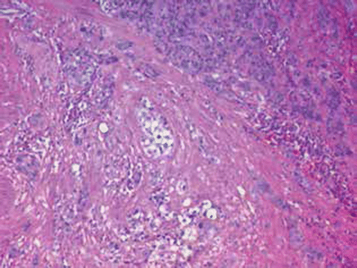
Figure shows a medium-power magnification view of the histology of the tumour in case study 1, showing the cords of the epithelioid cells in a hyalinised background, invading deep into the myometrium.
Figure 1: Medium power magnification view of the histology of the tumor in case 1, showing cords of epithelioid cells in a hyalinised background invading deep into the myometrium. Some of the epithelioid cells display clear cytoplasm. Mitotic activity can also be seen. Haematoxylin and eosin, 100 × magnification.
Case study 2
A 42-year-old gravia 3, para 3, presented in May 2005 with irregular and excessive vaginal bleeding. The gynaecological examination was unremarkable. A Pap smear was performed and revealed benign changes only. The laboratory results, including haemogram, coagulation panel and biochemical tests, were normal. The serum β-hcG was 10 u/l and the chest X-ray was normal. Hysteroscopy showed a normal uterine cavity, although histology of the curettings showed an ETT. The patient underwent TAH and BSO. The tumour was completely resected. The patient did not receive adjuvant therapy, and has remained clinically well and clear for eight years thereafter.
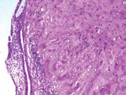
Figure shows a medium-power magnification view of the histology of the tumour in case study 2, showing a nodule of mostly hyalinised tumour.
Figure 2: Medium power magnification view of the histology of the tumor in case 2, showing a nodule of mostly hyalinised tumor. Haematoxylin and eosin, 100 × magnification.
Results
In the first case study, the tumour was located grossly in the lower uterine segment, and extended to both lateral excision margins. In the second case, there was no obvious macroscopic tumour on the cut section.
Microscopy showed that the serosal surface of the uterus was normal in both cases. Histology showed a tumour comprising sheets and nests of epithelioid cells with pleomorphic nuclei and relatively abundant weakly basophilic to eosinophilic cytoplasm. Hyaline material was present between the tumour cells. In the first case study, the tumour showed transmural infiltration of the uterus and involvement of the cervical stroma. In the second case study, minimal tumour remained after the initial curettage, and the residual tumour appeared to be confined to the endometrium.
Imunohistochemical staining in the first case showed positivity with the epithelial marker cell adhesion molecule 5.2 (CAM 5.2). In addition, there was positive staining with cytokeratin (CK) 1, vimentin, human placental lactogen (HPL) and cluster of differentiation 10 (CD10). Placental alkaline phosphotase (PLAP) was focally positive. The hcG, smooth muscle actin (SMA) and desmin stains were negative. These findings were diagnostic of an ETT.
Imunohistochemical staining in the second case study showed that the hcG and HPL were focally positive. CAM 5.2 and PLAP were weakly positive. The Ki-67 proliferation index was 9%. The differential diagnosis included an ETT and the placental site nodule. In the light of a prolonged history and Ki-67 proliferation index of 9%, a diagnosis of an ETT was favoured.
Discussion
An ETT is a rare form of trophoblastic disease. Approximately 90 cases have been reported in the literature, including tumours termed atypical choriocarcinoma and nodules of intermediate trophoblast.Citation1
Clinically, patients with this neoplasm are in the reproductive age group, aged 15-48 years (on average, 36.1 years old).Citation2 Only two cases have been reported after menopause.Citation3 Abnormal vaginal bleeding is the most commonly presenting symptom.
The serum level of β-hcG is almost always slightly raised in patients with an ETT, but this was not the case in the first patient, in whom the level was negative. A high level of serum β-HcG in an ETT is reported to be associated with a large tumour mass and unusually high mitotic activity.Citation4
The uterus is the most common site for an ETT, frequently occurring in the lower segment. Extrauterine cases of ETTs are rare.Citation3 ETTs have been reported as originating in the broad ligament,Citation4 small bowel, lungCitation3 and gallbladder.Citation5 A few cases presenting with metastases at multiple sites have been described. Rarely, an ETT may coexist with choriocarcinoma or a placental site trophoblastic tumours (PSTT).Citation6
An ETT has the same clinical behaviour as a PSTT, but the treatment options may differ. Furthermore, both can be misdiagnosed. Hence, it is imperative that a distinction is made between the two entities, each of which has a different histological appearance.Citation7 An ETT has a nodular growth pattern with expansile borders, in contrast to the dissecting pattern of myometrial infiltration seen with a PSTT.
Histologically, an ETT is characterised by sheets, nests and cords of mildly to moderately pleomorphic epithelioid mononuclear cells, with moderate to abundant amounts of eosinophilic to clear the cytoplasm embedded in the eosinophilic fibrillary matrix. The cells of an ETT are smaller than those seen in a PSTT.Citation1 Areas of geographical necrosis are typical, but the mitotic count tend to be low, with an average of two mitotic figures per 10 high-power fields.Citation8 Immunohistochemically, an ETT typically shows positive labelling with CK18, p63 and human leukocyte antigen G definition. An ETT also shows focal expression of HPL, PLAP, inhibin, hcG and melanoma cell adhesion molecule, also known as CD146.Citation8
Our cases exhibited the usual gross, light microscopic and immunohistochemical features of an ETT. However, it may be mistaken for squamous carcinoma of the cervix because of its epithelioid growth pattern and associated hyalinised material that resembles keratin.Citation3
It is suggested in the literature that a hysterectomy is the treatment of choice in the patients with disease confined to the uterus.Citation3 An ETT appears to be relatively chemoresistant, unlike choriocarcinoma. Although the optimal chemotherapy regimen for a PSTT and an ETT remains to be defined, the current clinical impression is that a platinum-containing regimen is the treatment of choice.Citation9
Curettage with adjuvant chemotherapy is a good option, especially in those desirous of future childbearing.Citation9 Radiation therapy is also useful in providing local control and palliation of recurrent disease.Citation3 However, there was no apparent response to either chemotherapy or radiotherapy in the patient in the first described case study. The course of her disease was an indolent, slowly progressive one over eight years, which was in contrast to the more aggressive course reported in a case study by Jordan et al.Citation10 Apparently, our second patient was cured by TAH and BSO.
Conclusion
We report these two cases because of the rarity of an ETT and the diagnostic dilemma caused for the unwary pathologist. These tumours have been labelled as squamous carcinoma, and have to be differentiated from PSTTs. An awareness of the histological features of an ETT is essential if misdiagnosis is to be avoided, as it represents a tumour which is primarily treated by surgery, rather than by other modalities. These cases were largely typical of those that have been reported in the literature with regard to ETTs.
References
- Fadare O, Parkash V, Carcangiu ML, Hui P. Epithelioid trophoblastic tumor: clinicopathological features with an emphasis on uterine cervical involvement. Mod Pathol. 2006;19(1):75–82.
- Shih IM, Kurman RJ. Epithelioid trophoblastic tumor: a neoplasm distinct from choriocarcinoma and placental site trophoblastic tumor simulating carcinoma. Am J Surg Pathol. 1998;22(11):1393–1403.
- Coulson LE, Kong CS, Zaloudek C. Epithelioid trophoblastic tumor of the uterus in a postmenopausal woman: a case report and review of the literature. Am J Surg Pathol. 2000;24(11):1558–1562.
- Kuo KT, Chen MJ, Lin MC. Epithelioid trophoblastic tumor of the broad ligament: a case report and review of the literature. Am J Surg Pathol. 2004;28(3):405–409.
- Macdonald MC, Palmer JE, Hancock BW, Tidy JA. Diagnostic challenges in extrauterine epitheloid trophoblastic tumours: a report of two cases. Gynecol Oncol. 2008;108(2):452–454.
- Shen DH, Khoo US, Ngan HYS, et al. Coexisting epithelioid trophoblastic tumour and choriocarcinoma following a chemo-resistant hydatidiform mole. Arch Pathol Lab Med. 2003;127(7):e291–e293.
- Moutte A, Doret M, Hajri T, et al. Placental site and epithelioid trophoblastic tumours: diagnostic pitfalls. Gynecol Oncol. 2013;128(3):568–572.
- Shih IM, Mazur MT, Kurman RJ. Gestational trophoblastic tumors and related tumor-like lesions. In: Kurman RJ, Ellenson LH, Ronnett BM, editors. Blaustein’s pathology of the female genital tract. 6th ed.. New York, NY: Springer; 2011. p. 1075–1135.
- Kim SJ. Placental site trophoblastic tumour. Best Pract Res Clin Obstet Gynaecol. 2003;17(6):969–984.
- Jordan S, Randall LM, Karamurzin Y, et al. Differentiating squamous cell carcinoma of the cervix and epithelioid trophoblastic tumor. Int J Gynecol Cancer. 2011;21(5):918–922.