
Abstract
Isolated metastasis to the iliopsoas muscle is rare and is often misdiagnosed as psoas abcess, especially when it is the only presenting feature in the absence of a known primary tumour. A 52-year-old woman presented to us with a clinicoradiological diagnosis of psoas abscess. Initially, the patient was managed as a case of tubercular psoas abscess. However, later on, when the disease had not responded to antitubercular therapy, a cytological examination of aspirated material was performed, which confirmed it as being malignant in origin. While searching for primary malignancy, a small growth on the cervix was noticed. A biopsy and histopathological examination confirmed the diagnosis of squamous cell carcinoma of the cervix. A final diagnosis of squamous cell carcinoma of the cervix with isolated iliopsoas muscle metastasis was proposed. This case highlights an unusual site of metastasis in an asymptomatic primary tumour which was diagnosed retrospectively.
Introduction
The iliopsoas compartment may be affected by several pathologies, including infection, tumours and haemorrhage. Isolated metastasis to the iliopsoas muscle is an uncommon presentation and is often misdiagnosed as an abscess.Citation1,2 It is also rare to find iliopsoas lesions as the only presenting feature in the absence of a known primary tumour.Citation3 We describe a rare case of a metastatic iliopsoas abscess arising from the squamous cell carcinoma of the cervix, in which a diagnosis of carcinoma cervix was made retrospectively.
Case history
A 52-year-old multiparous, postmenopausal female with a past history of subtotal hysterectomy for symptomatic fibroid seven years previously presented with complaints of left-sided abdominal pain extending to her back and left leg for two months. She also complained of a lump in the abdomen and inability to extend her left leg over the previous 15 days. There was no significant history of loss of appetite and weight loss.
On examination, her general condition appeared to be normal. Her abdomen was distended with minimal free fluid. There was a tender cystic mass in the left side of the abdomen affecting the left lumbar, and umbilical and left iliac quadrants, and measuring approximately 15 x 15 cm. Contrast-enhanced computed tomography of the abdomen was performed, which revealed a 16 x 15 x 12 cm cystic swelling involving the left iliacus and left psoas muscle, with peripheral enhancement and filled with dense fluid. Areas of thickening and necrosis were identified in the cyst wall. A diagnosis of a psoas abscess was implied. Ultrasound-guided aspiration was performed, which yielded 2 000 ml of straw- to chocolate brown-coloured fluid. Fluid was sent for culture, for Gram and acid-fast bacilli (AFB) staining, as well as for biochemical investigations. Although the AFB smear was negative, the patient was placed on antitubercular therapy because of the diagnosis of tubercular psoas abscess, as this disease is endemic and is the most common cause of psoas abscess in our country.Citation4
Figures detail the investigations made with respect to the patient.
Figure 1: The contrast-enhanced computed tomography scan demonstrates a large area of low attenuation involving the left iliopsoas muscle (arrow)
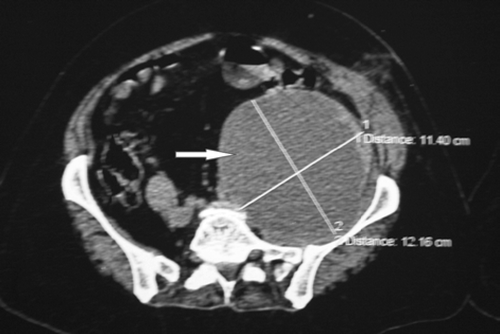
Figure 2: Coronal reconstruction demonstrates an area of low attenuation in the iliopsoas musculature on the left side (arrow)
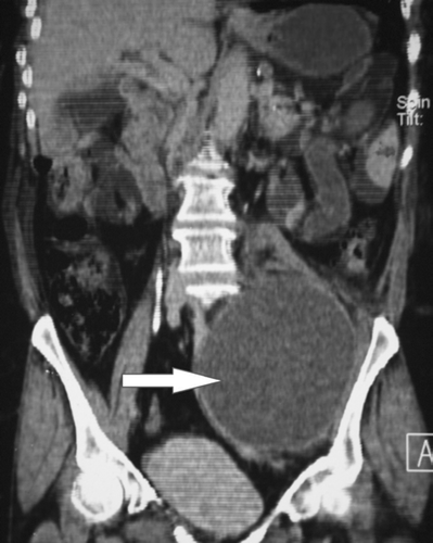
Figure 3: The microscopic appearance of aspirate from the psoas abscess, showing sheets of malignant squamous cells with a hyperchromatic nucleus and conspicuous nucleoli (inset), using haematoxylin and eosin staining
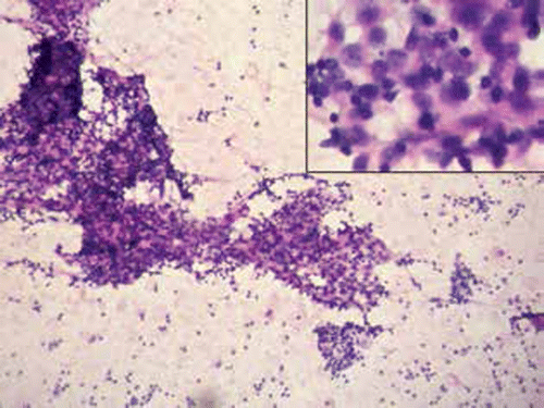
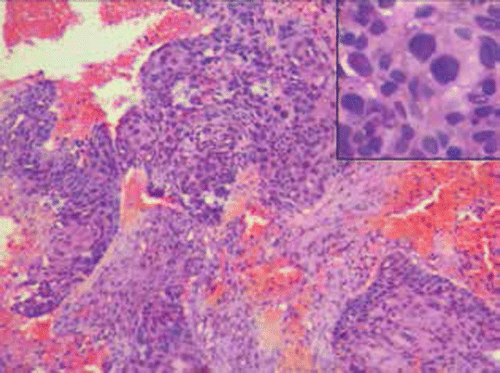
Figure 4: A histological examination of the cervical biopsy shows strips of malignant squamous cells with intracytoplasmic keratinisation (inset) along with area of necrosis and haemorrhage, using haematoxylin and eosin staining
After two months, the patient returned with the same presentation. There was no response to the antitubercular therapy. On examination, a cystic mass was palpated and noticed in the left iliac fossa. An ultrasound-guided aspiration was repeated, which showed 650 ml of haemorrhagic fluid. At this time, a cytological evaluation was also performed, together with biochemical investigations.
The cytological smear revealed occasional sheets and singly scattered malignant squamous cells in a lymphocyte-rich haemorrhagic fluid background. Hence, a diagnosis of metastatic squamous cell carcinoma was made. A thorough workup of the patient was carried out to determine the primary site of the malignancy. A speculum examination revealed an unhealthy cervical stump with an irregular surface and a small growth of 2.5 × 2 cm confined to the cervix. There was no discharge or bleeding, nor vaginal and parametrial involvement, and no rectal and bladder extension. No other metastatic site was identified in this patient. Her human immunodeficiency virus status was negative. A biopsy from the growth was taken, which revealed malignant squamous cell nests infiltrating into the stroma. Hence a final diagnosis of squamous cell carcinoma of the cervical stump with metastasis to the iliopsoas was made. Palliative chemotherapy was offered to the patient.
Discussion
Although the skeletal muscles account for nearly 50% of the total body weight and have a rich blood supply, the incidence of the skeletal muscle metastases is reported to be less than 1% of metastases of haematogenous origin.Citation1 The reasons for the rarity of metastatic involvement of the skeletal muscle are still unclear, but several hypotheses have been postulated. Dislodgement of the tumour cells by blood turbulence and muscle contraction, an unfavourable acidic pH, the effect of protease inhibitors inhibiting cell invasion and the anti-tumour activity of lymphocytes and/or natural killer cells within the skeletal muscle are examples of suggested hypotheses.Citation1,2,5,6
Skeletal muscle involvement from cervical carcinoma is extremely rare. It is usually documented in the context of an advanced-stage tumour or metastatic disease,Citation7,9or in severely immunocompromised patients, i.e. in patients with acquired immune deficiency sydnrome.Citation5,6 A case reported by Devendra and TayCitation9 described muscle involvement without bone involvement in advanced carcinoma cervix. Cases reported by Bar-Dayan et alCitation7 and others Citation10,11 also found associated bone involvement. The primary tumour was diagnosed simultaneously in these two case reports. Contrary to this, George and LaiCitation11 and Kalra et alCitation12 reported on a metastatic malignant psoas abscess two years after the completion of radiotherapy for squamous cell carcinoma cervix.
Our case is unique for many reasons, namely the unusual and only site of metastasis, an absence of history pertaining to the primary lesion, a diagnosis of cervical carcinoma being made in retrospect while searching for the primary malignancy, and finally, because the primary cervical malignancy was limited to the cervix, without loco regional extension.
Establishing the diagnosis of skeletal muscle metastasis is difficult, especially when the patient does not have a history of previous malignancy or clinical features relating to primary malignancy, as occurred in our case. The presentation as a psoas abscess, with no history of a primary lesion and high endemicity of tuberculosis, limited initial consideration of this case to that of a metastatic psoas abcess. It is probable that early cytological assessment of the aspirated material should have been performed in this case to avoid delayed diagnosis.
This case highlights that metastasis from squamous cell carcinoma of the cervix can present as an isolated psoas abscess, clinically and radiologically. This case demonstrates certain difficulties in diagnosis, even with the newer imaging modalities. High suspicion of metastasis with cytological evaluation of the aspirated material should be performed before making a diagnosis of tuberculosis and instituting antitubercular therapy, even in regions that are endemic to tuberculosis.
References
- Sudo A, Ogihara Y, Shiokawa Y, et al. Intramuscular metastasis of carcinoma. Clin Orthop Relat Res. 1993;296:213–217.
- Yang WT, Yeo W, Metreweli C. Imaging of iliopsoas metastasis. Clin Radiol. 1999;54(2):85–89.
- Singh AK, Gervais DA, Hahn PF, et al. Neoplastic iliopsoasmasses in oncology patients: CT findings. Abdom Imaging. 2008;33(4):493–497.
- Mallick IH, Thoufeeq MH, Rajendran TP. Iliopsoas abscess. Postgrad Med J. 2004;80(946):459–462.
- Schwartz LB, Carcangiu ML, Braham L, et al. Rapidly progressive squamous cell carcinoma of the cervix coexisting with human immunodeficiency virus infection: clinical opinion. Gynecol Oncol. 1991;41(3):255–258.
- Singh GS, Aikins JK, Deger R, et al. Metastatic cervicalcancer and pelvic inflammatory disease in an AIDS patient. Gynecol Oncol. 1994;54(3):372–376.
- Bar-Dayan Y, Fishman A, Levi Z, et al. Squamous cell carcinomaof the cervix with psoas abscess-like metastasis in an HIV-negative patient. Isr J Med Sci. 1997;33(10):674–676.
- Pathy S, Jayalakshmi S, Chander S, et al. Carcinoma cervix with metastases to deltoid muscle. Clin Oncol (R Coll Radiol). 2002;14(6):447–448.
- Devendra K, Tay SK. Metastatic carcinoma of the cervix presenting as a psoas abscess in an HIV-negative woman. Singapore Med J. 2003;44(6):302–303.
- Sonawane DV, Jagtap SA, Mathesul AA, et al. Psoas abscess like metastasis mimicking Koch’s spine. Clin Cancer Investig J. 2012;1:103–105.
- George J, Lai FM. Metastatic cervical carcinoma presenting as psoas abscess with osteoblastic and lytic bony metastasis. Singapore Med J. 1995;36(2):224–227.
- Kalra N, Aiyappan S, Nijhawan R, et al. Metastatic carcinoma of cervix mimicking psoas abscess on imaging: a case report. J Gynecol Oncol. 2009;20(2):129–131.