
ABSTRACT
A better understanding of immune-related adverse events is essential for the early detection and appropriate management of these phenomena. We conducted an observational study of cases recorded at the French reference center for hypereosinophilic syndromes and in the French national pharmacovigilance database. Thirty-seven reports of eosinophilia induced by treatment with immune checkpoint inhibitors (ICIs) were included. The median [range] time to the absolute eosinophil count (AEC) peak was 15 [4─139] weeks. The median AEC was 2.7 [0.8─90.9] G/L. Eosinophil-related manifestations were reported in 21 of the 37 cases (57%). If administered, corticosteroids were always effective (n = 10 out of 10). Partial or complete remission of eosinophilia was obtained in some patients not treated with corticosteroids, after discontinuation (n = 12) or with continuation (n = 4) of the ICI. The AEC should be monitored in ICI-treated patients. If required by oncologic indications, continuation of ICI may be an option in asymptomatic hypereosinophilic patients, and in corticosteroid responders.
Introduction
Immunosuppressive molecules are markedly overexpressed in the microenvironment of both solid and hematologic tumors, which thereby promotes immune escape. However, these molecules can be specifically targeted by immune checkpoint inhibitors (ICIs), such as ipilimumab (a monoclonal antibody against the cytotoxic T-lymphocyte antigen 4 (CTLA4)), nivolumab, and pembrolizumab (both of which target programmed cell death protein 1 (PD-1) or its ligand (PD-L1)).Citation1 These drugs have been approved for the treatment of several cancers, including melanoma, non-small-cell lung cancer, urothelial carcinoma, renal cell carcinoma, squamous cell carcinoma of the head and the neck, and/or Hodgkin’s lymphoma.Citation2 However, ICIs are also associated with frequent and potentially organ- or life-threatening immune-related adverse events (irAEs), which generally mimic autoimmune or inflammatory conditions; indeed, up to 90% of patients treated with ipilimumab and up to 70% of those treated with PD-1/PD-L1 antibodies experience at least one irAE).Citation3,Citation4 The early diagnosis and prompt management of irAEs are essential. Although an effective ICI may not have to be discontinued after a mild irAE, specific treatments and/or discontinuation of the ICI must be considered in the most severe cases.Citation5
In a recent retrospective single-center study, the prevalence of immune-related blood eosinophilia (an absolute eosinophil count (AEC) greater than 0.5 G/L) in patients treated with anti-PD1 or anti-PD-L1 drugs was 2.8%, and the median [range] peak AEC was 1.0 [0.6─5.6] G/L.Citation6 Although drug-induced eosinophilia (and thus, in theory, all other eosinophilic disorders) can be associated with eosinophil-induced organ damage, these cases of immune-related blood eosinophilia (Eo-ir) had a favorable outcome, and required neither specific treatment nor ICI discontinuation.Citation6
At the French national reference center for hypereosinophilic syndromes (CEREO), we were solicited for several patients with severe, well-documented, eosinophil-induced adverse events (Eo-irAEs) and organ dysfunction. The objective of the present study was to describe the characteristics and outcomes of patients with moderate-to-severe eosinophilia (defined in this study as an AEC >1G/L) and/or Eo-irAEs reported in CEREO’s database and the French national pharmacovigilance database (FPVD).
Results
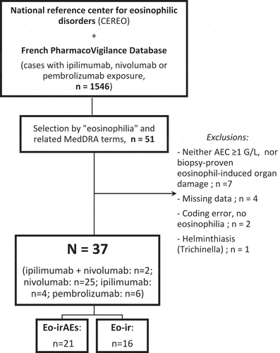
Thirty-seven patients were included in the study (): 25 were treated with nivolumab, 6 with pembrolizumab, 4 with ipilimumab, and 2 with a combination of nivolumab and ipilimumab (1 case with the two drugs concomitantly, and 1 case with a switch from ipilimumab to nivolumab).
Figure 1. Flow chart showing the case selection process. AEC: absolute eosinophil count; Eo-irAEs: eosinophil-induced adverse events; Eo-ir: immune-related blood eosinophilia
The indications were non-small-cell lung cancer (n = 18), melanoma (n = 18), and Hodgkin’s lymphoma (n = 1), the median [range] age at Eo-ir or Eo-irAE onset was 68 [33─84] years, and the male:female ratio was 2.7:1.
Before ICI initiation, 7 patients (19%) already displayed eosinophilia (an AEC between 0.5 and 1.5 G/L), 28 patients did not display eosinophilia, and this information was missing for 2 patients.
Twenty-one patients (57%) had an Eo-irAE, 12 others (32%) had an Eo-ir, and data enabling the classification of an event as an Eo-irAE was missing for 4 patients (11%). The patients’ individual data are given in and the characteristics are summarized in .
Table 1. Case-series with patients’individual data
Table 2. Characteristics of the eosinophil-induced immune-related adverse events
In the cohort as a whole, the median times to new-onset eosinophilia and to the AEC peak were respectively 6 [1─52] and 15 [4─139] weeks after ICI initiation. The median AEC was 2.7 [0.8─90.9] G/L, although 2 patients had an AEC peak <1 G/L but proven tissue eosinophilia on biopsy.
The data on the AEC before ICI initiation are categorized in . The median peak AECs did not differ when comparing patients with Eo-ir and those with Eo-irAEs (3.3[1.2─90.9] and 2.5[0.8─8.7] G/L, respectively, p = .15). The Eo-irAEs affected the skin (n = 10), lung (2 cases of eosinophilic pneumonia and 2 of eosinophilic bronchiolitis), kidneys (4 nephritis), liver (2 cholangitis), and heart (1 myocarditis). With regard to the severity of the Eo-irAEs, there were 1 grade IV case, 6 grade III cases, 8 grade II cases and 7 grade I cases. No deaths were attributable to Eo-irAEs (). The grade IV case was a maculopapular rash with laryngeal edema. The evolution was quickly favorable with corticosteroids. The other skin side effects were: maculopapular rashes (n = 3), eczematiform rashes (n = 2), lichenoid rashes (n = 2), bullous pemphigoid-like eruption and eosinophilic fasciitis (n = 1 each).
Overall, the ICI was discontinued in 26 of the 37 cases (70%); 13 of these (50%) were due to an Eo-ir or an Eo-irAE ().
The median length of follow-up after ICI initiation was 63 [7─300] weeks (n = 35). Nine deaths were reported during this period but none were attributable to Eo-irAEs.
Ten patients received corticosteroids for Eo-ir or Eo-irAEs; complete (n = 9) or partial (n = 1) disease remission was observed in all cases. Moreover, partial (n = 6) or complete (n = 10) remission of eosinophilia were reported in 16 other patients who did not receive corticosteroids, including 4 for whom ICI was continued. However, data on the time from corticosteroids onset to remission were not available for the great majority of cases. Lastly, 6 patients showed prolonged long-term eosinophilia (lasting for at least 6, 7, 59, 68 and 144 weeks after time of AEC peak) despite ICI discontinuation. Finally, 19 of 29 patients with Eo-ir or Eo-irAE were good responders to ICI (unknown outcome in n = 8), including 5 who kept stable (n = 4) or increased (n = 1) AEC ().
Table 3. The AEC outcome, depending on the clinical response to ICI
Discussion
Here, we report on the largest yet series of patients with moderate-to-severe Eo-ir and Eo-irAEs. Our results suggest that the AEC should be closely monitored during the course of ICI. We also reported on patients with a favorable outcome despite persistent blood eosinophilia, and we discuss below how to manage patients with Eo-ir or Eo-irAEs.
Considering the time to new-onset eosinophilia (median [range]: 6 weeks [1─52] or 1.4 months [0.2─12]), our results suggest that monthly monitoring of AEC is warranted during a course of treatment with an ICI. Moreover, this time to onset was shorter in our study than in Berrnard-Teissier et al.’s retrospective observational study of 26 cases with a normal AEC at baseline and an AEC >0.5G/L 3 [0.6─31.3] months after ICI initiation.Citation6 Although the time course of eosinophilia onset has yet to be characterized, one can reasonably hypothesize that moderate-to-severe eosinophilia may have an early onset. Similarly, the median time to the AEC peak observed in our study (3.4 months [1─32.4]), was shorter than that observed by Berrnard-Teissier et al. (6.4 months [1.4─32]). Fifty percent patients (13/26, see ) developed another irAE before or at the same time of Eo-irAE. The time to onset of moderate-to-severe eosinophilia reported in our study is in line with that described for other irAEs that typically arise within a few weeks or months of ICIinitiation.Citation7
Given our stringent inclusion criteria, our objective here was to describe the patients with the most severe ICI-induced AEs; according to Bernard-Teissier et al. these patients may account for up to 1–2% of people treated with ICIs.Citation6 We found that a high proportion of these patients developed Eo-irAEs (57%) – suggesting that although blood eosinophilia is highly unusual,Citation6 it should not be neglected by attending physicians because severe eosinophil-related organ dysfunction is likely to occur. In the present study, we chose to include patients (n = 7) with an AEC >0.5 G/L at baseline (i.e. before ICI initiation), only one developed an Eo-irAE. This suggests that an elevated AEC at baseline is not associated with more severe eosinophilia during treatment with ICIs. Furthermore, an elevated eosinophil count prior to treatment was associated with longer overall survival in several studies.Citation8–Citation12 Since there was a trend toward a higher median AEC in asymptomatic patients than in patients with Eo-irAEs (3.3[1.2─90.9] and 2.5[0.8─8.7] G/L, respectively), our case-series suggests that AECs and new-onset Eo-irAEs are not correlated. Hence, a high AEC alone is not an index of severity. Moreover, this observation is also supported by reports of an association between elevated eosinophil counts, better clinical responses and longer overall survival in several types of advanced cancer; this association might be stronger and more clinically relevant for patients treated with ICIs than with conventional chemotherapy.Citation8–Citation11,Citation13-Citation17 Considering that eosinophils can have a role in the response against cancer,Citation17–Citation20 an elevated AEC might be a marker of effectiveness in some patients. Further research is needed to determine the mechanisms involved in Eo-ir, the clinical significance of high blood eosinophilia on cancer outcome, and whether eosinophils are involved in ICIT effectiveness or just a reactive “biological” phenomenon.Citation17,Citation21
Given its retrospective design, our study had several inherent limitations: misclassification, missing data, and the risk of selection bias due to the FPVD’s self-reporting procedure (emphasizing the most symptomatic cases). However, this case series enabled to consider differential diagnoses and the management of Eo-irAEs.
When eosinophilia occurs during treatment with an ICI, differential diagnoses must be considered: another medication, helminthiasis (mainly toxocara) and atopic disease, for example. Furthermore, the AEC, clinical symptoms, electrocardiogram, and laboratory markers of heart/kidney/liver status must be closely monitored. In previous reports, eosinophilia sometimes resolved spontaneously.Citation6 Hence, in patients with Eo-ir but no evidence of eosinophil-related organ dysfunction, we suggest that ICIs can be continued with caution as long as the patients are closely monitored for at least 6 months. In contrast, we observed 7 cases of severe (grades 3 and 4) organ damage (myocarditis, eosinophilic pneumonia, cholangitis, skin rash, eosinophilic fasciitis, and nephritis) and 15 cases of mild-to-moderate (grades 1 and 2) organ damageCitation22 (). Interestingly, high remission rates were obtained when corticosteroids were given – even when the ICI was not discontinued (n = 3). Corticosteroids are the usual first-line treatment for both reactive eosinophilic disorders and irAEs.Citation2,Citation7,Citation23 Our results suggest that corticosteroids constitute an effective treatment for moderate-to-severe eosinophilia, even though the dosage was not specified in the pharmacovigilance reports. Phillips et al. recently reported a large cohort of 285 patients with immune-related cutaneous AEs, including 7 (2.4%) who were refractory to corticosteroids. Increased AEC, serum IL-6, Il-10 and IgE levels were associated with corticosteroid-refractory adverse events and with grade 3 or greater cutaneous AEs, but the direct accountability of eosinophils was not assessed in these exceptional cases.Citation24 Even if some severe cutaneous AEs like drug reaction with eosinophilia and systemic symptoms (DRESS) require high-dose corticosteroids,Citation25,Citation26 multiple recent reports of Eo-irAEs like eosinophilic fasciitisCitation27-Citation30 or eosinophilic granulomatosis with polyangiitisCitation31 suggest that topical or low-dose oral corticosteroids, with or without CS-sparing treatments, can give excellent results. Taking account these data, and given that in our work (i) Eo-ir and Eo-irAEs accounted for half of all ICI discontinuations and (ii) no deaths were directly attributable to eosinophil-organ damage, we suggest that the initiation of corticosteroids and the maintenance of the ICI might be an effective therapeutic strategy in patients with moderate-to-severe eosinophilia and whose cancer is under control. Although most international guidelines recommend higher doses of corticosteroids (from 0.5 to 2 mg/kg/day) for other irAEs,Citation5,Citation32,Citation33 eosinophilia and eosinophil-induced organ dysfunction typically respond quickly to corticosteroids, and some non-severe cases respond to low doses. Progressive corticosteroid tapering may be warranted after 1 or 3 weeks, depending on the severity and the initial clinical response. Hence, considering that high-dose corticosteroids could reduce the ICI’s effectiveness, it would be possible to reach a dose of 10 mg/d.Citation34 In the other hand, early use of steroids was associated with worse clinical outcomes and remarkable modulation of peripheral blood immune cells (including the decrease of the AEC), which could contribute to restraining the activation of antitumour immunity.Citation35 Eosinophilic heart involvement can be asymptomatic, whereas myocarditis is a life-threatening complication. The electrocardiogram and serum levels of troponin and brain-natriuretic peptide should be monitored every 2 or 4 weeks, and echochardiography should be performed at least at when hypereosinophilia is diagnosed. Cardiac MRI should be considered in the event of doubt or if myocarditis is suspected.
Conclusion
Taken as a whole, our results suggest that moderate-to-severe eosinophilia can occur soon after ICI initiation and can lead to severe eosinophilic-related organ damage. Further prospective studies are warranted, in order to assess, the risk factors to develop Eo-ir or Eo-irAE, the long-term outcomes of Eo-irAEs and to better define the optimal management of these complications. Lastly, given the ICIs’ potency against cancer, our observations suggest that asymptomatic blood eosinophilia and Eo-irAEs (grade ≤3) do not necessarily constitute sufficient grounds for treatment discontinuation.
Methods
Data source
The CEREO and FPVD databases were searched for cases of moderate-to-severe eosinophilia or Eo-irAEs. Briefly, the FPVD has recorded all adverse drug reactions spontaneously notified to France’s 31 regional pharmacovigilance centers since 1985.Citation36 Indeed, French legislation requires healthcare professionals to report all adverse drug reactions to their regional pharmacovigilance center. Although patient consent is not required, the records remain fully anonymous. Next, each adverse drug reaction report is analyzed by pharmacologists with expertise in the field. Causality is assessed according to the French methodCitation37 which is based on both intrinsic imputability (i.e. cross-checking against chronologic and semiologic criteria) and extrinsic imputability (i.e. based on literature data). Lastly, the case is recorded in the database after being coded according to the Medical Dictionary for Regulatory Activities (MedDRA) classification.
Case selection
The FPVD was searched up until November 1st, 2017, whereas the CEREO’s records were searched up until January 15th, 2019.
In the FPVD, cases were selected using logical combinations of the MedDRA preferred terms “eosinophilia”, “eosinophil count abnormal”, “eosinophil count increased”, “eosinophil percentage abnormal”, “eosinophil percentage increased”, “eosinophilic cellulitis”, “eosinophilic fasciitis”, “eosinophilic pustular folliculitis”, “eosinophilic pustulosis”, “drug reaction with eosinophilia and systemic symptoms”, “eosinophilia myalgia syndrome”, “allergic eosinophilia”, “pulmonary eosinophilia”, “eosinophilic pleural effusion”, “eosinophilic bronchitis”, “eosinophilic pneumonitis”, “eosinophilic pneumonitis acute”, “eosinophilic pneumonitis chronic”, “gastroenteritis eosinophilic”, “eosinophilic colitis”, “eosinophilic oesophagus”, “hepatic infiltration eosinophilic”, “eosinophilic myocarditis”, “eosinophilic cystitis”, “eosinophilic granulomatosis with polyangiitis”, “meningitis eosinophilic”, “panniculitis eosinophilic”, “hypereosinophilic syndrome” AND “ipilimumab”, “nivolumab” or “pembrolizumab” exposure; only cases where an adverse reaction to the drugs were “suspected” were selected.Citation38
Patients were included if at least one AEC after initiation of ICI therapy (nivolumab, pembrolizumab or ipilimumab) was >1 G/L and/or eosinophil-induced organ damage was confirmed on biopsy.Citation39 We excluded patients with other likely etiologies for eosinophilia (e.g. helminthiasis) and/or missing data.
Data collection
For each case, we noted the patient’s demographic and clinical characteristics (age, gender, neoplastic disease, and length of follow-up), data regarding the ICI (dose, duration of treatment, the best anti-tumor response during treatment (according to the oncologist), potential discontinuation and other irAEs,) and history of eosinophilia (time to onset and to peak, confirmed or suspected Eo-irAEs and their outcomes) were recorded. Data were collected from FPVD reports, and missing data were extracted from corresponding medical charts by each regional pharmacovigilance coordinator. After careful analysis of both the patient’s medical charts and the chronologic relationship between blood eosinophilia and onset of organ dysfunction, the adverse drug reaction were classified either as Eo-ir (i.e. no organ dysfunction was attributed to eosinophilia) or Eo-irAEs (i.e. organ dysfunction was considered to have been induced by proven tissue eosinophilia and/or potentially induced by eosinophils after a chart review of the organ dysfunction and the presence of a consistent chronologic relationship between blood eosinophilia and the onset of organ dysfunction).
Ethics
According to French legislation, formal approval by an investigational review board is not required for this type of study (performed here by the French Pharmacovigilance Network).
Statistics
Quantitative variables were quoted as the median [range], and qualitative variables were quoted as the number (percentage). Median values were compared using a Wilcoxon rank sum test with continuity correction. All tests were two-tailed, and the threshold for statistical significance was set to p < .05. All statistical analysis were performed using R software via R studio (R version 3.4.0., The R Foundation for Statistical Computing, Vienna, Austria).
Conflicts of interest
ABC reports personal fees or honoraria from BMS, MSD, Astra-Zeneca, and Roche. LM reports honoraria, consulting or advisory fees from Roche, BMS, Novartis, MSD, Amgem, Pierre Fabre, Sanofi, Merck, and Pfizer; and travel or accommodation expenses from BMS, Novartis, MSD, and Pierre Fabre, MG reports personal fees or honoraria from Astra-Zeneca, and JEK and GL report personal fees or honoraria from GSK and Astra-Zeneca.
Disclosure of Potential Conflicts of Interest
The others authors declare that they have no conflict of interest.
Acknowledgments
The authors would like to thank all the physicians who contributed adverse drug reaction reports to the French pharmacovigilance system.
References
- Mellman I, Coukos G, Dranoff G. Cancer immunotherapy comes of age. Nature. 2011;480(7378):480–9. doi:10.1038/nature10673.
- Champiat S, Lambotte O, Barreau E, Belkhir R, Berdelou A, Carbonnel F, Cauquil C, Chanson P, Collins M, Durrbach A, et al. Management of immune checkpoint blockade dysimmune toxicities: a collaborative position paper. Ann Oncol. 2016;27(4):559–574. doi:10.1093/annonc/mdv623.
- Michot JM, Bigenwald C, Champiat S, Collins M, Carbonnel F, Postel-Vinay S, Berdelou A, Varga A, Bahleda R, Hollebecque A, et al. Immune-related adverse events with immune checkpoint blockade: a comprehensive review. Eur J Cancer. 2016;54:139–148. doi:10.1016/j.ejca.2015.11.016.
- Reck M, Rodríguez-Abreu D, Robinson AG, Hui R, Csőszi T, Fülöp A, Gottfried M, Peled N, Tafreshi A, Cuffe S, et al. Pembrolizumab versus chemotherapy for PD-L1–positive non–small-cell lung cancer. 2016 Oct 8. [accessed 2019 Feb 22]. https://www.nejm.org/doi/10.1056/NEJMoa1606774?url_ver=Z39.88-2003&rfr_id=ori%3Arid%3Acrossref.org&rfr_dat=cr_pub%3Dwww.ncbi.nlm.nih.gov.
- Brahmer JR, Lacchetti C, Schneider BJ, Atkins MB, Brassil KJ, Caterino JM, Chau I, Ernstoff MS, Gardner JM, Ginex P, et al. Management of immune-related adverse events in patients treated with immune checkpoint inhibitor therapy: American society of clinical oncology clinical practice guideline. J Clin Oncol. 2018;36(17):1714–1768. doi:10.1200/JCO.2017.77.6385.
- Bernard-Tessier A, Jeanville P, Champiat S, Lazarovici J, Voisin A-L, Mateus C, Lambotte O, Annereau M, Michot J-M. Immune-related eosinophilia induced by anti-programmed death 1 or death-ligand 1 antibodies. Eur J Cancer. 2017;81:135–137. doi:10.1016/j.ejca.2017.05.017.
- Postow MA, Sidlow R, Hellmann MD. Immune-related adverse events associated with immune checkpoint blockade. N Engl J Med. 2018;378(2):158–168. doi:10.1056/NEJMra1703481.
- Delyon J, Mateus C, Lefeuvre D, Lanoy E, Zitvogel L, Chaput N, Roy S, Eggermont AMM, Routier E, Robert C. Experience in daily practice with ipilimumab for the treatment of patients with metastatic melanoma: an early increase in lymphocyte and eosinophil counts is associated with improved survival. Ann Oncol. 2013;24(6):1697–1703. doi:10.1093/annonc/mdt027.
- Schindler K, Harmankaya K, Postow MA, Frantal S, Bello D, Ariyan CE, Michielin OA, Hoeller C, Pehamberger H, Wolchok JD. Pretreatment levels of absolute and relative eosinophil count to improve overall survival (OS) in patients with metastatic melanoma under treatment with ipilimumab, an anti CTLA-4 antibody. J Clin Oncol. 2013;31(15_suppl):9024. doi:10.1200/jco.2013.31.15_suppl.9024.
- Martens A, Wistuba-Hamprecht K, Foppen MG, Yuan J, Postow MA, Wong P, Romano E, Khammari A, Dreno B, Capone M, et al. Baseline peripheral blood biomarkers associated with clinical outcome of advanced melanoma patients treated with ipilimumab. Clin Cancer Res. 2016;22(12):2908–2918. doi:10.1158/1078-0432.CCR-15-2412.
- Weide B, Martens A, Hassel JC, Berking C, Postow MA, Bisschop K, Simeone E, Mangana J, Schilling B, Giacomo AMD, et al. Baseline biomarkers for outcome of melanoma patients treated with pembrolizumab. Clin Cancer Res. 2016;22(22):5487–5496. doi:10.1158/1078-0432.CCR-16-0127.
- Hude I, Sasse S, Bröckelmann PJ, Tresckow BV, Momotow J, Engert A, Borchmann S. Leucocyte and eosinophil counts predict progression-free survival in relapsed or refractory classical Hodgkin lymphoma patients treated with PD1 inhibition. Br J Haematol. 2018;181(6):837–840. doi:10.1111/bjh.14705.
- Gaba L, Victoria I, Pineda E, Fernandez A, Aya F, Prat A, Arance AM. Changes in blood eosinophilia during anti-PD1 therapy as a predictor of long term disease control in metastatic melanoma. J Clin Oncol. 2015;33(15_suppl):9069. doi:10.1200/jco.2015.33.15_suppl.9069.
- Gebhardt C, Sevko A, Jiang H, Lichtenberger R, Reith M, Tarnanidis K, Holland-Letz T, Umansky L, Beckhove P, Sucker A, et al. Myeloid cells and related chronic inflammatory factors as novel predictive markers in melanoma treatment with ipilimumab. Clin Cancer Res. 2015;21(24):5453–5459. doi:10.1158/1078-0432.CCR-15-0676.
- Umansky V, Utikal J, Gebhardt C. Predictive immune markers in advanced melanoma patients treated with ipilimumab. Oncoimmunology. 2016;5:6. doi:10.1080/2162402X.2016.1158901.
- Moreira A. Eosinophilic count as a biomarker for prognosis of melanoma patients and its importance in the response to immunotherapy. Immunotherapy. 2017;9(2):115–121. doi:10.2217/imt-2016-0138.
- Simon SCS, Utikal J, Umansky V. Opposing roles of eosinophils in cancer. Cancer Immunol Immunother. 2018 Oct 9. doi:10.1007/s00262-018-2255-4.
- Carretero R, Sektioglu IM, Garbi N, Salgado OC, Beckhove P, Hämmerling GJ. Eosinophils orchestrate cancer rejection by normalizing tumor vessels and enhancing infiltration of CD8+ T cells. Nat Immunol. 2015;16(6):609–617. doi:10.1038/ni.3159.
- Reichman H, Karo-Atar D, Munitz A. Emerging roles for eosinophils in the tumor microenvironment. Trends Cancer. 2016;2(11):664–675. doi:10.1016/j.trecan.2016.10.002.
- Gatault S, Legrand F, Delbeke M, Loiseau S, Capron M. Involvement of eosinophils in the anti-tumor response. Cancer Immunol Immunother. 2012;61(9):1527–1534. doi:10.1007/s00262-012-1288-3.
- Kahn JE, Groh M, Lefèvre G. (A critical appraisal of) Classification of hypereosinophilic disorders. Front Med. 2017:4. doi:10.3389/fmed.2017.00216.
- Terminology Criteria for Adverse Events (CTCAE). Version 5.0. U.S. Department of Health and Human Services. National Institutes of Health. National Cancer Institute. Novembre 27, 2017.
- Belum VR, Benhuri B, Postow MA, Hellmann MD, Lesokhin AM, Segal NH, Motzer RJ, Wu S, Busam KJ, Wolchok JD, et al. Characterisation and management of dermatologic adverse events to agents targeting the PD-1 receptor. Eur J Cancer. 2016;60:12–25. doi:10.1016/j.ejca.2016.02.010.
- Phillips GS, Wu J, Hellmann MD, Postow MA, Rizvi NA, Freites-Martinez A, Chan D, Dusza S, Motzer RJ, Rosenberg JE, et al. Treatment outcomes of immune-related cutaneous adverse events. J Clin Oncol. 2019 Jun 19:JCO.18.02141. doi:10.1200/JCO.18.02141.
- Mirza S, Hill E, Ludlow S, Nanjappa S. Checkpoint inhibitor-associated drug reaction with eosinophilia and systemic symptom syndrome. Melanoma Res. 2017;27(3):271–273. doi:10.1097/CMR.0000000000000326.
- Lu J, Thuraisingam T, Chergui M, Nguyen K. Nivolumab-associated DRESS syndrome: A case report. JAAD Case Rep. 2019;5(3):216–218. doi:10.1016/j.jdcr.2018.11.017.
- Toussaint F, Hammon M, Erdmann M, Moreira A, Kirchberger MC, Schuler G, Schett G, Heinzerling L. Checkpoint inhibitor-induced eosinophilic fasciitis following high eosinophilia associated with complete response. Rheumatology. 2019;58(10):1875–1877. doi:10.1093/rheumatology/kez164.
- Khoja L, Maurice C, Chappell M, MacMillan L, Al-Habeeb AS, Al-Faraidy N, Butler MO, Rogalla P, Mason W, Joshua AM, et al. Eosinophilic fasciitis and acute encephalopathy toxicity from pembrolizumab treatment of a patient with metastatic melanoma. Cancer Immunol Res. 2016;4(3):175–178. doi:10.1158/2326-6066.CIR-15-0186.
- Lidar M, Giat E, Garelick D, Horowitz Y, Amital H, Steinberg-Silman Y, Schachter J, Shapira-Frommer R, Markel G. Rheumatic manifestations among cancer patients treated with immune checkpoint inhibitors. Autoimmun Rev. 2018;17(3):284–289. doi:10.1016/j.autrev.2018.01.003.
- Andrés‐Lencina -J-J, Burillo‐Martínez S, Aragón‐Miguel R, Calleja‐Algarra A, Rodríguez‐Peralto J-L, Ortiz‐Romero P-L, Gargallo‐Moneva V. Eosinophilic fasciitis and lichen sclerosus in a patient treated with nivolumab. Australas J Dermatol. 2018;59(4):e302–e304. doi:10.1111/ajd.12836.
- Roger A, Groh M, Lorillon G, Pendu CL, Maillet J, Arangalage D, Tazi A, Lebbe C, Baroudjian B, Delyon J. Eosinophilic granulomatosis with polyangiitis (Churg-Strauss) induced by immune checkpoint inhibitors. Ann Rheum Dis. 2019;78(8):e82–e82. doi:10.1136/annrheumdis-2018-213857.
- Haanen JBAG, Carbonnel F, Robert C, Kerr KM, Peters S, Larkin J, Jordan K. Management of toxicities from immunotherapy: ESMO Clinical Practice Guidelines for diagnosis, treatment and follow-up. Ann Oncol. 2017;28(suppl_4):iv119–iv142. doi:10.1093/annonc/mdx225.
- Puzanov I, Diab A, Abdallah K, Bingham CO, Brogdon C, Dadu R, Hamad L, Kim S, Lacouture ME, LeBoeuf NR, et al. Managing toxicities associated with immune checkpoint inhibitors: consensus recommendations from the Society for Immunotherapy of Cancer (SITC) toxicity management working group. J Immunother Cancer. 2017;5. doi:10.1186/s40425-017-0300-z.
- Maxwell R, Luksik AS, Garzon-Muvdi T, Hung AL, Kim ES, Wu A, Xia Y, Belcaid Z, Gorelick N, Choi J, et al. Contrasting impact of corticosteroids on anti-PD-1 immunotherapy efficacy for tumor histologies located within or outside the central nervous system. Oncoimmunology. 2018;7:12. doi:10.1080/2162402X.2018.1500108.
- Fucà G, Galli G, Poggi M, Lo Russo G, Proto C, Imbimbo M, Ferrara R, Zilembo N, Ganzinelli M, Sica A, et al. Modulation of peripheral blood immune cells by early use of steroids and its association with clinical outcomes in patients with metastatic non-small cell lung cancer treated with immune checkpoint inhibitors. ESMO Open. 2019;4:1. doi:10.1136/esmoopen-2018-000457.
- Vial T. French pharmacovigilance: missions, organization and perspectives. Therapie. 2016;71(2):143–150. doi:10.1016/j.therap.2016.02.029.
- Théophile H, Dutertre J-P, Gérardin M, Valnet-Rabier M-B, Bidault I, Guy C, Haramburu F, Hillaire-Buys D, Méglio C, Arimone Y. Validation and reproducibility of the updated french causality assessment method: an evaluation by pharmacovigilance centres & pharmaceutical companies. Thérapie. 2015;70(5):465–476. doi:10.2515/therapie/2015028.
- Brown EG, Wood L, Wood S. The medical dictionary for regulatory activities (MedDRA). Drug Saf. 1999;20(2):109–117. doi:10.2165/00002018-199920020-00002.
- Simon H-U, Rothenberg ME, Bochner BS, Weller PF, Wardlaw AJ, Wechsler ME, Rosenwasser LJ, Roufosse F, Gleich GJ, Klion AD. Refining the definition of hypereosinophilic syndrome. J Allergy Clin Immunol. 2010;126(1):45–49. doi:10.1016/j.jaci.2010.03.042.