
ABSTRACT
Bacille de Calmette et Guerin (BCG) is the only licensed tuberculosis vaccine to prevent severe tuberculosis. The adverse events of BCG vaccination, including local reactions, lymphadenitis, osteomyelitis, tuberculid, and disseminated infection, have been reported. Two infants presented erythema at the inoculation site of BCG after the resolution of Kawasaki disease (KD). They received BCG vaccination 1 week and 6 weeks before the KD onset, respectively. Intravenous immunoglobulin improved the KD activity, however the skin rash of BCG inoculation site extended to the face and extremities days 24 and 10 after the KD onset, respectively. Both bacteriological study and interferon-γ release assay were negative for Mycobacterium tuberculosis infection. These patients were diagnosed as having tuberculid after KD. The skin lesions gradually disappeared without antibiotic therapy over 2 months. The development of tuberculid in these patients might be associated with the remnant immune activation of KD.
Introduction
Bacille de Calmette et Guerin (BCG) vaccine is the only available vaccine for the prevention of severe and disseminated tuberculosis.Citation1 Local reactions, lymphadenitis, osteomyelitis, tuberculid (erythema induratum, lichen scrofulosorum, and papulonecrotic tuberculid), and disseminated infection have been reported as the adverse events following BCG vaccination.Citation1
Kawasaki disease (KD) is an acute, febrile, and systemic vasculitis primarily occurring in infants and young children.Citation2 The incidence of KD is different among ethnic groups, and the highest incidence is reported in Japan (239/100,000 < 5 y of age).Citation2 The most serious complication is coronary arterial lesion (CAL).Citation2 Intravenous immunoglobulin (IVIG) and oral aspirin are effectively used during the acute phase of KD to reduce inflammation, particularly CAL.Citation2,3 Prior to the introduction of IVIG, 20–30% of cases progressed to CAL, with 2% of mortality rate.Citation3 The etiology of KD is still unknown, however it may be caused by an infectious agent that precipitates an excessive auto-inflammation in genetically predisposed individuals.Citation2-4 Additionally, the changes at the inoculation site of BCG, including erythema or ulceration, are often observed during the acute phase of KD.Citation5,6 BCG triggers apoptosis of tumor cells and promotes T cell activation responsible for long-term anti-tumor defense. These suggest that BCG response may be connected with the pathophysiology of KD.
Tuberculid including erythema induratum, lichen scrofulosorum, and papulonecrotic tuberculid, is one of the frequent adverse events of BCG vaccination. Affected patients present with dull red or bluish red nodules (erythema induratum), pin head-size, skin-colored, to erythematous lichenoid, firm, follicular or perifollicular papules (lichen scrofulosorum), and papulonodular lesions (papulonecrotic tuberculid).Citation7 The skin lesions usually emerge from 10 d to 2 months after vaccination.Citation8 The mechanism of tuberculid is considered the skin reactions against BCG vaccine and/or immune responses to degenerated dead bacilli or antigenic fragments,Citation8,9 because the lesions are negative for bacteria. However, there is little information about the detailed pathophysiology of tuberculid. It is critical to distinguish tuberculid from disseminated BCG infection or cutaneous tuberculosis. Neither antibiotics nor anti-tuberculous therapy is needed for the treatment of tuberculid.
There are no reports about a relationship between tuberculid and KD. We herein first report 2 infants who developed tuberculid during the convalescent phase of KD.
Patient presentation
Patient 1
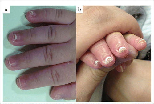
A 5 month-old Japanese male was hospitalized because of 5-days' high fever, erythema of the trunk, and conjunctival hyperemia. On admission, he had redness of lip, and swelling of feet and hands. The previously healthy boy was born to healthy parents having unremarkable family history. He received BCG vaccination intradermally at the left upper limb one week before the onset of KD. Complete blood counts showed leukocytes 22.2 × 10⁹ /L, hemoglobin 11.1 g/dL, and platelets 416 × 109 /L. Blood chemistries revealed increased levels of C-reactive protein (CRP) (1.31 mg/dL, reference range [rr]: 0.01–0.14), and normal levels of aspartate transaminase (AST) 31 U/L (rr: 13–33), alanine transaminase (ALT) 15 U/L (rr: 8–42), albumin 4.2 g/dL (rr: 3.7–5.2), and sodium138 mmol/L (rr: 135–148). Under the diagnosis of KD, IVIG (2 g/kg) and aspirin (30 mg/kg, p.o.) were started on the 5th day of illness. The treatment led to the defervescence promptly, and sheet-like desquamation on the 9th day, respectively. On the day 25, small papules in the face and trunks, along with redness and crust formation at the inoculation site of BCG were observed (). Skin biopsy revealed infiltration of inflammatory cells, ulceration, and crust formation without granuloma and leukocytoclastic vasculitis (). Both bacteriological examination from skin lesions and interferon-γ release assay (IGRA) against Mycobacterium tuberculosis were negative. These results suggested that he had papulonecrotic tuberculid associated with BCG inoculation. The skin lesions disappeared without antibiotic therapy over 2 months. His nails became whitish, and were peeled off 45 d after KD onset (). These manifestations completely resoluted 73 d after the onset of KD. He is alive and well without CAL one year after the hospital discharge.
Figure 1. Rash of cheek and BCG inoculation site on day 43 in patient 1 (a), and cheek (b) and BCG inoculation site (c) on day 23 of KD onset in patient 2.
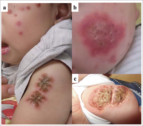
Figure 2. Histopathological finding of skin biopsy from patients 1 (H & E; a: × 100, b: × 400) and 2 (H & E; c: × 100, d: × 400).
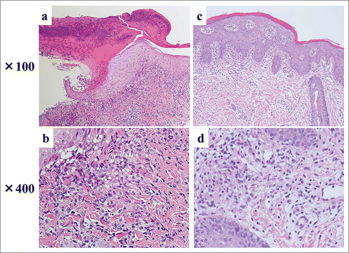
Figure 3. The changes of nails on day 45 of KD onset in patient 1 (a) and in convalescent phase KD of patient 2 (b).
Patient 2
A 5 month-old Japanese male was hospitalized because of fever, redness at the BCG inoculation site, and redness of the lips. The previously healthy boy was born to healthy parents having unremarkable family history. He received BCG vaccination intradermally at the left upper limb before 6 weeks of the KD onset. On admission, he showed neither redness nor ulceration at BCG inoculation site. Complete blood counts showed leukocytes 14.4 × 10⁹ /L, hemoglobin 10.4 g/dL, and platelets 274 × 109 /L. Blood chemistries revealed increased levels of AST 298 U/L (rr: 12–34) and ALT 61 U/L (rr: 5–43), CRP (18.14 mg/dL, rr: 0.01–0.14), and procalcitonin (1.58 ng/mL, rr: 0.00–0.10), low levels of sodium (132 mmol/L, rr: 137–147). On the day 4th of illness, he had conjunctival hyperemia, erythema of the trunk of the body, and swelling of feet and hands. Under the diagnosis of KD, IVIG (2 g/kg) and oral aspirin (30 mg/kg) were started on the 5th day of illness, but not led to the defervescence. Repeated IVIG on the 7th day of illness led to the complete resolution of KD symptoms. On the 10th day of KD, erythema in bilateral cheeks and blood clot and crust formation at the inoculation site of BCG were observed (). Skin biopsy revealed infiltration of inflammatory cells, acanthosis, and epithelioid granuloma without caseous necrosis (). Both bacteriological examination from skin lesions and IGRA were negative. These results suggested that he had lichen scrofulosorum. The skin lesions were gradually disappeared without administration of antibiotics over 2 months. His nails changed during tuberculid (). He is alive and well without CAL one year after the hospital discharge.
Discussion
Skin complications including tuberculid are occasional adverse events of BCG vaccination. The incidence of tuberculid was extremely low, because the infants with tuberculid after BCG vaccination were reported to be 10–20 per 1 million inoculations in Japan.Citation10 In our infants, the onsets of tuberculid were observed 10 d (patient 1) or 24 d (patient 2) after onset of KD. Redness, crust formation, and ulceration of BCG inoculation site are occasionally observed during acute phase in KD patients.Citation5,6,11 The mechanism of changes at BCC inoculation sites remains unclear, however Siresi, et al. Citation12 reported that T cell activation, and cross-reactivity between specific epitopes of mycobacterial and human Heat Shock Protein (HSP) may be involved in the changes of BCG inoculation site. In present cases, subsequent immune activation of KD might be a trigger of the onset of tuberculid. The changes at BCG inoculation site in young infants with KD could be linked to the pathophysiology of tuberculid.
Both present cases received BCG vaccination 1.5 months (patient 1) or one week (patient 2) before the onset of KD, respectively. The relationship between the development of KD and vaccination is controversial. Previous reports have shown that KD was developed after the vaccinations of BCG, diphtheria–pertussis–tetanus (DPT), hepatitis A, hepatitis B, influenza, and rotavirus.Citation4,13-18 On the other hand, vaccinations do not increase the risk of KD .Citation18,19 In our infants, we could not prove the relationship between BCG inoculation and the onset of KD.
Both infants with tubeculid showed changes of the color and hypertrophy of nails and their nails were peered off since the convalescent phases of KD, as shown in . There are no reports about changes of the nails in infants with tuberculid. The mechanism of the nail changes in patients with tuberculid with KD is still unknown. However, it may be similar to sheet-like desquamation in KD patients, because they started after the convalescent phase of KD. These nail changes may be a specific symptom in infants with tuberculid associated with KD.
There have been several reports about tuberculid after BCG vaccination.Citation8-10,20-22 All children with tuberculid after BCG were reported to be previously healthy. On the other hand, disseminated BCG infection, which we need to distinguish from tuberculid, often appears under the impaired immune system.Citation23 The patients' age were 3, 6, 6, 8, 9, 9 month-old, 2 and 7 year-old, respectively.Citation8-10,20-22,24,25 In Japan, children generally received BCG vaccination before 1 y old. In addition, 80% of KD cases occur between the ages 6 months and 4 y.Citation1 Our observations in these cases suggested a possible association among the age of BCG vaccination, the KD onset, and the development of tuberculid.
We herein first report two 5-month-old infants with tuberculid after resolution of KD symptoms. The present patients suggest there may be relationships between the onset of tuberculid and the pathophysiology of KD.
Abbreviations
| BCG | = | Bacille de Calmette et Guerin |
| IVIG | = | intravenous immunoglobulin |
| KD | = | Kawasaki disease |
| IGRA | = | interferon-γ release assay |
Disclosure of potential conflicts of interest
No potential conflicts of interest were disclosed.
Contributions to authorship
H.Y., S.H., and S.O. were the principal investigators taking primary responsibility for the paper and wrote the paper. H.Y., H.O., Y.A., M.H., R.K., N.O., Y.O., S.O., and M.H. treated the patients.
Funding
This study was supported by a Grant-in-Aid for Scientific Research from the Ministry of Education, Culture, Sports, Science and Technology of Japan.
References
- Dierig A, Tebruegge M, Krivec U, Heininger U, Ritz N; Paediatric Tuberculosis Network European Trials group (ptbnet). Current status of Bacille Calmette Guérin (BCG) immunisation in Europe - A ptbnet survey and review of current guidelines. Vaccine 2015; 33(38):4994-9; PMID:26151543; http://dx.doi.org/10.1016/j.vaccine.2015.06.097
- Greco A, De Virgilio A, Rizzo MI, Tombolini M, Gallo A, Fusconi M, Ruoppolo G, Pagliuca G, Martellucci S, de Vincentiis M. Kawasaki disease: an evolving paradigm. Autoimmun Rev 2015; 14:703-9; PMID:25882057; http://dx.doi.org/10.1016/j.autrev.2015.04.002
- Jamieson N, Singh-Grewal D. Kawasaki disease: A clinician's update. Int J Pediatr 2013; 2013:645391; PMID:24282419; http://dx.doi.org/10.1155/2013/645391
- Bonetto C, Trotta F, Felicetti P, Alarcón GS, Santuccio C, Bachtiar NS, Brauchli Pernus Y, Chandler R, Girolomoni G, Hadden RD, et al. Vasculitis as an adverse event following immunization - Systematic literature review. Vaccine 2015 [Epub ahead of print]; pii: S0264-410X(15)01292-X; PMID:26398442; http://dx.doi.org/10.1016/j.vaccine.2015.09.026
- Gorman KM, Gavin PJ, Capra L. Bacillus calmette-guérin scar erythema: “Haloing” the diagnosis in Kawasaki disease. J Pediatr 2015; 167:774; PMID:26205182; http://dx.doi.org/10.1016/j.jpeds.2015.06.043
- Uda K, Hataya H. Ulceration at bacillus calmette-guérin inoculation site in a patient with Kawasaki disease. J Pediatr 2015; 167(5):1167-7.e1; PMID:26342720; http://dx.doi.org/10.1016/j.jpeds.2015.08.018
- Sethuraman G, Ramesh V. Cutaneous tuberculosis in children. Pediatr Dermatol 2013; 30:7-16; PMID:23173930; http://dx.doi.org/10.1111/j.1525-1470.2012.01794.x
- Muto J, Kuroda K, Tajima S. Papular tuberculides post-BCG vaccination: case report and review of the literature in Japan. Clin Exp Dermatol 2006; 31:611-2; PMID:16716184; http://dx.doi.org/10.1111/j.1365-2230.2006.02083.x
- Takada T, Yamashita T, Sato M, Kawakami Y, Tominaga A, Ono I, Jimbow M, Tsutsumi H, Jimbow K. Papulonecrotic tuberculid-like eruptions after Bacille Calmette-Guerin vaccination. J Dermatol 2007; 34:183-8; PMID:17291299; http://dx.doi.org/10.1111/j.1346-8138.2007.00246.x
- Ishigaki M, Oishi Y, Asou S. Two cases of Papulonecrotic tuberculid after Bacille Calmette-Guerin (BCG) vaccinatation. Shonika Rinsho 2012; 65:989-96; (Japanese)
- Uehara R, Igarashi H, Yashiro M, Nakamura Y, Yanagawa H. Kawasaki disease patients with redness or crust formation at the Bacille Calmette-Guérin inoculation site. Pediatr Infect Dis J 2010; 29:430-3; PMID:20032807; http://dx.doi.org/10.1097/INF.0b013e3181cacede
- Sireci G, Dieli F, Salerno A. T cells recognize an immunodominant epitope of heat shock protein 65 in Kawasaki disease. Mol Med 2000; 6:581-90; PMID:10997339
- Miron D, Fink D, Hashkes PJ. Kawasaki disease in an infant following immunisation with hepatitis B vaccine. Clin Rheumatol 2003; 22:461-3; PMID:14677029; http://dx.doi.org/10.1007/s10067-003-0785-3
- Shimada S, Watanabe T, Sato S. A Patient with Kawasaki Disease Following Influenza Vaccinations. Pediatr Infect Dis J 2015; 34:913; PMID:26376188; http://dx.doi.org/10.1097/INF.0000000000000713
- Seo JH, Yu JJ, Ko HK, Choi HS, Kim YH, Ko JK. Diagnosis of incomplete kawasaki disease in infants based on an inflammation at the bacille calmette-guérin inoculation site. Korean Circ J 2012; 42:823-9; PMID:23323120; http://dx.doi.org/10.4070/kcj.2012.42.12.823
- Lai CC, Lee PC, Wang CC, Hwang BT, Meng CC, Tsai MC. Reaction at the bacillus Calmette-Guérin inoculation site in patients with Kawasaki disease. Pediatr Neonatol 2013; 54:43-8; PMID:23445742; http://dx.doi.org/10.1016/j.pedneo.2012.10.003
- Yin S, Liubao P, Chongqing T, Xiaomin W. The first case of Kawasaki disease in a 20-month old baby following immunization with rotavirus vaccine and hepatitis A vaccine in China: A case report. Hum Vaccin Immunother 2015; 11:2740-3; PMID:26158590; http://dx.doi.org/10.1080/21645515.2015.1050571
- Hua W, Izurieta HS, Slade B, Belay ED, Haber P, Tiernan R, Woo EJ, Iskander J, Braun MM, Ball R. Kawasaki disease after vaccination: reports to the vaccine adverse event reporting system 1990–2007. Pediatr Infect Dis J 2009; 28:943-7; PMID:19755926; http://dx.doi.org/10.1097/INF.0b013e3181a66471
- Abrams JY, Weintraub ES, Baggs JM, McCarthy NL, Schonberger LB, Lee GM, Klein NP, Belongia EA, Jackson ML, Naleway AL, et al. Childhood vaccines and Kawasaki disease, Vaccine Safety Datalink, 1996–2006. Vaccine 2015; 33:382-7; PMID:25444786; http://dx.doi.org/10.1016/j.vaccine.2014.10.044
- Park YM, Kang H, Cho SH, Cho BK. Lichen scrofulosorum-like eruption localized to multipuncture BCG vaccination site. J Am Acad Dermatol 1999; 41:262-4; PMID:10426899; http://dx.doi.org/10.1016/S0190-9622(99)70059-9
- Figueiredo A, Poiares-Baptista A, Branco M, da Mota HC. Papular tuberculids post-BCG vaccination. Int J Dermatol 1987; 26:291-4; PMID:3610434; http://dx.doi.org/10.1111/j.1365-4362.1987.tb00191.x
- Evans RG, Warner J. Lichen scrofulosorum following B.C.G. Arch Dis Child 1967; 42:448; PMID:4951645; http://dx.doi.org/10.1136/adc.42.224.448
- Zaïem A, El Ferjani S, Lakhoua G, Sahnoun R, Badri T, Kastalli S, Daghfous R, Lakhal M, El Aidli S. Probable disseminated BCG infection in a 10-month-old child after BCG vaccination. Hum Vaccin Immunother 2014; 10:2081-2; PMID:25424819; http://dx.doi.org/10.4161/hv.28913
- Karte K, Wollina U. Postvaccination inflammatory papules of the skin–think of Bacille Calmette-Guérin (BCG). Pediatr Dermatol 1998; 15:73-4; PMID:9496818; http://dx.doi.org/10.1046/j.1525-1470.1998.1998015073.x
- Atasoy M, Aliağaoğlu C, Erdem T, Ozdemir S, Aktaş A. Scrofuloderma following BCG vaccination. Pediatr Dermatol 2005; 22:179-80; PMID:15804315; http://dx.doi.org/10.1111/j.1525-1470.2005.22222.x