
ABSTRACT
1 patient with SSPE at 4 y. He had had measles and measles encephalitis at 7.5 months. In China, the first and the second measles immunizations are recommended at 8 months and at 18–24 months, respectively. We recommend above immunizations should be given separately at 6 months and at 12–15 months.
A 4–year–old boy was admitted to our hospital in October, 2016, with a 25–day history of right angular salivation, and a 4–day history of frequent falling in walking, unsteady gait and sitting, having difficulty maintenance in raising head but with no seizure. Behavior, speech, mind, and mental status were normal. His pupillary light reflex was normal. Power and tension of muscle of his 4 limbs, knee reflex, and babinski sign were normal. There was no visible facial asymmetry. Finger–to–nose test was negative. Electromyography reveals he has right central facial palsy. Brain magnetic resonance imaging (MRI) showed temporal horn of the left lateral ventricle was slightly larger than the contralateral side (). Spinal MRI was normal. MR spectroscopy in hippocampi revealed bilateral hippocampus sclerosis[NAA/(Cho+Cr) on the left, 0.57, NAA/(Cho+Cr) on the right, 0.58, nr > 0.72]. Electroencephalogram (EEG) showed δ, θ waves rhythm of high amplitude paroxysmal activity in the every lead, particularly over the frontal, central and parietal areas, and intermixed with sharp–and–slow–wave discharge at interval between 7–11 seconds (). The patient had CSF examination. CSF pressure, cell count, protein, and glucose were normal. Antibodies of autoimmune encephalitis(NMDA–R–Ab, CASPR2–Ab, AMPA1–R–Ab, AMPA2–R–Ab, LGI1–Ab, GABA2–R–Ab), neuromyelitis optica spectrum disorders, paraneoplastic syndrome and anti–ganglioside in CSF were negative. CSF analysis showed oligoclonal IgG bands and specific oligoclonal IgG bands were positive. Oigoclonal IgG bands in serum were probable positive. Cellular immunity was normal. Humoral immunity showed normal IgA, elevated IgG12.20 g/L (5–10.6), increased IgM1.49 g/L (0.44–1.44). The patient was assumed to have special autoimmune encephalitis. Thus, he was treated with intravenous immune globulin, challenging dosage of methylprednisolone sodium succinate, vidarabine, and levetiracetam.
Figure 1. MRI T2 section and EEG of the patient. (a) MRI on admission, (b) MRI after the first month in hospital, (c) MRI after the second months, (d) EEG on admission, (e) EEG after the second months.
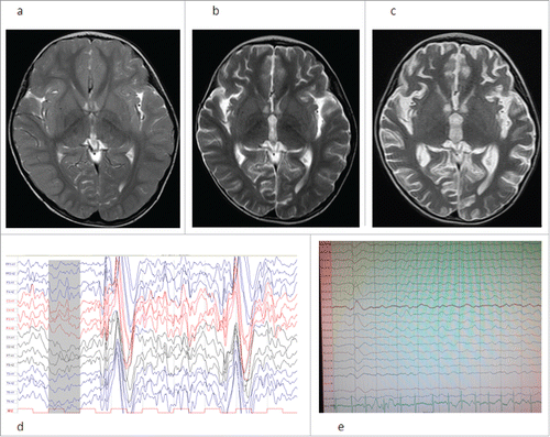
After his first week in hospital, the patient deteriorated rapidly, progressive myoclonus and spasm, tremor, hypologia, and fidgetiness were found. A second EEG showed high amplitude slow waves of paroxysmal activity in the every lead, occasionally intermingled with sharp waves. On the 28th day, only seizures were controlled by combined therapy with valproate and levetiracetam. After the first month, light coma, bradycardia, hypermyotonia, brisk reflexes, positive babinski sign were noted, and hypostatic pneumonia occurred. A second brain MRI showed the brain atrophy was evident, in which cinerea atrophy was severer ().
After the second months, he has light coma, opisthotonos, ocular bobbing, and abolition of pupillary light reflex. A third brain MRI shows atrophy of cinerea and white matter deteriorated rapidly (), hyperintense signal of pons and brachium pontis on T–weighted, and FLAIR images. A third EEG shows a background with high–amplitude δ activity of irregularity, with unresponsive to stimuli and, paroxysmal activity of periodic high amplitude slow waves which are obviously over the left frontal, parietal, and posterior temporal lobes, at interval between 4–10 seconds (). The second CSF examination shows CSF pressure, cell count, and glucose are normal, protein is mildly elevated, RT–PCR and Real–Time PCR for measles RNA are negative, CSF measles IgG is 4445.1mIU/mL (nr < 200). Serum measles IgG is 4263.3 mIU/mL (nr < 200).The IgG index (CSF IgG/ serum IgG) is 1.04. Measles IgM is negative in both CSF and serum. He had had measles and measles encephalitis at 7.5 months of age without previous immunization and had a complete recovery. Measles immunizations had been received at age 20 months and at 2 y of age. He frequently suffered upper respiratory infection within 1.5 y after measles, once every half a month. The diagnosis of subacute sclerosing panencephalitis (SSPE) of the patient was identified, based on clinical features, measles antibodies in the CSF, and EEG. The patient was treated with ribavirin and cimetidine for 1 month. After the treatment, his condition is in stable status with light coma, rigidity, and existence of pupillary light reflex.
SSPE is a chronic persistent infection of the central nervous system (CNS) caused by an altered form of the measles virus.Citation1 Most patients have a history of measles infection before the age of 2.Citation2 SSPE is mainly characterized by mental and behavioral changes, motor symptoms, and seizures. SSPE is a usually fatal disease. The reason of our patient developing SSPE might be the mutant measles virus leading to chronic infection of CNS with immune disturbances after measles. There is no exact cure in the treatment of SSPE. The course of the patient is rapidly progressive, due to uncertain pathogenesis or the treatment with methylprednisolone as glucocorticoid in earlier period. His IgG index value indicates measles antibody synthesis within CNS and destruction of blood–brain barrier.
The best way of eradication of SSPE is measles immunizations. SSPE is very rare in China with widespread vaccination now. In the schedule of Chinese national immunization program, the first and the second measles immunizations are recommended at 8 months and at 18–24 months, respectively. However, there is no measles vaccination coverage between 6 and 8 months in vulnerable infancy period and there is often a disappearance of maternal antibodies, as occurs similarly in many other countries and regions. Therefore, we recommend above measles immunizations should be given separately at 6 months and at 12–15 months. Credible strategies had been confirmed.Citation3–5 High levels of measles immunity are necessary. Other special populations such as reproductive women, immunocompromised children and travelers should also be adequately protected from measles.
Disclosure of potential conflicts of interest
The authors report no conflict of interest.
References
- Colpak AI, Erdener SE, Ozgen B, Anlar B, Kansu T. Neuro–ophthalmology of subacute sclerosing panencephalitis: two cases and a review of the literature. Curr Opin Ophthalmol. 2012;23(6):466–71. doi:10.1097/ICU.0b013e328358b196. PMID:23047165
- Yalaz K, Anlar B, Renda Y, Aysun S, Topcu M, Ozdirim E. Subacute sclerosing panencephalitis in Turkey: epidemiological features. J Trop Pediatr. 1988;34:301–5. doi:10.1093/tropej/34.6.301. PMID:3221414
- Gans H, Yasukawa L, Rinki M, DeHovitz R, Forghani B, Beeler J, Audet S, Maldonado Y, Arvin AM. Immune responses to measles and mumps vaccination of infants at 6, 9, and 12 months. J Infect Dis. 2001;184(7):817–26. doi:10.1086/323346. PMID:11528592
- Carson MM, Spady DW, Beeler JA, Krezolek MP, Audet S, Pabst HF. Follow– up of infants given measles vaccine at 6 months of age: antibody and CMI responses to MMRII at 15 months of age and antibody levels at 27 months of age. Vaccine. 2005;23(25):3247–55. doi:10.1016/j.vaccine.2005.01.092. PMID:15837229
- Gans HA, Yasukawa LL, Alderson A, Rinki M, DeHovitz R, Beeler J, Audet S, Maldonado Y, Arvin AM. Humoral and cell–mediated immune responses to an early 2–dose measles vaccination regimen in the United States. J Infect Dis. 2004;190(1):83–90. doi:10.1086/421032. PMID:15195246