
 ?Mathematical formulae have been encoded as MathML and are displayed in this HTML version using MathJax in order to improve their display. Uncheck the box to turn MathJax off. This feature requires Javascript. Click on a formula to zoom.
ABSTRACT
Women's health is seriously threatened by breast cancer. Taking advantage of efficient diagnostic instruments to identify the disease is very meaningful in prolonging life. As a cheap noninvasive radiation-free technology, Near-infrared Spectroscopy is suitable for general breast cancer examination. A discrimination method of breast cancer is presented using the deference between absorption coefficients and applied to construct a blood oxygen detection device based on Modified Lambert-Beer theory. Combined with multi-wavelength multi-path near-infrared sensing technology, the proposed method can quantitatively distinguish the normal breast from the abnormal one by measuring the absorption coefficients of breast tissue and the blood oxygen saturation. An objective judgment about the breast tumor is made according to its high absorption of near-infrared light. The phantom experiment is implemented to show the presented method is able to recognize the absorption differences between phantoms and demonstrates its feasibility in the breast tumor detection.
Introduction
As a malignant disease, breast cancer threatens people's health, especially for females.Citation1,2 The most efficient way of controlling breast cancer and enhancing survival rates is to diagnose it as soon as possible. Therefore, the development of simple stable detecting technology for early stage breast tumor has become one of the most important aspects of breast cancer diagnosis and treatment.Citation3 Recently, some of imaging technologies have been adopted in the clinic detection, which are normally expensive, bulky, radioactive and only suitable for applications in hospital. The current technologies' drawbacks make the detection inconvenient and restrain the general examinations basically.Citation4 With this in mind, it is of great significance to develop a portable, noninvasive, low-cost, and popularized detection method for this disease.
According to the current trend of breast cancer detection technology, information-based mode is now replacing the traditional symptom mode gradually. Namely, the judgment is expected to be given based on the biomedical or functional information shown in the early stage of breast cancer.Citation1,5 Near-infrared Spectroscopy (NIRS) coincides with this trend, which exploits the principle that biological tissue, to some extent, can absorb and scatter light. On the basis of intensities of input and output light, the quantitative information of the content that may absorb or scatter light will be achieved. This technology can test the functional change of breast tissue if the structural change appears. Combined with imaging methods, we can intuitionally obtain the tissue structure and physiological status with NIRS.Citation2,6,7 Additionally, NIRS also has other advantages such as noninvasive methodology, indolence, and high-resolution as well as capability of precise tumor location. For these reasons, it turns out to be one of the primary developing directions of early-stage breast tumor detection.Citation8 An array sensor composed by continuous NIRS lights and a breast tumor detection system is constructed based on Modified Lambert-Beer theory in our study. The near-infrared light may also be influenced by chromophores other than hemoglobin such as, myoglobin, melanin and cytochrome. As skin color is also determined by the presence of chromophores, it is plausible that NIRS signal quality may be affected by dark skin pigmentation.Citation9 Considering this phenomenon, a phantom experiment is implemented, which use Intralipid and India Ink to simulate the absorbance and scattering of NIRS light respectively. The experimental result shows that the proposed device is able to measure the absorption coefficients of the phantom tissue and the blood oxygen saturation, so that the concerned canceration case can be detected.
Theory
The breast cancer detection is investigated based on steady Near-infrared spectrum technology. The continuous LED working on the band of near-infrared is used in the experiment. According to Modified Lambert-Beer theory, the optical attenuation is caused by absorption of certain mediums in the human tissue. Thus the concentrations of these mediums can be calculated with the transmitted intensity.Citation10 Considering the grading of hemoglobin concentration in breast tumor, the resolution of blood oxygen concentration should be no less than 10 mol/L to make sure early-stage tumor to be identified by the system.
Modified lambert-beer theory
Traditional Lambert-Beer theory is not appropriate for highly scattering mediums, for example, breast tissue. In the case of existence of diffusion, photon motions between 2 fixed points are random, which, in large scale, present a banana-shaped propagation path. The length of the path is considerably larger than ,
being the distance between light source and detector. Based on this feature, Deloy modified the original Lambert-Beer theory by averaging light path and differential path,Citation11 and got
(1)
(1) where,
stands for optical density.
and
respectively represent input and output light intensities.
(Differential Path length Factor) denotes the function of light wavelength, which is generally unknown and can be estimated by Monte Carlo simulation.
and
respectively represent the absorption coefficient and molality of the ith material.
denotes the attenuation factor synthesising all the background factors that may disturb the absorption and scattering. G is usually regarded as constant when the light source and detector are the same.
Measurement theory of blood oxygen
Compared with absorption, scattering occurs more frequently in human organs and tissues at infrared wavelengths, which enhances the migration of photons greatly. And the optical absorption ability of water is much less than that of hemoglobin so that the absorption ability of biological tissue mostly depends on the molalities of oxyhemoglobin and deoxyhemoglobin. If two or more near-infrared lights with different wavelengths are available, the molalities can be resolved based on Modified Lambert-Beer theory. Additionally, the greater the differences between these light sources are, the higher the detecting precision is.12 in Equation Equation(1)
(1)
(1) can be calculated in practice based on multiple parameters. However,
can be regarded as constant at the certain tissue location. Assume
below is the optical density.
(2)
(2)
and
respectively represent the extinction coefficients of oxyhemoglobin and deoxyhemoglobin. While
and
respectively represent their molalities. For the near-infrared lights with wavelengths of
and
, differences of optical densities gathered by 2 individual detectors can be expressed as
(3)
(3) where,
and
represents the distances between detectors and light sources. The blood oxygen saturation
is calculated using Equation Equation(4)
(4)
(4) .
(4)
(4)
Based on the relations among molality, absorption coefficient and extinction coefficient, we can achieve the extinction coefficient of near-infrared light with the equation below.(5)
(5)
The difference between the absorption coefficients for the measuring spot and the healthy breast tissue used as a reference reflects the diversity of their optical properties. If the difference is obvious, we can draw the conclusion that there is likely a breast lesion on the spot with greater absorption coefficient. Therefore, the difference of absorption coefficients can be applied as the criteria to detect breast cancer.
Data acquisition system
Probe structure
As for near-infrared light, breast tissue can be regarded as a semi-infinite transmission medium. According to the path parsing function of near-infrared light with a vertical incidence, the deepest point it can reach situates halfway between light and detector and its depth is times length of
, the distance from light to detector. In order to meet the requirement of multi-depth detection, a probe with 8 light sources and 7 electro-optical detectors, as shown in , is applied in the test system. For the convenience of operation, the probe is designed to work independently of the control circuit.
Figure 1. Layout diagram of near-infrared probe.
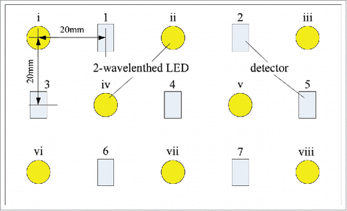
The yellow circles in stand for LED lights, which can emit near-infrared light of 735 nm, 805 nm, and 850 nm. The rectangular marks represent the electro-optical detectors, OPT101. The distances from the center of any light to its adjacent detectors are unified to 20 mm. Considering all the combination of light sources, the reachable depths of measurement are respectively 7.1 mm, 15.8 mm, 21.2 mm, 25.5 mm, and 29.2 mm beneath the skin.
Composition of sampling system
The NIRS-based blood oxygen detection system is designed and produced in the study composed of a near-infrared probe, a sampling circuit, and a signal processing computer. The structure diagram and the picture of the real product are respectively shown in and . The probe is in charge of emitting and receiving the near-infrared light. As the kernel component of the sampling circuit shown in , the microprocessor is responsible for collecting signals besides filter, amplification, analog-to-digital conversion, data caching, and communication with the computer. The mission of the computer includes analyzing and displaying the received digital signals.
Figure 2. NIRS-based blood oxygen sampling system.
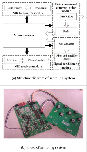
Taking advantage of the time-sharing technology, the microprocessor drives the light sources and chooses the detecting channels in sequence. The A/D integrated circuit converts the amplified filtered analog voltage signals into digital ones. Coordinating with latch chip, the data storage module may save the resource of the I/O parallel port tremendously. The signals collected are sent to the communication module and then uploaded to the computer at its command.
Experiment and analysis
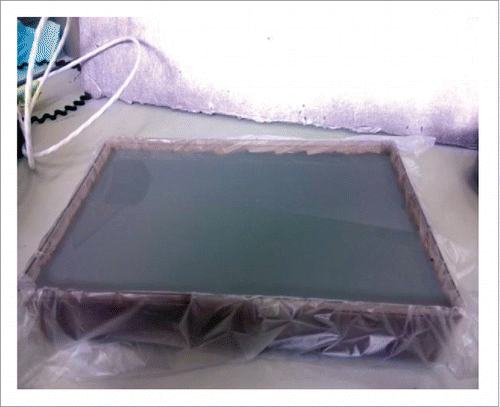
The feasibility of the presented theory and the designed system remain to be testified during clinical trials by recruiting volunteers. Phantom experiment can be regarded as an excellent way to pre-check the performance. One of the best features of phantom experiment is that human tissue of certain absorption and scattering coefficients could be simulated by adjusting the proportions of the components in phantoms. Intralipid-10% is used to control the scattering property of a phantom sample. In patients with dark skin pigmentation, the blood oxygen saturation should be interpreted with caution, as melanin clearly interferes with the quality of the reflected NIRS signal.Citation9 For emulating the effect, India Ink is applied as an absorber to control the phantom tissue' absorption coefficient. In addition, gelatine is chosen as the coagulator to simulate tumor tissue.
For better result, Intralipid and India Ink need to be mixed with each other under the condition of high temperature. Thus Brownian movement is relatively forceful so that the components are evenly distributed in the solution before solidifying. Then the solution is put in an incubator chamber at 5 degrees, where it is solidified under accelerated-cooling conditions. At the same time, the solution is also dripped into a quartz cuvette for calibration. After that, a plastic film is applied to cover the surface of the solidification for retaining water, preventing the air and bacteria from entering, and prolonging the storage time. Up to now, the tissue phantom shown in has been produced. The mold is a 20 cm long, 13 cm wide, and 7 cm high rectangular solid. The area that the probe needs to be placed on is a 9 cm long, 5 cm wide rectangular. According to the banana-shaped light beam theory, the mold is deep enough for the propagation of the light.
Figure 3. Tissue phantom.
Based on the research of breast tumor detection at Connecticut University, we made up several samples shown in using the absorption coefficient of 735 nm near-infrared light as a reference. Wherein, the samples possess 2 kinds of scattering coefficients (6 cm−1 and 3 cm−1) and 4 kinds of absorption coefficients (0.0200 cm−1, 0.0500 cm−1, 0.1000 cm−1 and 0.2000 cm−1) respectively. Samples No.1 and No. 5 are the phantoms of normal breast tissue, which act as the references. According to the feature of high absorption of near-infrared light, tumor is simulated by samples No. 4 and No. 8.
Table 1. Theoretical absorption and scattering coefficients of phantoms.
Each sample of phantom is repeatedly metered with the Fourier infrared spectrometer and the mean value serves as the actual measurement of its absorption coefficient, which is listed in . Obviously, the measured absorption coefficients of 735 nm near-infrared light conform to their theoretical ones. The absorbances are positively correlated with the concentrations of India Ink and does not change with the variations of Intralipid and gelatine. The differences between theoretical absorption coefficients and measured ones are derived from the preparation operation of the sample solution.
Table 2. Measured coefficients of phantoms.
The phantoms are divided into 2 sample groups: No. 1 through 4 belong to the low scattering coefficient group. No. 5 through 8 belong to high scattering coefficient group. The experimental procedure is shown as following.
Taking sample No. 1 as the study subject, drive LED i to emit near-infrared light with light intensity of | |||||
Calculate absorption coefficients | |||||
Light up LED ii, and repeat step (a) and (b) until all 8 lights are lit. | |||||
| (d) | Based on the recorded light intensity, compute the average absorption coefficients, | ||||
Taking sample No. 2 as a target, repeat steps (a) through (d) until all 8 phantoms are traversed once, then absorption coefficients, | |||||
The differences between measured spot and reference one are achieved by subtracting the absorption coefficients of No. 2 to No. 4 with that of No. 1, we will obtain | |||||
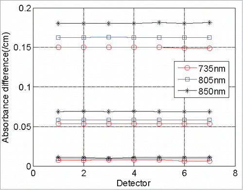
The specific test results of 2 kinds of phantoms are shown in . Apparently, absorbance differences obtained by the detectors from lights of the same wavelength are identical for a certain phantom. Compared with the data in , the measurement errors with maximum of 0.0003 cm−1 achieved can ensure that the presented system is capable of measuring the absorption coefficients precisely. Furthermore, in the case of uniform concentrations of Intralipid and India Ink, the absorbance of 735 nm near-infrared light is always the greatest one among the 3 of them and that of 850 nm near-infrared light is the least. The result coincides with the measurement of spectrograph. For the same concentration of Intralipid and wavelength of near-infrared lights, the higher the concentration of India Ink is, the bigger the absorbance is. Within the error range permitted, absorbance is linearly correlated with the concentration of India Ink. Meanwhile, the absorbance differences between measured results and standard ones are consistent under the condition of the same wavelength of light and concentration of India Ink but different concentrations of Intralipid. It is testified that the effect of Intralipid on absorbance is tiny. As can be seen from the curves in , the concentrations of Intralipid in phantoms are easy to tell, which means that the presented method and system in this paper can provide quantified differences of blood oxygen between healthy and diseased breasts, based on which breast tumor can be detected reliably.
Figure 4. Measured results of low scattering phantoms.
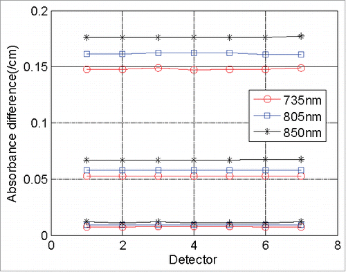
Figure 5. Measured results of high scattering phantoms.
Conclusion
A noninvasive breast tumor detection method based on Modified Lambert-Beer theory is presented in this paper. The multi-wavelength multi-path probe is designed by using continuous near-infrared light, as well as a blood oxygen detection system. To evaluate the performance of the proposed method and system, phantoms of 2 kinds of scattering coefficients and 4 kinds of absorption coefficients are prepared. Using the phantom of normal tissue as a reference, the differences of other samples' absorption coefficients are achieved, which can be used as the indicators to detect the diseased breast. It is manifested by the experiment that the presented method is able to measure the concentration of India Ink precisely. The accuracy is high enough to tell the difference between the healthy breast and the unhealthy one in blood oxygen. The experiment results show that the research of this paper is very promising and highly anticipated for in vivo detection of early-stage breast cancer.
Disclosure of potential conflicts of interest
No potential conflicts of interest were disclosed.
Authors' contributions
Dr. Dan Liu and Dr. Xin Liu were responsible for the system construction. Dr. Qisong Wang was in charge of the coding. Dr. Yan Zhang performed the experiments for the presented data. Dr. Dan Liu coordinated and drafted the manuscript. Jingyang Lu polished the English translation. All authors edited the manuscript and approved the final version.
Funding
This work is sponsored by National Natural Science Foundation of China (GN: 61301012 and 61401117), and the Fundamental Research Funds for the Central Universities (Grant No. HIT. IBRSEM. 201304).
References
- Poellinger A, Grosenick D. Optical imaging in mammography. Comprehensive Biomed Phys 2014; 4:345-62; http://dx.doi.org/10.1016/B978-0-444-53632-7.00426-3
- Korb ML, Hartman YE, Zinn KR, Bland KI, Rosenthal EL. Use of near-infrared imaging to improve surgical resection of breast cancer. J Am College Surg 2013; 217:S126-7; PMID:24360117; http://dx.doi.org/10.1016/j.jamcollsurg.2013.07.292
- Poellinger A, Martin JC, Ponder SL, Freund T, Hamm B, Bick U, Diekmann F. Near-infrared laser computed tomography of the breast: first clinical experience. Acad Radiol 2008; 15:1545-53; PMID:19000871; http://dx.doi.org/10.1016/j.acra.2008.07.023
- Erickson SJ, Godavarty A. Hand-held based near-infrared optical imaging devices: a review. Med Eng Phys 2009; 31:495-509; PMID:19054704; http://dx.doi.org/10.1016/j.medengphy.2008.10.004
- Sampath L, Kwon S, Hall MA, Price , Muraca EMS. Detection of cancer metastases with a dual-labeled near-infrared/positron emission tomography imaging agent. Transl Oncol 2010; 3(5):307-17; PMID:20885893; http://dx.doi.org/10.1593/tlo.10139
- Zhu S, Zhang JT, Janjanam J, Bi JH, Vegesna G, Tiwari A, Luo FT, Wei JJ, Liu H Y. Highly water-soluble, near-infrared emissive BODIPY polymeric dye bearing RGD peptide residues for cancer imaging. Analytica Chimica Acta 2013; 758:138-44; PMID:23245906; http://dx.doi.org/10.1016/j.aca.2012.10.026
- Peidaee P, Almansour N, Shukla R, Pirogova E. The cytotoxic effects of low intensity visible and infrared light on human breast cancer (MCF7) cells. Comput Struct Biotechnol J 2013; 6(7): e201303015; PMID:24688723; http://dx.doi.org/10.5936/csbj.201303015
- Hirosawa N, Sakamoto Y, Katayama H, Tonooka S, Yano K. In vivo investigation of progressive alterations in rat mammary gland tumors by near-infrared spectroscopy. Anal Biochem 2002; 305:156-65; PMID:12054444; http://dx.doi.org/10.1006/abio.2002.5649
- Wassenaar BE, Van den Brand JGH. Reliability of Near-Infrared spectroscopy in people with dark skin pigmentation. J Clin Monitoring Comput 2005; 19:195-9; PMID:16244841; http://dx.doi.org/10.1007/s10877-005-1655-0
- Cui X, Signe B, Bryant DM, Glover GH, Reiss AL. A quantitative comparison of NIRS and fMRI across multiple cognitive tasks. Neuro Image 2011; 54(4):2808-21; PMID:21047559
- Xu T, Zhang CP, Wang XY, Zhang LS, Tian JG. Measurement and analysis of light distribution in Intralipid-10% at 650 nm. Appl Opt 2003; 42(28):5777-84; PMID:14528943; http://dx.doi.org/10.1364/AO.42.005777