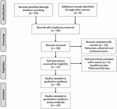
2,091
Views
5
CrossRef citations to date
0
Altmetric
Review

Clinical significance of long noncoding RNA MNX1-AS1 in human cancers: a meta-analysis of cohort studies and bioinformatics analysis based on TCGA datasets

, , , &
Pages 875-885 | Received 03 Nov 2020, Accepted 26 Jan 2021, Published online: 09 Mar 2021

ABSTRACT

MNX1-AS1 expression has been proposed to be abnormally upregulated in multiple human malignancies and be linked with the survival outcome of patients. However, relevant conclusions were yielded based on the limited samples. Therefore, we herein implemented a meta-analysis of the published cohort studies to further decipher the relationship of MNX1-AS1 level to prognosis and clinicopathological features in various cancers. Additionally, using The Cancer Genome Atlas (TCGA) datasets we carried out a bioinformatics analysis to make a further evaluation on the prognostic value of MNX1-AS1 expression. The results of meta-analysis indicated elevated MNX1-AS1 level closely correlated with poorer overall survival (OS) (HR = 1.97, 95% CI, 1.73–2.24; P < 0.00001), and disease-free survival (DFS) (HR = 2.24, 95% CI, 1.48–3.38; P = 0.0001) in cancers, which was confirmed by the bioinformatics analysis. Besides, it was observed the upregulated MNX1-AS1 level was significantly related to invasion depth, disease stage, tumor metastasis, and differentiation. Collectively, high MNX1-AS1 level correlated with poor survival outcome and aggressive clinicopathological characteristics in various cancers, suggesting that MNX1-AS1 may be applied as a prognostic marker and even a therapeutic target. Nevertheless, more high-quality studies designed with a large sample size should be conducted to further determine the clinical role of MNX1-AS1 in specific cancer types.

Graphical abstract

Introduction

As the incidence and mortality of cancers continuously rise, cancer has become an intractable global public health problem undoubtedly [Citation1]. Although huge advancement in surgical intervention, chemotherapy, and radiotherapy, cancer patients, especially at an advanced stage, still had a much unsatisfactory prognosis [Citation2]. Molecular-targeted therapy has been considered to be a promising therapeutic modality for cancers [Citation3,Citation4]. Therefore, it is very meaningful to identify the molecular biomarkers that correlate with cancer progression and prognosis, so as to provide a theoretical basis for developing individualized treatment target.

Noncoding RNAs such as microRNA, circular RNA, and long noncoding RNA (lncRNA) account for a significant proportion of the human genome [Citation5]. The amount of evidence shows lncRNAs are involved in multiple physio-pathologic processes, such as development, inflammatory response, tissue regeneration, ischemia reperfusion injury, cardiovascular diseases, and cancers [Citation5,Citation6]. In malignancies, numerous lncRNAs are abnormally expressed, which epigenetically and transcriptionally influences expressions of oncogenes and tumor suppressors, suggesting lncRNAs may be developed as the prognostic indicators and treatment targets [Citation7]. For example, long intergenic noncoding RNA01134 was substantially upregulated in hepatocellular carcinoma and it was tightly linked with poor patient outcome [Citation8]. In colon cancer, DNAJC3-AS1 was significantly downregulated, which could promote cancer cell migration, invasion, and epithelial-mesenchymal transition (EMT) through regulating the miR-214-3p/LIVIN [Citation9]. Inversely, LncRNA FLANC level was found to be elevated in colorectal cancer and FLANC could sustain activation of STAT3 signaling by prolonging its phosphorylation, which resulted in VEGFA upregulation to promote angiogenesis, thereby accelerating tumor progression [Citation10]. LncRNA PART1 expression was dramatically reduced in gastric cancer tissues and this dysregulation closely correlated with the unfavorable prognosis of patients who underwent surgery [Citation11]. Further investigation disclosed PART1 exerted a tumor-suppressive effect through upregulating PLZF expression, which recruited EZH2 to induce epigenetic PDGFB silencing, ultimately counteracting PI3K/Akt signaling transduction [Citation11].

MNX1 antisense RNA 1 (MNX1-AS1) was firstly discovered as a highly expressed gene in colorectal malignant tumor, which is termed CCAT5 as well [Citation12]. Recently, a growing number of studies suggested that MNX1-AS1 was aberrantly expressed in diverse malignancies, including cholangiocarcinoma [Citation13], esophagus cancer [Citation14], breast carcinoma [Citation15–17], gastric carcinoma [Citation12,Citation18,Citation19], bladder carcinoma [Citation20], ovarian carcinoma [Citation21,Citation22], cervical cancer [Citation23], osteosarcoma [Citation24,Citation25], laryngeal cancer [Citation26], lung cancer [Citation27–29], prostate cancer [Citation30], hepatocellular carcinoma [Citation15], glioblastoma [Citation31] and colon adenocarcinoma [Citation32]. Moreover, aberrant MNX1-AS1 expression has been proposed to correlated with cancer patient survival, indicating its potential as prognostic marker and therapeutic target [Citation12,Citation15,Citation18,Citation23,Citation26].

Nevertheless, the previous studies were restricted by small sample sizes, in which the clinical significance of MNX1-AS1 level in cancer might be overestimated. Therefore, in this study, we conducted a meta-analysis of the published literature and bioinformatics analysis using TCGA datasets to further evaluate the clinical role of MNX1-AS1 expression in cancers.

Methods

Search strategy

‘MNX1-AS1 or CCAT5’ in combination with cancer-related words including ‘cancer,’ ‘tumor,’ ‘carcinoma,’ ‘neoplasm’ ‘adenocarcinoma,’ and ‘malignancy’ were applied to search relevant articles in Chinese National Knowledge Infrastructure (CNKI), PubMed, and Web of Science (WOS) databases from inception to October 2020. Additionally, we also examined the reference lists of the relevant articles to identify eligible studies that could be omitted by electronic search.

Inclusion and exclusion criteria

Studies that conform to the following items simultaneously were selected [Citation1]: the patients should be definitely diagnosed with cancers by histopathology [Citation2]; the association of MNX1-AS1 with overall survival (OS) or disease-free survival (DFS) was provided regardless of its statistical significance [Citation3]; MNX1-AS1 expression in was detected using polymerase chain reaction (PCR) or in situ hybridization (IHS) [Citation4]; the studies were written in English or Chinese language.

The following conditions were used to exclude the ineligible studies [Citation1]: the articles were not original articles, such as review articles, editorial comments, and meeting abstracts [Citation2]; the studies enrolled the overlapping patient population. In this situation, the latest study was included [Citation3]. The HRs with 95% (CIs) could not be obtained, although the association of MNX1-AS1 expression with survival outcome in cancer patients was explored [Citation4]; the studies analyzed the association of MNX1-AS1 expression with survival outcome in cancer patients based on RNA sequencing data from The Cancer Genome Atlas (TCGA) and Gene Expression Omnibus (GEO) datasets [Citation5]; the studies focused on the irrelevant topics [Citation6]; the studies were conducted in animal models.

Data collection and quality evaluation

Two investigators collected data from selected articles independently. Any discordance was eliminated via discussion among all the researchers. The collected data included: the first author, the year of publication, region, cancer type, the number of patients, the sex ratio of patients, the age of patients, detection method, antibody type, cutoff of high MNX1-AS1 expression, the percent of patients at TNM III–IV stage, the number of patients with metastasis, the hazard ratios (HRs) with 95% confidence intervals (CIs) for OS and DFS, as well as the analysis method of assessing the prognostic value of MNX1-AS1. HRs from the multivariate analysis were preferentially chosen. If the included studies did not provide HRs, we tried to apply Engauge Digitizer version 4.1 to calculate HRs with CIs from Kaplan-Meier curves [Citation33]. The methodological quality of selected studies was assessed using Newcastle-Ottawa Scale (NOS) system [Citation34]. In this assessment system, three aspects (subject selection, comparability of the subject, and clinical outcome) are evaluated and a score ranging from 0 to 9 may be given to a study. In this meta-analysis, we defined that the studies with more than the mean score of all studies had a high-quality methodology.

Bioinformatics analysis of TCGA data

Gene Expression Profiling Interactive Analysis (GEPIA), which is supported by TCGA databases, was used for further evaluating the associations of MNX1-AS1 level with OS and DFS. K-M method and log-rank test were employed to perform survival outcome analysis, and the relevant statistical results such as HRs and P values were displayed by K-M curve as previously described [Citation35].

Statistical analysis

The Review Manager V 5.3 Software (Cochrane Collaboration, London, U.K.) and STATA V 12.0 Software (Stata, College Station, TX) were used for performing statistics analysis in this study. The pooled HRs with 95% CIs were calculated to analyze the association of MNX1-AS1 expression with survival outcomes, and the association between MNX1-AS1 level and clinicopathological parameters was estimated using the pooled odds ratios (ORs) with 95% CIs. We tested the heterogeneity with the help of Cochran’s Q test and the Higgins I-squared statistic, and P value < 0.05 or I2 > 50% suggested heterogeneity was statistically significant. When significant heterogeneity was tested, we conducted the pooled analysis using the random effects model. On the contrast, the fixed effects model was selected [Citation36]. Sensitivity analysis was performed via sequential deletion of a single included study to test if the overall pooled estimation was stable. Egger’s linear regression test and Begg’s rank test were used to assess publication bias [Citation37–39]. P values in this study were two-sided, and P < 0.05 indicated there was a statistical significance.

Results

Numerous studies have suggested that increased MNX1-AS1 expression might correlate with unfavorable survival outcome in cancer patients. However, the conclusions in those studies may be limited by small sample sizes, probably overestimating the clinical significance of MNX1-AS1 expression in cancer patients. Hence, herein we performed a meta-analysis of the previous studies and bioinformatics analysis based on TCGA datasets to further evaluate the clinical significance of MNX1-AS1 expression in cancers.

Study search and selection

A total of 84 relevant articles were retrieved from the initial search of PubMed, WOS, and CNKI databases. First, 54 repetitive records were removed. Then, we checked the titles and abstracts of the remained articles and further excluded 16 records for retracted papers, editorials, and irrelevant topics. Next, we carefully reviewed the rest of the articles by full text. In this step, we deleted four articles since they provided unavailable data [Citation40] or data from TCGA or CEO [Citation12,Citation26,Citation27]. Finally, 10 articles were chosen for this meta-analysis [Citation12,Citation15,Citation17–19,Citation22,Citation23,Citation27–29]. The flow diagram of study search and selection is summarized in .

Figure 1. Flow diagram of searching and selecting eligible studies

Figure 1. Flow diagram of searching and selecting eligible studies

Main characteristics of selected studies

The main characteristics of selected studies are shown in . All the included studies were published in recent five years and designed with sample sizes from 43 to 177. MNX1-AS1 level was detected through qRT-PCR in nine studies, and by ISH in one study. All these studies were performed in China. Six types of cancer were studied in these studies, including gastric carcinoma, lung carcinoma, hepatocellular carcinoma, breast cancer, cervical cancer, and ovarian carcinoma. Association of MNX1-AS1 level with OS was evaluated in 10 studies, and its prognostic value for DFS was reported in two studies. HRs with CIs were produced by multivariate analysis in five included studies, whereas the other studies only provided survival curves and thereby we calculated HRs with CIs using Engauge Digitizer version 4.1 (Supplement 1). NOS scores of the included studies ranged from 6 to 8.

Table 1. The main characteristics of included cohort studies

Association of MNX1-AS1 level with survival outcome

Ten studies comprising 1119 cancer patients investigate the link between MNX1-AS1 level and OS. Because no significant heterogeneity was identified (I2 = 37%, P = 0.12), we conducted the pooled analysis using fixed effects model. The merged HR implied that high MNX1-AS1 level was closely connected with worse OS (HR = 1.97, 95% CI, 1.73–2.24; P < 0.00001) (). Besides, only two studies with 269 patients were included to assess the correlation of MNX1-AS1 expression with DFS. Consistent with OS, it was found increased MNX1-AS1 expression was linked with unfavorable DFS (HR = 2.24, 95% CI: 1.48–3.38; P = 0.0001) ().

Figure 2. Forest plot illustrated the prognostic significance of MNX1-AS1 level for overall survival (a) and disease-free survival (b)

Figure 2. Forest plot illustrated the prognostic significance of MNX1-AS1 level for overall survival (a) and disease-free survival (b)

Association of MNX1-AS1 expression with clinicopathologic parameters

Aggressive clinicopathologic characteristics closely correlate with cancer patient survival. Therefore, we further evaluated the connection of MNX1-AS1 expression with clinicopathologic parameters of cancer patients. As summarized in , increased MNX1-AS1 level was related to advanced TNM stage (OR = 5.21, 95% CI: 2.39–11.36) (), invasion depth (OR = 3.00, 95% CI: 1.31–6.86) (), lymphatic metastasis (OR = 3.70, 95% CI: 2.07–6.61) (), distant metastasis (OR = 3.99, 95% CI: 2.41–6.60) () and poor differentiation (OR = 1.62, 95% CI: 1.15–2.28) (). Nevertheless, no significant links of MNX1-AS1 level with age (OR = 1.26, 95% CI: 0.91–1.72) and sex (OR = 1.02, 95% CI: 0.71–1.47) were observed ().

Table 2. The correlation between clinicopathological characteristics and MNX1-AS1 expression

Figure 3. Forest plot illustrated the associations of MNX1-AS1 level with tumor TNM stage (a) and invasion depth (b)

Figure 3. Forest plot illustrated the associations of MNX1-AS1 level with tumor TNM stage (a) and invasion depth (b)

Figure 4. Forest plot illustrated the associations of MNX1-AS1 level with lymphatic metastasis (a), distant metastasis (b) and tumor differentiation (c)

Figure 4. Forest plot illustrated the associations of MNX1-AS1 level with lymphatic metastasis (a), distant metastasis (b) and tumor differentiation (c)

Sensitivity analysis and publication bias

Sensitivity analysis was implemented via deletion of a single excluded study in each step. It was observed HRs with CIs for OS did not alter significantly after deleting any eligible study, suggesting the robustness of our combined result (). Egger’s linear regression test and Begg’s rank test were used to assess publication bias. The results showed that P values from the two tests were above 0.05 (Supplement 2), indicating there was no significant publication bias. Collectively, these results suggested that our pooled analysis was relatively reliable.

Figure 5. Kaplan-Meier survival curves generated from Gene Expression Profiling Interactive Analysis (GEPIA) illustrated the prognostic significance of MNX1-AS1 level for overall survival (a) and disease-free survival (b) in cancers

Figure 5. Kaplan-Meier survival curves generated from Gene Expression Profiling Interactive Analysis (GEPIA) illustrated the prognostic significance of MNX1-AS1 level for overall survival (a) and disease-free survival (b) in cancers

Validation based on TCGA cohort

Next, we performed bioinformatics analysis using GEPIA to further verify the results of the prognostic value of MNX1-AS1 expression in the meta-analysis above.

The K-M curves labeled with HRs and P values indicated elevated MNX1-AS1 level was connected with the dismal OS () and DFS (). Overall, the bioinformatics analysis based on TCGA datasets further validates the findings in the meta-analysis.

Figure 6. Sensitivity analysis for the pooled estimation of MNX1-AS1 level and OS in cancers

Figure 6. Sensitivity analysis for the pooled estimation of MNX1-AS1 level and OS in cancers

Discussion

Previous studies have reported that MNX1-AS1 level was abnormally upregulated in diverse malignancies, and it was closely related to survival outcome and clinicopathological features of patients. However, the previous studies were restricted by small sample sizes, in which the clinical significance of MNX1-AS1 level in cancer might be overestimated. Therefore, in this study, we implemented a meta-analysis of the published literature and bioinformatics analysis using TCGA datasets to further evaluate the clinical role of MNX1-AS1 expression in cancers. In this meta-analysis, 10 studies comprising 1119 cancer patients were included and the combined results suggested higher MNX1-AS1 level closely correlated with shorter OS and DFS, which was further supported by the bioinformatics analysis. In addition, the combined results showed that increased MNX1-AS1 level was related to multiple aggressive clinicopathologic features, including TNM stage, lymphatic metastasis, distant metastasis, invasion depth, and poor differentiation.

Numerous researchers have tried to decipher the molecular mechanisms accounting for the protumor roles of MNX1-AS1. Lv et al. [Citation21] reported MNX1-AS1 could contribute to ovarian cancer cell proliferation and invasion via upregulation of Bcl-2 family proteins (Bcl-2 and Bax), cyclin-dependent kinase, and cyclin D. Gao et al. [Citation31] demonstrated MNX1-AS1 could enhance the proliferative ability and invasive capacity of glioblastoma cells through targeting miR-4443. In cervical cancer, MNX1-AS1 could promote the proliferation, and suppress the apoptosis of cervical cancer cells via activation of the MAPK signaling pathway [Citation23]. Ye et al. [Citation32] uncovered that MNX1-AS1 could increase colon adenocarcinoma cell growth and metastasis in vitro by regulating the miR-218-5p/SEC61A1 signaling axis. Wang et al. [Citation20] revealed MNX1-AS1 could modulate RAB1A level via inhibiting miR-218-5p to facilitate bladder cancer progression. Similarly, Ji et al. [Citation15] demonstrated that MNX1-AS1 could also target miR-218-5p to increase COMMD8 level in hepatocellular carcinoma cells, consequently accelerating malignant cell growth, motility and invasion in vitro. In lung cancer, MNX1-AS1 was capable of promoting tumor cell proliferation, mobility, and invasion by regulating the miR-527/BRF2 signaling axis [Citation28]. In breast cancer, Cheng et al. [Citation16] first proposed MNX1-AS1 acted as a functional oncogene that could induce the epithelial-mesenchymal transition of cancer cells via activation of AKT/mTOR signaling pathway and upregulation of its natural sense transcript MNX1. Besides, a most recent study revealed MNX1-AS1 could enhance the aggressiveness of triple-negative breast cancer cells through interacting with Stat3 and increasing its phosphorylation in a p-JAK-dependent manner [Citation17]. In prostate cancer, MNX1-AS1 was proved to facilitate cancer cell proliferation, migration, and invasion, but its molecular mechanisms remain unknown [Citation30]. Ma et al. reported MNX1-AS1 was able to foster invasion and metastasis of gastric cancer cells by downregulating CDKN1A [Citation19]. Additionally, Shuai et al. [Citation12] also found upregulation of MNX1-AS1 could promote gastric cancer cell proliferation, motility and invasion and further uncovered that MNX1-AS1 can sponge miR-6785-5p to enhance BCL2 expression in gastric cancer cells [Citation12]. In osteosarcoma, MNX1-AS1 could accelerate osteosarcoma cell proliferation and invasion via downregulating KISS1 [Citation25]. Besides, Wu et al. [Citation24] suggested that MNX1-AS1 could enhance tumor cell epithelial-mesenchymal transition through upregulating MNX1 expression. Autologously, Li et al. uncovered that MNX1-AS1 could also enhance MNX1 transcription, which then activated Ajuba/Hippo signaling pathway in intrahepatic cholangiocarcinoma cells, consequently facilitating tumor progression [Citation13]. In esophagus cancer, MNX1-AS1 was also demonstrated to facilitate the proliferative and invasive capacities of tumor cells through regulating the miR-34a/SIRT1 signaling axis [Citation14]. Collectively, the evidence mentioned above strongly supports that MNX1-AS1 may play a protumor role in human cancers.

Some shortcomings in this meta-analysis should be considered. First, some studies did not report HRs directly. Thus, we calculated HRs manually from Kaplan-Meier survival curves, which may cause operating errors unavoidably. Second, all studies were performed in China. Hence, the results of this meta-analysis may not reflect the clinical significance of MNX1-AS1 expression in other ethnic population. Future studies are needed to determine this issue. Third, the number of studies focusing on a specific cancer type was limited, so the pooled analysis was not conducted in this regard. Obviously, the clinical significance of MNX1-AS1 expression in a specific cancer type should be further validated in more cohort studies. Fourth, this meta-analysis only included studies published in English or Chinese, which might contribute to a bias. Fifth, overall analysis using the GEPIA tool showed that high MNX1-AS1 expression was correlated with worse OS and DFS of cancer patients, whereas the association between MNX1-AS1 and survival outcome was not observed in several cancer types when each type of cancer was analyzed individually. This inconsistency may be partly attributed to the limitation of small sample size, the differences in sequencing technology, and the diversity of tumor genetic backgrounds. Therefore, more studies should be done to further determine the role of MNX1-AS1 in specific cancer types. Sixth, although the current study suggested that MNX1-AS1 may be a potential therapeutic target for cancer, a series of further studies are needed to confirm this point. First of all, more homogeneous clinical studies with larger samples should be conducted to further determine the clinical significance of MNX1-AS1 in each cancer type; In addition, the roles of MNX1-AS1 in cancers and the corresponding mechanisms should be fully elucidated using in vitro cell model and in vivo animal models, such as MNX1-AS1-knockout mice and tumor organoid model; Furthermore, the anticancer effects of MNX1-AS1-targeting drugs should be evaluated in the patient-derived tumor xenograft (PDX) model and tumor organoid model. Next, cautious and stepwise clinical trials may be considered to assess the efficacy of MNX1-AS1-targeting drugs in cancer patients. Seventh, many methodologies are available to evaluate publication bias [Citation39,Citation41–44], but only Egger’s linear regression test and Begg’s rank test were applied in our meta-analysis. Thus, our publication bias evaluation may be not enough reliable, though both Egger’s linear regression test and Begg’s rank test indicated no significant publication bias in the current meta-analysis.

Conclusion

In summary, high MNX1-AS1 level correlated with poor survival outcome of cancer patients, suggesting that MNX1-AS1 may be exploited as a prognostic marker and even a therapeutic target. However, more high-quality studies designed with a large sample size are demanded to further determine the clinical role of MNX1-AS1 in specific cancer types.

Highlights

Increased MNX1-AS1 level correlated with worse overall survival in cancers

Elevated MNX1-AS1 level correlated with poor disease-free survival in cancers

High MNX1-AS1 expression was related to tumor metastasis

Author contributions

Hai-peng Liu designed this study. Kang Chen and Gan Jian-xin Gan extracted the data and analyzed the data; Kang Chen and Ze-ping Huang drafted the manuscript. Kang Chen and Jun Liu revised the manuscript. All authors have read and confirmed the final manuscript.

Ethics approval and consent to participate

This study is a meta-analysis and bioinformatics analysis, so it is unnecessary to obtain Ethical Approval.

Supplemental material

Supplemental Material

Download ()

Disclosure statement

All authors have declared no conflict of interest exists for this study.

Supplementary material

Supplemental data for this article can be accessed here.

Additional information

Funding

The study was supported by the Training Program of Lanzhou University Second Hospital Cuiying postgraduate supervisor in 2019 (NO. CYDSPY201907).

References

  • Siegel RL, Miller KD, Jemal A. Cancer statistics, 2018. CA Cancer J Clin. 2018;68(1):7–30.
  • Chen W, Zheng R, Baade PD, et al. Cancer statistics in China, 2015. CA Cancer J Clin. 2016;66(2):115–132. .
  • Guan LY, Lu Y. New developments in molecular targeted therapy of ovarian cancer. Discov Med. 2018;26(144):219–229.
  • Yan C, Wang J, Yang Y, et al. Molecular biomarker-guided anti-angiogenic targeted therapy for malignant glioma. J Cell Mol Med. 2019. DOI:10.1111/jcmm.14417
  • Ghafouri-Fard S, Shoorei H, Taheri M. Non-coding RNAs participate in the ischemia-reperfusion injury. Biomed Pharmacothe. 2020;129:110419.
  • Balamurali D, Stoll M. Non-Coding RNA Databases in Cardiovascular Research. Noncoding RNA. 2020;6(3). DOI:10.3390/ncrna6030035
  • Zhang C, Ren X, Zhang W, et al. Prognostic and clinical significance of long non-coding RNA SNHG12 expression in various cancers. Bioengineered. 2020;11(1):1112–1123. .
  • Zheng S, Guo Y, Dai L, et al. Long intergenic noncoding RNA01134 accelerates hepatocellular carcinoma progression by sponging microRNA-4784 and downregulating structure specific recognition protein 1. Bioengineered. 2020;11(1):1016–1026.
  • Han B, Ge Y, Cui J, et al. Down-regulation of lncRNA DNAJC3-AS1 inhibits colon cancer via regulating miR-214-3p/LIVIN axis. Bioengineered. 2020;11(1):524–535.
  • Pichler M, Rodriguez-Aguayo C, Nam SY, et al. Therapeutic potential of FLANC, a novel primate-specific long non-coding RNA in colorectal cancer. Gut. 2020;69(10):1818–1831. .
  • Han H, Wang S, Meng J, et al. Long noncoding RNA PART1 restrains aggressive gastric cancer through the epigenetic silencing of PDGFB via the PLZF-mediated recruitment of EZH2. Oncogene. 2020;39(42):6513–6528. .
  • Shuai Y, Ma Z, Liu W, et al. TEAD4 modulated LncRNA MNX1-AS1 contributes to gastric cancer progression partly through suppressing BTG2 and activating BCL2. Mol Cancer. 2020;19(1):6. .
  • Li F, Chen Q, Xue H, et al. LncRNA MNX1-AS1 promotes progression of intrahepatic cholangiocarcinoma through the MNX1/Hippo axis. Cell Death Dis. 2020;11(10):894.
  • Chu J, Li H, Xing Y, et al. LncRNA MNX1-AS1 promotes progression of esophageal squamous cell carcinoma by regulating miR-34a/SIRT1 axis. Biomed Pharmacothe. 2019;116:109029.
  • Ji D, Wang Y, Sun B, et al. Long non-coding RNA MNX1-AS1 promotes hepatocellular carcinoma proliferation and invasion through targeting miR-218-5p/COMMD8 axis. Biochem Biophys Res Commun. 2019;513(3):669–674.
  • Cheng Y, Pan Y, Pan Y, et al. MNX1-AS1 is a functional oncogene that induces EMT and activates the AKT/mTOR pathway and MNX1 in breast cancer. Cancer Manag Res. 2019;11:803–812.
  • Li J, Li Q, Li D, et al. Long non-coding RNA MNX1-AS1 promotes progression of triple negative breast cancer by enhancing phosphorylation of Stat3. Front Oncol. 2020;10:1108.
  • Zhang W, Huang L, Lu X, et al. Upregulated expression of MNX1-AS1 long noncoding RNA predicts poor prognosis in gastric cancer. Bosn J Basic Med Sci. 2019;19(2):164–171.
  • Ma JX, Yang YL, He XY, et al. Long noncoding RNA MNX1-AS1 overexpression promotes the invasion and metastasis of gastric cancer through repressing CDKN1A. Eur Rev Med Pharmacol Sci. 2019;23(11):4756–4762.
  • Wang J, Xing H, Nikzad AA, et al. Long noncoding RNA MNX1 antisense RNA 1 exerts oncogenic functions in bladder cancer by regulating miR-218-5p/RAB1A axis. J Pharmacol Exp Ther. 2020;372(3):237–247. .
  • Lv Y, Li H, Li F, et al. Long noncoding RNA MNX1-AS1 knockdown inhibits cell proliferation and migration in ovarian cancer. Cancer Biother Radiopharm. 2017;32(3):91–99.
  • Li AH, Zhang HH. Overexpression of lncRNA MNX1-AS1 is associated with poor clinical outcome in epithelial ovarian cancer. Eur Rev Med Pharmacol Sci. 2017;21(24):5618–5623.
  • Liu X, Yang Q, Yan J, et al. LncRNA MNX1-AS1 promotes the progression of cervical cancer through activating MAPK pathway. J Cell Biochem. 2019;120(3):4268–4277.
  • Wu F, Zhong Y, Lang XB, et al. MNX1-AS1 accelerates the epithelial-mesenchymal transition in osteosarcoma cells by activating MNX1 as a functional oncogene. Eur Rev Med Pharmacol Sci. 2019;23(19):8194–8202.
  • Zhang YX, Cui HX, Liu L, et al. Long non-coding RNA MNX1-AS1 promoted osteosarcoma proliferation and invasion via inhibiting KISS1. Eur Rev Med Pharmacol Sci. 2019;23(14):6045–6052.
  • Zhang G, Fan E, Zhong Q, et al. Identification and potential mechanisms of a 4-lncRNA signature that predicts prognosis in patients with laryngeal cancer. Hum Genomics. 2019;13(1):36. .
  • Yang R, Wang L, Han M. MNX1-AS1 is a novel biomarker for predicting clinical progression and poor prognosis in lung adenocarcinoma. J Cell Biochem. 2018;20(5):7222-7228.
  • Liu H, Han L, Liu Z, et al. Long noncoding RNA MNX1-AS1 contributes to lung cancer progression through the miR-527/BRF2 pathway. J Cell Physiol. 2019;234(8):13843–13850.
  • Liu G, Guo X, Zhang Y, et al. Expression and significance of LncRNA MNX1-AS1 in non-small cell lung cancer. Onco Targets Ther. 2019;12:3129–3138.
  • Li Z, Wang F, Zhang S. Knockdown of lncRNA MNX1-AS1 suppresses cell proliferation, migration, and invasion in prostate cancer. FEBS Open Bio. 2019;9(5):851–858.
  • Gao Y, Xu Y, Wang J, et al. lncRNA MNX1-AS1 promotes glioblastoma progression through inhibition of miR-4443. Oncol Res. 2019;27(3):341–347.
  • Ye Y, Gu B, Wang Y, et al. E2F1-mediated MNX1-AS1-miR-218-5p-SEC61A1 feedback loop contributes to the progression of colon adenocarcinoma. J Cell Biochem. 2019;120(4):6145–6153.
  • Tierney JF, Stewart LA, Ghersi D, et al. Practical methods for incorporating summary time-to-event data into meta-analysis. Trials. 2007;8(1):16.
  • Andreas S. Critical evaluation of the Newcastle-Ottawa scale for the assessment of the quality of nonrandomized studies in meta-analyses. Eur J Epidemiol. 2010;25(9):603–605.
  • Ding J, Cao J, Chen Z, et al. The role of long intergenic noncoding RNA 00511 in malignant tumors: a meta-analysis, database validation and review. Bioengineered. 2020;11(1):812–823.
  • Gao X, Zhang L, Wei Y, et al. Prognostic value of XIAP level in patients with various cancers: a systematic review and meta-analysis. J Cancer. 2019;10(6):1528–1537. .
  • Egger M, Davey Smith G, Schneider M, et al. Bias in meta-analysis detected by a simple, graphical test. BMJ (Clin Res Ed). 1997;315(7109):629–634.
  • Begg CB, Mazumdar M. Operating characteristics of a rank correlation test for publication bias. Biometrics. 1994;50(4):1088–1101.
  • Lin L, Chu H, Murad MH, et al. Empirical comparison of publication bias tests in meta-analysis. J Gen Intern Med. 2018;33(8):1260–1267. .
  • Ji D, Wang Y, Sun B, et al. Long non-coding RNA MNX1-AS1 promotes hepatocellular carcinoma proliferation and invasion through targeting miR-218-5p/COMMD8 axis. Biochem Biophys Res Commun. 2019. DOI:10.1016/j.bbrc.2019.04.012.
  • Furuya-Kanamori L, Barendregt JJ, Doi SAR. A new improved graphical and quantitative method for detecting bias in meta-analysis. Int J Evid Based Healthc. 2018;16(4):195–203.
  • Lin L. Graphical augmentations to sample-size-based funnel plot in meta-analysis. Res Synth Methods. 2019;10(3):376–388.
  • Lin L. Hybrid test for publication bias in meta-analysis. Stat Methods Med Res. 2020;29(10):2881–2899.
  • Lin L, Chu H. Quantifying publication bias in meta-analysis. Biometrics. 2018;74(3):785–794.