
ABSTRACT
Nanocrystalline materials are radiation-tolerant materials’ candidates due to their high defect sink density. Here, nanocrystalline iron films were irradiated with 10 keV helium ions in situ in a transmission electron microscope at elevated temperatures. Grain-size-dependent bubble density changes and denuded zone occurrence were observed at 700 K, but not at 573 K. This transition, attributed to increased helium–vacancy migration at elevated temperatures, suggests that nanocrystalline microstructures are more resistant to swelling at 700 K due to decreased bubble density. Finally, denuded zone formation had no correlation with grain size and misorientation angle under the conditions studied.
GRAPHICAL ABSTRACT
IMPACT STATEMENT
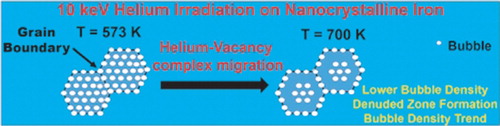
Denuded zone formation and bubble density/swelling vs. grain size trends were shown to occur over a threshold temperature in helium-irradiated nanocrystalline iron.
Many future nuclear energy systems require advanced engineering materials capable of withstanding severe environmental conditions, particularly high irradiation doses and high temperatures [Citation1]. Energetic ions create high numbers of point defects (vacancies and interstitials) during the initial collision cascade. Most of these defects recombine in tens of picoseconds, leaving a subset of vacancies and interstitials that become freely migrating defects (FMDs). These FMDs can diffuse, recombine, cluster, or be absorbed by sinks such as grain boundaries [Citation2]. Clustering of these defects can lead to the formation of more complex defect structures, such as vacancy loops, interstitial loops, voids, and bubbles, which all can potentially degrade the mechanical properties [Citation3,Citation4] of the irradiated material and ultimately cause failure [Citation1]. To mitigate these detrimental effects, recent studies have investigated novel engineering materials with high densities of defect sinks, such as grain boundaries [Citation5–7] or structural interfaces [Citation8]. Grain boundaries are thought to act as defect sinks and, in particular, to enhance point defect annihilation by absorbing interstitials and then re-emitting them back to recombine with nearby vacancies [Citation9]. They can also operate as sinks for interstitial impurities, such as helium [Citation10] as in the case of plasma-facing components in fusion systems which are exposed to high-flux and high-fluence helium irradiation [Citation11]. For these reasons nanocrystalline materials, with their high density of grain boundaries, are considered candidates for radiation-tolerant materials.
Several studies have suggested that nanocrystalline materials have higher radiation tolerances than their coarse-grained counterparts by demonstrating that they have lower defect densities after exposure to the same levels of irradiation [Citation12,Citation13]. Studies have also investigated the relationship between grain boundary character and sink efficiency, which is often correlated to denuded zone formation [Citation14], or the formation of a void- and defect-free zone near the grain boundary. However, the effect of temperature on bubble-denuded zone formation and bubble concentration in a nanocrystalline microstructure has yet to be explored. In this study, in situ, low-energy helium implantation was performed in a transmission electron microscope (TEM) on free-standing nanocrystalline iron films at two distinct temperatures. Both defect generation and helium trapping occur at the irradiation conditions in this study and grain boundaries are supposed to play a role in defect annihilations and helium trapping. The different results from these two experiments revealed evidence of a temperature threshold for denuded zone formation in nanocrystalline Fe. Additionally, through the analysis of a large set of grain boundaries, it is shown that denuded zone formation had no correlation with grain size and grain boundary misorientation angle.
Nanocrystalline iron films (approximately 100 nm in thickness) [Citation15] were prepared by sputter-depositing 99.9% pure iron target onto a <100> NaCl substrate held at 573 K, and subsequently floated in water onto molybdenum TEM grids using the procedure described elsewhere [Citation16]. The samples were then annealed in situ at 873 K in a JEOL 2100 Lab6 TEM using a Gatan 628 single-tilt heating stage to reach an equilibrium microstructure. In situ irradiation, executed at the In-situ Ion Irradiation TEM (I3TEM) facility at Sandia National Laboratories [Citation17], was performed with approximately 10 keV helium ions at calibrated temperatures of 573 and 700 K and an incident angle of 60°. These parameters give a projected range of up to 80 nm as calculated by the ‘detailed calculation with full damage cascade’ option in Stopping Range of Ions in Matter (SRIM), a Monte Carlo-based collision computer code (version 2013) [Citation18]. The ion dose rate and the final dose were nominally 8.74 × 10¹⁷ m−2 s−1 and 2.8 × 10²¹ m−2, respectively. The majority of the in situ images and videos were taken under Fresnel imaging condition, which provides an accurate and rapid method to characterize bubble density and an estimate of the bubble size [Citation19]. In addition, initial and post microstructural characterization was performed in the 2100 JEOL TEM utilizing automated crystallographic orientation mapping (ACOM) analysis produced by NanoMEGAS ASTAR precession electron diffraction system that is integrated into the I3TEM [Citation20]. The ACOM analysis was performed using 5 nm spot size and 2 nm step size.
Fresnel TEM images and corresponding inverse pole figure (IPF) maps of nanocrystalline irons films after irradiation with 10 keV helium at 573 and 700 K are shown in Figure . Analysis of the IPF maps concluded that the films contain 81% (by number of grain boundaries) high-angle grain boundaries as measured by the ‘Grain Boundary Quick Map’ tool in TSL®OIM Analysis 7.0 software. While bubbles are present in the grain matrices, the grain boundaries are also decorated with bubbles (Figure ) which demonstrates helium trapping by grain boundaries. Formation of bubbles in Figure can be illustrated as follows: due to the low energy for interstitial helium diffusion (0.06 eV) [Citation21], helium is expected to migrate freely unless it is trapped by the pre-existing defects or those generated (FMDs) in the matrix and along the grain boundaries, which are known to be effective helium-trapping sites [Citation10]. In the matrix, helium–vacancy complexes are assumed to be generated on the order of 10 picoseconds [Citation22] and have been observed or modeled to grow into bubbles by emitting interstitial atoms or by absorbing neighboring vacancy and helium atoms [Citation23,Citation24].
Figure 1. (a) and (b) Overfocused bright field TEM images of 10 keV helium irradiation on nanocrystalline iron at calibrated temperatures of 573 and 700 K, respectively. The inset figure in (b) demonstrates one denuded zone measurement (c) and (d) the corresponding [001] IPF maps.
![Figure 1. (a) and (b) Overfocused bright field TEM images of 10 keV helium irradiation on nanocrystalline iron at calibrated temperatures of 573 and 700 K, respectively. The inset figure in (b) demonstrates one denuded zone measurement (c) and (d) the corresponding [001] IPF maps.](/cms/asset/c7cedcb1-9a5e-492f-af79-9b23b5220045/tmrl_a_1243591_f0001_c.jpg)
Although bubble formation occurred in the grain matrix and the grain boundaries, the irradiation tolerance of the nanocrystalline samples to bubble formation can be studied. The resistance of grain boundaries to bubble formation in the grain matrix was correlated to the formation of denuded zones near the grain boundaries, which is a function of grain boundary sink efficiency [Citation8]. Experimentally, insight about grain boundary sink efficiency can be gained through examining denuded zone formation in the irradiated samples. Nanocrystalline iron samples irradiated at 573 K showed no denuded zone formation and uniform distribution of bubbles (average size of 195 bubbles counted is 2.1 nm with a standard deviation of 0.36 nm) throughout the grain matrix and at the grain boundaries (e.g. Figure (a)). The sample irradiated with a similar dose and dose rate, but at 700 K demonstrated denuded zone formation along the grain boundaries, different bubble densities for different grain sizes, and a larger average bubble size (average size of 313 bubbles counted is 3 nm with a standard deviation of 0.24 nm) in the grain matrix. To examine the effect of this behavior on the irradiation tolerance of the material, the average bubble density vs. grain size was plotted for both cases (Figure ). In the 700 K case, where denuded zones are formed, an increase of bubble density vs. grain size (area) was observed. In the 573 K irradiation case, there was no trend observed and the average bubble density was 0.129/nm2, which is 2.15 times higher than the saturation value (approximately 0.06/nm2) in the 700 K case, demonstrating the dependence of bubble density and bubble density vs. grain size trend formation on temperature. In both irradiation cases, a trend in bubble size trend as a function of grain size was not found.
Figure 2. Areal bubble density (N°/nm2) vs. grain size (area) for 10 keV helium irradaition on nanocrystalline iron at (a) 573 and (b) 700 K. Red fitting in (b) demonstrates the trend in density change [Citation25].
![Figure 2. Areal bubble density (N°/nm2) vs. grain size (area) for 10 keV helium irradaition on nanocrystalline iron at (a) 573 and (b) 700 K. Red fitting in (b) demonstrates the trend in density change [Citation25].](/cms/asset/09c6b107-850e-40ab-bd98-68a3740a1cf4/tmrl_a_1243591_f0002_c.jpg)
Investigating the bubble density and size as a function of temperature can provide insight into the resistance to swelling, another important aspect of the radiation-tolerant material. Swelling can be described as the change in the volume of the material [Citation26] due to increased vacancies, voids, and bubbles. An implication of the TEM observable bubbles and cavities on swelling in this study is determined using the TEM Fresnel micrographs. Using the average bubble area and the average bubble density in the 573 K case,
(found using
, where
is the void density and
is the radius of the void) was found to be 0.63%. In the 700 K case, this value ranges from 0.0096% to 0.98% as a function of grain size. At higher temperature more damage (higher bubble density and size) is expected due to higher vacancy coalescence and faster helium–vacancy complex growth [Citation27]. However, the bubble density was shown to be smaller in the 700 K case. Swelling is also expected to increase with temperature in this temperature regime [Citation28], but the 700 K result demonstrated lower calculated or apparent swelling values particularly at small grain sizes. One can then conclude that the resistance to bubble formation and subsequently to swelling in nanocrystalline iron is temperature dependent. El-Atwani et al. [Citation29,Citation30] demonstrated lower bubble density in the nanocrystalline regime for another BCC material (tungsten) when denuded zones were present and the irradiation conditions permitted both vacancy generation and migration. In this study, vacancy generation is present due to the displacement damage from the 10 keV He and a similar mechanism might also be operational. According to SRIM, the vacancy production is 30 vacancies/ion. Due to the low vacancy migration energy in iron (0.67 eV) [Citation21], vacancy migration is expected to occur at both temperatures (573 and 700 K). However, the rapid formation of helium–vacancy complexes can affect the migration of the vacancies. It was reported [Citation31] that migration of helium–vacancy complexes can occur at temperatures higher than 573 K (the low temperature case in this study). Moreover, vacancies are predicted to be unstable at temperatures below 573 K and can only be stabilized through helium–vacancy complex formation [Citation31]. Therefore, one might expect a change in the efficiency in the migration of small helium–vacancy complexes in the 700 K irradiation case. Migration of these complexes to grain boundaries or their coalescence should lead to smaller number of bubble nucleation sites but higher probability for helium atoms to find existing complexes during irradiation. It was predicted through molecular dynamics simulations that as temperatures increase, helium–vacancy complex formation occurs faster in iron and less helium atoms are needed to emit vacancies (during bubble growth) despite the dissociation of small complexes [Citation27]. These arguments are in agreement with the results in this study.
The following equation, from [Citation8], is used to find the width of the denuded zone () :
(1)
where
is the vacancy sink reaction rate coefficient,
is defect production rate,
is vacancy diffusivity,
is the vacancy concentration, and
is the vacancy sink efficiency describing the ability of the sink to absorb supersaturated vacancies;
is described as the ratio of vacancy flux going into the sink to the vacancy flux going into a perfect sink. It can range from 0 (no vacancy absorption) to 1 (perfect sink).
From Equation (1), a reduction in will result in denuded zone shrinkage. Formation of helium–vacancy complexes affects the vacancy diffusivity and decreases it for large complexes. Helium-stabilized vacancies (small helium–vacancy complexes) will migrate more efficiently at 700 K. Thus, comparison of the 573 and 700 K experiments is reasonable. It should be mentioned that defect clusters can also be vacancy sinks which is ignored in the above equation [Citation8].
The relationships between denuded zone formation and grain boundary misorientation angle and grain size are studied and demonstrated in Figure . In Figure (a), denuded zone formation vs. grain size is plotted. The experimental data were partitioned into three categories: fully denuded grain boundaries surrounding a grain, at least half of the boundaries are denuded, and no denuded grain boundaries surrounding the grain. Denuded zone formation and size (the perpendicular distance from the denuded boundary to nearby bubbles as shown in Figure (b)) vs. misorientation angle are demonstrated in Figure (b) and 3(c), respectively. For Figure (b), boundaries were classified into fully denuded boundaries (the majority of the boundary has a bubble-free zone in the vicinity of the grain boundary), non-denuded boundaries (no bubble-free zone in the vicinity of the grain boundary), and partially denuded grain boundaries (about half of the boundary has a bubble-free zone in its vicinity). A typical TEM image for this case is shown in Figure (d). Both low-angle and high-angle boundaries can be denuded or non-denuded and no trend for denuded zone formation and denuded zone size as a function of misorientation angle was observed, suggesting an additional parameter is affecting the grain boundary sink efficiency. The term (vacancy sink efficiency of the boundary) in Equation (1) is shown to be dependent on other parameters, in addition to misorientation angle, such as grain boundary plane [Citation14].
Figure 3. (a) and (b) Denuded zone type vs. grain size and misorientation angle, respectively. (c) Denuded zone size (nm) vs. misorientation angle and (d) overfocused bright field TEM image showing half denuded grain boundaries (pointed by red arrows). All images are for the 700 K irradiation case.
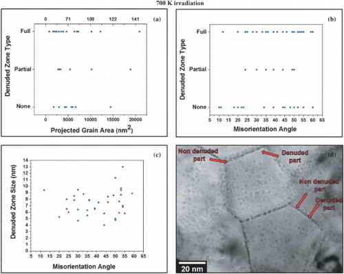
Considering misorientation angle alone (without considering the grain boundary plane) in this work demonstrated no correlation with denuded zone formation. Therefore, our results are in agreement that misorientation angle, grain size, and grain boundary type (high-angle or low-angle grain boundaries) are not the only factors affecting denuded zone formation.
In summary, low-energy helium implantation performed on free-standing nanocrystalline iron films in situ at 573 and 700 K demonstrated bubble density and area dependence on temperature. Denuded zone formation occurred at 700 K, which coincided with a trend in bubble density as a function of grain size; however, no direct correlation was observed between denuded zone formation and misorientation angle. Nanocrystalline iron was shown to have less bubble density and lower apparent swelling values at the temperature above which helium–vacancy complexes can migrate (over 573 K).
Acknowledgements
The authors thank Dr Brittany Muntifering and Dan Buller for their assistance with the experimental setup at SNL.
Disclosure statement
No potential conflict of interest exist between the authors.
Additional information
Funding
References
- Zinkle SJ, Busby JT. Structural materials for fission & fusion energy. Mater Today. 2009;12:12–19. doi: 10.1016/S1369-7021(09)70294-9
- Sizmann R. The effect of radiation upon diffusion in metals. J Nucl Mater. 1978;69–70:386–412. doi: 10.1016/0022-3115(78)90256-8
- Zinkle SJ, Was G. Materials challenges in nuclear energy. Acta Mater. 2013;61:735–758. doi: 10.1016/j.actamat.2012.11.004
- Byun T, Farrell K. Plastic instability in polycrystalline metals after low temperature irradiation. Acta Mater. 2004;52:1597–1608. doi: 10.1016/j.actamat.2003.12.023
- Beyerlein I, Caro A, Demkowicz M, et al. Radiation damage tolerant nanomaterials. Mater Today. 2013;16:443–449. doi: 10.1016/j.mattod.2013.10.019
- El-Atwani O, Gonderman S, Efe M, et al. Ultrafine tungsten as a plasma-facing component in fusion devices: effect of high flux, high fluence low energy helium irradiation. Nucl Fusion. 2014;54:083013-1–9. DOI:doi: 10.1088/0029-5515/54/8/083013.
- El-Atwani O, Suslova A, Novakowski T, et al. In-situ TEM/heavy ion irradiation on ultrafine-and nanocrystalline-grained tungsten: effect of 3 MeV Si, Cu and W ions. Mater Charact. 2015;99:68–76. doi: 10.1016/j.matchar.2014.11.013
- Beyerlein I, Demkowicz M, Misra A, et al. Defect-interface interactions. Prog Mater Sci. 2015;74:125–210. doi: 10.1016/j.pmatsci.2015.02.001
- Bai X-M, Voter AF, Hoagland RG, et al. Efficient annealing of radiation damage near grain boundaries via interstitial emission. Science. 2010;327:1631–1634. doi: 10.1126/science.1183723
- Singh BN, Foreman A. Calculated grain size-dependent vacancy supersaturation and its effect on void formation. Philos Mag. 1974;29:847–858. doi: 10.1080/14786437408222075
- Davis J, Barabash V, Makhankov A, et al. Assessment of tungsten for use in the ITER plasma facing components. J Nucl Mater. 1998;258–263:308–312. doi: 10.1016/S0022-3115(98)00285-2
- Nita N, Schaeublin R, Victoria M. Impact of irradiation on the microstructure of nanocrystalline materials. J Nucl Mater. 2004;329–333:953–957. doi: 10.1016/j.jnucmat.2004.04.058
- Chimi Y, Iwase A, Ishikawa N, et al. Accumulation and recovery of defects in ion-irradiated nanocrystalline gold. J Nucl Mater. 2001;297:355–357. doi: 10.1016/S0022-3115(01)00629-8
- Han W, Demkowicz M, Fu E, et al. Effect of grain boundary character on sink efficiency. Acta Mater. 2012;60:6341–6351. doi: 10.1016/j.actamat.2012.08.009
- Heintze C, Bergner F, Hernández-Mayoral M, et al. Irradiation hardening of Fe–9Cr-based alloys and ODS Eurofer: effect of helium implantation and iron-ion irradiation at 300° C including sequence effects. J Nucl Mater. 2016;470:258–267. doi: 10.1016/j.jnucmat.2015.12.041
- Vetterick G, Baldwin J, Misra A, et al. Texture evolution in nanocrystalline iron films deposited using biased magnetron sputtering. J Appl Phys. 2014;116:233503-1–6. DOI:doi: 10.1063/1.4904077.
- Hattar K, Bufford DC, Buller DL. Concurrent in situ ion irradiation transmission electron microscope. Nucl Instrum Meth B. 2014;338:56–65. doi: 10.1016/j.nimb.2014.08.002
- Ziegler JF, Ziegler MD, Biersack JP. SRIM – the stopping and range of ions in matter. Nucl Instrum Meth B. 2010;268:1818–1823. doi: 10.1016/j.nimb.2010.02.091
- Jenkins M. Characterisation of radiation-damage microstructures by TEM. J Nucl Mater. 1994;216:124–156. doi: 10.1016/0022-3115(94)90010-8
- Moeck P, Rouvimov S, Rauch E, et al. High spatial resolution semi-automatic crystallite orientation and phase mapping of nanocrystals in transmission electron microscopes. Cryst Res Technol. 2011;46:589–606. doi: 10.1002/crat.201000676
- Fu C-C, Willaime F. Ab initio study of helium in α− Fe: Dissolution, migration, and clustering with vacancies. Phys Rev B. 2005;72:064117-1–6. doi: 10.1103/PhysRevB.72.064117
- Deo CS, Okuniewski MA, Srivilliputhur SG, et al. Helium bubble nucleation in bcc iron studied by kinetic Monte Carlo simulations. J Nucl Mater. 2007;361:141–148. doi: 10.1016/j.jnucmat.2006.12.018
- Caspers L, Fastenau R, Van Veen A, et al. Mutation of vacancies to divacancies by helium trapping in molybdenum effect on the onset of percolation. Phys Status Solidi (a). 1978;46:541–546. doi: 10.1002/pssa.2210460218
- Wilson W, Bisson C, Baskes M. Self-trapping of helium in metals. Phys Rev B. 1981;24:5616–5624. doi: 10.1103/PhysRevB.24.5616
- El-Atwani O, Nathaniel J, Leff AC, et al. Identification of grain size regimes for He bubble formation: implications for swelling resistance. Unpublished.
- Mansur L. Theory and experimental background on dimensional changes in irradiated alloys. J Nucl Mater. 1994;216:97–123. doi: 10.1016/0022-3115(94)90009-4
- Stewart D, Osetskiy Y, Stoller R. Atomistic studies of formation and diffusion of helium clusters and bubbles in BCC iron. J Nucl Mater. 2011;417:1110–1114. doi: 10.1016/j.jnucmat.2010.12.217
- Little E, Stow D. Void-swelling in irons and ferritic steels: II. An experimental survey of materials irradiated in a fast reactor. J Nucl Mater. 1979;87:25–39.
- El-Atwani O, Hinks J, Greaves G, et al. In-situ TEM observation of the response of ultrafine- and nanocrystalline-grained tungsten to extreme irradiation environments. Sci Rep. 2014;4:4716-1–7. DOI:doi: 10.1038/srep04716.
- El-Atwani O, Hattar K, Hinks J, et al. Helium bubble formation in ultrafine and nanocrystalline tungsten under different extreme conditions. J Nucl Mater. 2015;458:216–223. doi: 10.1016/j.jnucmat.2014.12.095
- Borodin V, Vladimirov P. Diffusion coefficients and thermal stability of small helium–vacancy clusters in iron. J Nucl Mater. 2007;362:161–166. doi: 10.1016/j.jnucmat.2007.01.019