
Abstract
In recent decades, several scientists focused their process towards nanoparticles synthesis by using various sustainable approaches. Cocos nucifera (C. nucifera) was one of the versatile trees in tropical regions which also can act as a thrust quencher in all over the world. Cocos nucifera coir was one of the waste by-products in all coconut-refining industries and with the help C. nucifera coir, Palladium nanoparticles (Pd NPs) were synthesized. Green-synthesized spherical-shape Pd NPs were over layered by secondary metabolites from C. nucifera coir extract and with an average particle size of 62 ± 2 nm, which were confirmed by morphological analysis. Eco-friendly mediated Pd NPs were further subjected to several biological applications like larvicidal against Aedes aegypti (A. aegypti) and anti-feedent, ovicidal, and oviposition deterrent against agricultural pest Callasobruchus maculates (C. maculates) and compared with C. nuciferacoir methanolic extract, which results in LC50 value of 288.88 ppm and LC90 value of 483.06 ppm using LSD-Tukey’s test against dengue vector (A. aegypti). Cocos nucifera coir methanolic extract shows significant output while compared with Pd NPs towards anti-feedent assays; ovicidal activity and oviposition deterrent were discussed here.
Introduction
The only accepted species in Cocos genus was said to be Cocos nucifera (C. nucifera) that belongs to Arecaceae family. It is one of the most important fruit trees in tropical and sub-tropical regions which contains several by-products like coconut water, toddy, oil, etc. (Gao et al. Citation2014). It is endorsed with 51.9% of endosperm of water, 26.1% of fats, and 15.1% of traces of vitamins, carbohydrates, and several minerals which are most essential for day-to-day life (Ochoa-Velasco et al. Citation2014). This fruit contains major fractions of proteins like globulins, albumins, glutelins 1 and 2. This plant is utilized in the treatment of several biological applications like anti-oxidants, anti-malarial, antimicrobial, leishmanicidal, and insecticidal activities (Roopan and Elango Citation2015, Roopan et al. Citation2013). Various solvent extracts of C. nucifera coir were used as secondary metabolites for greener synthesis of metal nanoparticles, which play a major role nowadays and are to be exploited further in future.
The combination of two major fields like nanotechnology and biotechnology is said to be nanobiotechnology (Akhtar et al. Citation2013). Phyto or green-mediated syntheses of nanoparticles were considered under higher exploration technique to avoid toxic nature of nanoparticles related to environment (Kumar et al. Citation2014, Madhumitha et al. Citation2015, Citation2016, Madhumitha and Roopan Citation2013, Roopan et al. Citation2012, Surendra and Roopan Citation2016). This green synthesis of nanoparticles has several benefits by decreasing the usage of reducing agents like hydrogen gas or hydrazine, sodium borohydride, etc., which mostly result in advantages over environment (Nasrollahzadeh et al. Citation2016).
Transition metal nanoparticles were rated as one of the important metallurgies in periodic elements because of biocompatibility, greener approach, eco-friendly adoptable nature, and phyto-synthesizing property (Elango et al. Citation2015). Many metals like Pd, Au, Ag, Pt, and Cu were frequently utilized for the synthesis due to their colloidal stability, which is majorly utilized in several fields: catalysis, photo thermal therapy, optoelectronics, and labeling the biological substances. Here, we plan to synthesize palladium nanoparticles (Pd NPs) using agricultural wastes of C. nucifera coir due to its important applications like hydrogenation, low temperature in pollutants of automobile reduction, and also majorly as primary catalyst in many organic reactions (Chowdary et al. Citation2016).
There are several physio-chemical methods for fast and rapid synthesis of metal nanoparticles like hydrothermal, microwave, nano-micro-emulsion, sol–gel and sonochemical-mediated synthesis (Jia et al. Citation2009). But, when compared to physio-chemical methods which are available for Pd NPs synthesis, eco-friendly synthesis of Pd NPs plays a major role in environment by avoiding some toxic defects and also has several advantages like easy reaction setup, usage of non-toxic solvents, mild reaction progress, less cost, and suitable for biological and pharmaceutical applications (Nasrollahzadeh et al. Citation2015). These Pd NPs also play a major role in several medical applications without destructing the morphology and bind with single-strand DNA, which also has a property to act as a sensor for glucose-sensing instruments (Kalaiselvi et al. Citation2015). The green synthesis by using various plant sources does not require higher temperature maintenance and is an easiest process yet to be explored (Elango and Roopan Citation2015). Hence, we focused towards C. nucifera coir-mediated synthesis of Pd NPs, subjected to various applicational studies. The present study completely deals about the green synthesis of Pd NPs and applicational study towards various biological activities like anti-feedent, oviposition deterrent, and ovicidal activity by cow pea adult bruchid Callasobruchus maculates (C. maculates); further, it was tested against a dengue vector named Aedes aegypti (A. aegypti) (Erick and Padmanabhan Citation2015).
Materials and methods
Resources
Cocos nucifera coir was aggregated from Gudiyatham, Vellore district, Tamil Nadu, India, and its coordinates are 12.9397°N, 78.8644°E. The collected sources were further subjected for authentication process in Tamil Nadu Agricultural University (TNAU), Coimbatore, Tamil Nadu, India, with the authentication no: BSI/SRC/5/23/2013–14/Tech 1118 and kept for future reference. Palladium acetate was purchased from Sigma Aldrich Chemicals Ltd, Mumbai, India, and CH3OH from AVRA Laboratories, Hyderabad, India. Throughout this experimental process, double-distilled water was utilized without any modifications.
Methanolic extract collection of C. nucifera coir
Collected samples of C. nucifera coir were washed and cleaned with double-distilled water and subjected to air-drying in shade conditions at room temperature for 72 h. Further, it was grinded using mechanical mixer and powdered particles were stored in room temperature for future experimental process. Preliminarily collected fine-powdered materials were subjected for pet ether extraction for hydrocarbons removal. Once hydrocarbons were removed by checking thin-layer chromatography (TLC) method, further fine-powdered material was placed in room temperature for the evaporation of pet ether solvents for 2–3 h. After this process, samples were re-packed in a Soxhlet apparatus for methanol extraction and it was distilled and stored in a refrigerator for further process.
Bio-approach for the production of Pd NPs
We have followed the protocol of Elango and Roopan (Citation2015) with slight modifications for the synthesis of Pd NPs. About 100 ml of 1 mM palladium acetate solution was prepared using double-distilled water and placed on stirrer under room temperature for one hour. 20 mL of methanolic extract (1000 ppm) was collected and mixed with 80 mL of palladium acetate solution, then placed under 60° C for 4 h with the help of water bath and observed using UV–Vis at 1 h interval. Once the highest absorbance was confirmed by UV–Vis spectroscopy, further collected sample was subjected for centrifugation for 30 min at 3000 rpm and further pellets were placed in muffle furnace for 2 h at 350 °C for calcination process.
Characterization
Here, the agricultural waste-mediated synthesis of Pd NPs were monitored and confirmed by various analytical techniques like UV–Vis spectroscopy (UV-1800-Schimadzu, Shimadzu Analytica, Mumbai, India), Bruker D8 make XRD for the analysis of crystalline data of sample, Bruker Alpha T-FT-IR for the functional group analysis, Philips make TEM for structural analysis; to identify the phytoconstituents, samples were analyzed using GCMS, Zeta potential (Horiba Nanoparticle Analyzer, Horiba Corp., Chennai, India).
Pd NPs activity against larvae’s
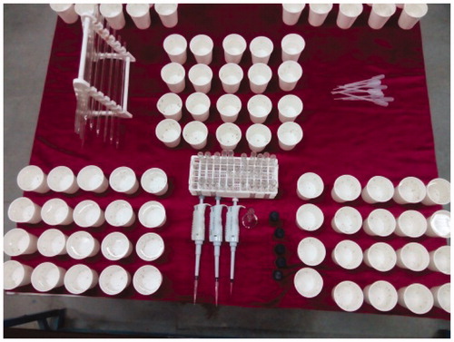
Aedes aegypti dengue vector were segregated from stagnant water area near Cooum river, Saidapet, Chennai, its coordinates are 13.0681°N, 80.2858°E and were identified by Dr. Kuppusamy Elumalai, Department of Advanced Zoology and Biotechnology, Government Arts College (Autonomous), Nandanam, Chennai. With various concentrations like 100, 200, 300, 400, 500 ppm, synthesized Pd NPs were subjected towards 20 larvae which were placed in 10 mL of distilled water. This experimental setup was processed using re-cyclable paper cups, as shown in with five replicates. Each test having a control contains distilled water and larva in the absence of Pd NPs. We identified mortality percentage, dose–response bioassay for various concentrations ranging 100, 200, 300, 400, and 500 ppm. With the help of LSD Tukey’s test, we have also identified the LC50 and LC90 values reported here.
Figure 1. Experimental setup for larvicidal activity.
Adulticidal bioassay of Callasobruchus maculates(C. maculates) by anti-feedent method
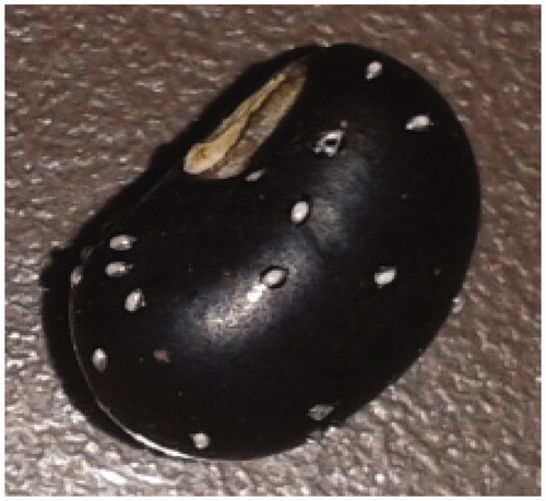
We have collected C. maculates from infected cow pea seeds. The collected pests were processed before emergence and transferred it into 35 mm petri dishes which contain 100 cow pea seeds, as shown in . We further separated the two groups in a set of three, in which one group of petri dishes was coated with extract and another with Pd NPs in the presence of seed and pests with different concentrations like 1.25, 2.5, 5, 10, and 20 mg/L. In each petri dish, we introduced 10 pairs of adults. Dishes will be separated and placed in laboratory (28 ± 2 °C, 75 ± 10% (rh) conditions at 12 h dark and 12 h light. Mortality rate was observed 1, 3, 5, 7, and 14 d (Erick and Padmanabhan Citation2015, Roopan et al. Citation2013). Further, we utilized distilled water as a control. The mortality of adults was observed from the 24 h of exposure and the results were reported with replicates of five. We compared this process with C. nucifera coir methanolic extract and reported.
Figure 2. Adulticidal bioassay (anti-feedent method).
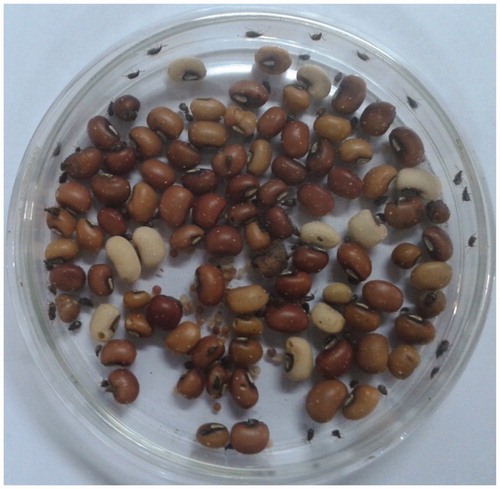
Ovicidal activity using Vigna unguiculata (cow pea) seeds on C. maculates eggs
Everyone knows that ovicidal means nothing but to kill eggs of insects, mites, nematodes, etc. Here, we collected C. maculates egg from cow pea seeds, as shown in , and placed in petri dishes with various concentrations of green-synthesized Pd NPs and C. nucifera coir methanolic extracts (1.25, 2.5, 5, 10, and 20 mg/L). We placed the petri dishes under laboratory conditions and recorded the ovicidal rate of the eggs at different intervals of time (24, 48, 72, and 96 h) with five replicates by following the protocol of Adedire et al. (Krishnaraju et al. Citation2006) with slight modifications.
Figure 3. Collection of C. maculates egg for ovicidal activity.
Oviposition deterrent activity of C. maculates on Vigna unguiculata
We processed oviposition deterrent activity using cow pea seeds against C. maculates egg by following the protocol of Prajapati et al. (Su et al. Citation1998) with slight modifications with different concentrations of Pd NPs at 1.25, 2.5, 5, 10, and 20 mg/L, then later compared with C. nucifera coir methanolic extract. Five replications were studied and are reported here.
Results and discussion
Pd NPs analysis results
Pd NPs–UV–visible spectroscopy
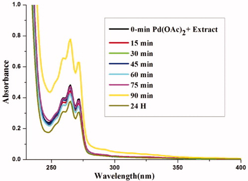
Reaction progress of conversion of Pd(OAc)2 to Pd NPs was observed using UV–Vis spec at different time intervals. Further progress was noted at the wavelength of 200–800 nm. Result in peak exactly at 264 nm and clearly indicates the formation of Pd NPs by surface plasmon resonance. The optimized absorbance for nanoparticle formation was said to be 90 min as clearly shown in .
Figure 4. UV–visible spectrum of Pd NPs at various intervals.
Pd NPs-XRD pattern
clearly depicts the XRD crystalline pattern of prepared Pd NPs at 60 °C for 90 min. Obtained XRD pattern was matched with Joint Committee Powder Diffraction Standards (JCPDS) data and the resulted number was 89–4897, which clearly reveals that green-synthesized Pd NPs were crystalline in nature. (***) indicates the presence of some organic compounds in the extract. The observed result was identified as d= 2.24061 with 2θ plane= 40.048. With the help of the Scherer equation, the particle sizes average was found to be 64.43 nm (Prajapati et al. Citation2005).
Figure 5. XRD pattern of Pd NPs.
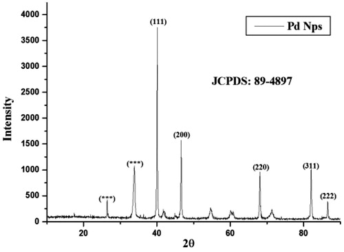
D =kλ/β cosθ where the particle size is denoted by D, (0.94) constant of Scherrer denoted by k, λ value can be evacuated from the equation derived by Bragg's (2d sin θ=nλ), λ is the wave length, β is the width half and maximum full, and θ is the angle of diffraction.
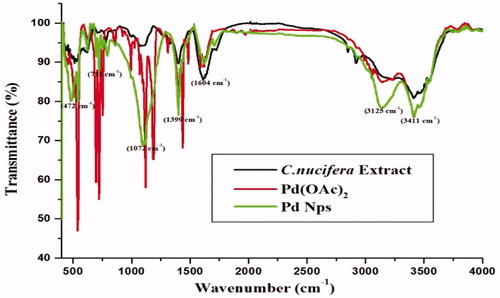
Pd NPs-FT-IR
FT-IR was done for palladium salt precursor, C. nucifera coir methanolic extract, and green synthesized Pd-NPs. The results clearly state that stretching of palladium precursor and C. nucifera coir methanolic extract at 3411 cm−1 is because of the hydroxyl groups (–OH). Then, researchers identified a stretching exactly at 1604 cm−1 due to the presence of C = O and also we noticed that in the peak range of 3125 cm−1 two different peaks are separated which resulted in the Pd NPs formation, as illustrated in .
Figure 6. FT-IR spectrum of Pd NPs.
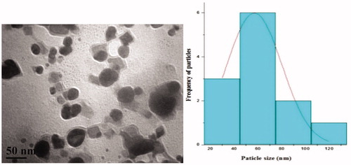
TEM analysis
We processed the synthesized sample for its morphological studies which was performed using transmission electron microscope and its results show that the particles were spherical in shape, the average size was 62 ± 2 nm as shown in . Here, we are able to conclude that the methanolic extract of C. nucifera was used to convert Pd (OAC)2 into Pd NPs as a reducing agent. With the help of Image J software, histogram was calculated and the resultant particle size was averaged to be 60 nm.
Figure 7. TEM and particle size histogram of Pd NPs.
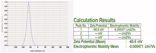
Pd NPs-Zeta potential
Pd NPs were processed in Horiba nanoparticles analyzer to identify the stability which resulted in −60.6 mV of high stability, as shown in .
Figure 8. Zeta potential of Pd NPs.
GC-MS prediction of C. nucifera coir
We processed our C. nucifera coir methanolic extract for gas chromatography and mass spectroscopy analysis to identify the secondary metabolites for the formation of Pd (OAc)2 to Pd NPs as a reducing agent. We have identified that 3-methoxy cinnamic acid has higher area percentage compared with other secondary metabolites. So, we confirmed that 3-methoxy cinnamic acid can be used as a reducing agent for the conversion of Pd (OAc)2 to Pd NPs. These GC-MS processes were already studied and our research group has already been reported in another work (Elango and Roopan Citation2015).
Pd NPs-larvicidal activity
With different concentrations like 100–500 ppm, larvicidal activity of Pd NPs was tested in the presence of larvae causing dengue A. aegypti. The results revealed are shown in .
Table 1. Larvicidal activity of synthesized Pd NPs against dengue vector A. aegypti.
Anti-feedent activity of Pd NPs against C. maculates
Mortality of the adults treated with nanoparticles was recorded under laboratory conditions after 1, 3, 5, 7, and 14 d. Here, we kept distilled water as a control and replicated five times for optimization of different concentrations of 1.25, 2.5, 5, 10, and 20 mg/L were studied for both Pd NPs and C. nucifera methanolic extract and reported in . We finally concluded that: compared with Pd NPs, C. nucifera coir methanolic extract shows better anti-feedent activity (Kumari et al. Citation2015, Petla et al. Citation2012).
Table 2. Anti-feedent activity of Pd NPs and C. nucifera methanolic extract using C. Maculates.
Ovicidal activity of Pd NPs
Ovicidal activity of the synthesized Pd NPs and C. nucifera coir methanolic extract is compared and is reported in , . The obtained result clearly indicates that Pd NPs have less efficiency to kill eggs of C. maculates compared with C. nucifera coir methanolic extract.
Table 3. (a) Ovicidal activity of Pd NPs.
Oviposition deterrent activity against gravid ofC. maculates
Cocos nucifera coir methanolic extract and Pd NPs were tested for its oviposition deterrent activity against gravid of C. maculates, as reported in . Hence, we identified that C. nucifera coir methanolic extract has high efficacy of oviposition deterrent activity while compared with Pd NPs from the obtained results.
Table 4. Oviposition deterrent activity using Pd NPs and C. nucifera coir methanolic extract against C. maculates.
Conclusion
This article summarizes that the C. nucifera coir mediated the synthesized Pd NPs of 62 ± 2 nm average size with spherical shape. This methodology is one of the cheapest methods for synthesizing Pd NPs. Further larvicidal activity using Pd NPs against dengue vector (Aedes aegypti) resulted in LC50 value of 288.88 ppm and LC90 value of 483.06 ppm using LSD-Tukey’s test and shows 98% of mortality rate in 500 ppm of Pd NPs. We conclude that C. nucifera coir methanolic extract shows better output while compared with Pd NPs towards anti-feedent assay, ovicidal activity, and oviposition deterrent.
Acknowledgements
Elango express his gratitude towards DBT for Research Assistant fellowship. Dr. S. M. Roopan prompts his thank to the Department of Biotechnology, Government of India, for providing the research grant to carry out this work (DBT-RGYI scheme no. BT/PR6891/GBT/27/491/2012). Prof. M. Valan Arasu and Naif Abdullah Al-Dhabi encompass their heartfelt obligation to the Deanship of Scientific Research (King Saud University) for its finance to Prolific Research Group (PRG-1437–28).
Disclosure statement
The authors report no conflicts of interest. The authors alone are responsible for the content and writing of this article.
Additional information
Funding
References
- Akhtar MS, Panwa J, Yun YS. 2013. Biogenic synthesis of metallic nanoparticles by plant extracts. ACS Sustainable Chem Eng. 1:591–602.
- Chowdary NR, MacGregor-Ramiasa M, Zilm P, Majewski P, Vasilev K. 2016. ‘Chocolate’ silver nanoparticles: synthesis, antibacterial activity and cytotoxicity. J Colloid Interface Sci. 482:151–158.
- Elango G, Kumaran SM, Kumar SS, Muthuraja S, Roopan SM. 2015. Green synthesis of SnO2 nanoparticles and its photocatalytic activity of phenolsulfonphthalein dye. Spectrochim Acta a Mol Biomol Spectrosc. 145:176–180.
- Elango G, Roopan SM. 2015. Green synthesis, spectroscopic investigation and photocatalytic activity of lead nanoparticles. Spectrochim Acta a. 139:367–373.
- Erick ON, Padmanabhan MN. 2015. Green chemistry focus on optimization of silver nanoparticles using response surface methodology (RSM) and mosquitocidal activity: Anopheles stephensi (Diptera: Culicidae). Spectrochim Acta a. 149:978–984.
- Gao L, Sun R, Liang Y, Zhang M, Zheng Y, Li D. 2014. Cloning and functional expression of a cDNA encoding stearoyl-ACP Δ9-desaturase from the endosperm of coconut (Cocos nucifera L.). Gene 549:70–76.
- Jia L, Zhang Q, Li Q, Song H. 2009. The biosynthesis of palladium nanoparticles by antioxidants in Gardenia jasminoides Ellis: long lifetime nanocatalysts for p-nitrotoluene hydrogenation. Nanotechnology 20:385601.
- Kalaiselvi A, Roopan SM, Madhumitha G, Ramalingam C, Elango G. 2015. Synthesis and characterization of palladium nanoparticles using Catharanthus roseus leaf extract and its application in the photo-catalytic degradation. Spectrochim Acta a. 135:116–119.
- Krishnaraju AV, Rao TVN, Sundararaju D, Vanisree M, Tsay H-S, Subbaraju GV. 2006. Biological screening of medicinal plants collected from eastern Ghats of India using Artemia salina (Brine Shrimp Test). Int J Appl Sci Eng. 4:115–125.
- Kumar DA, Palanichamy V, Roopan SM. 2014. Green synthesis of silver nanoparticles using Alternanthera dentata leaf extract at room temperature and their antimicrobial activity. Spectrochim Acta a. 127:168–171.
- Kumari AS, Venkatesham M, Ayodhya D, Veerabhadram G. 2015. Green synthesis, characterization and catalytic activity of palladium nanoparticles by xanthan gum. Appl Nanosci. 5:315–320.
- Madhumitha G, Elango G, Roopan SM. 2015. Bio-functionalized doped silver nanoparticles and its antimicrobial studies. J Sol-Gel Sci Technol. 73:476–483.
- Madhumitha G, Elango G, Roopan SM. 2016. Biotechnological aspects of ZnO nanoparticles: overview on synthesis and its applications. Appl Microbiol Biotechnol. 100:571–581.
- Madhumitha G, Roopan SM. 2013. Devastated crops: multifunctional efficacy for the production of nanoparticles. J Nanomater. 2013:1–12.
- Nasrollahzadeh M, Sajad SM, Rostami-Vartoonii A, Alizadeh M, Bagherzadeh M. 2016. Green synthesis of the Pd nanoparticles supported on reduced graphene oxide using barberry fruit extract and its application as a recyclable and heterogeneous catalyst for the reduction of nitroarenes. J Colloid Interface Sci. 466:360–368.
- Nasrollahzadeh M, Sajadi SM, Maham M. 2015. Green synthesis of palladium nanoparticles using Hippophae rhamnoides Linn leaf extract and their catalytic activity for the Suzuki–Miyaura coupling in water. J Mol Catal a. 396:297–303.
- Ochoa-Velasco CE, Cruz-Gonzalez M, Guerrero-Beltran JA. 2014. Ultraviolet-C light inactivation of Escherichia coli and Salmonella typhimurium in coconut (Cocos nucifera L.) milk. Food Sci Emerg Technol. 26:199–204.
- Petla RK, Vivekanandhan S, Misra M, Mohanty AK, Satyanarayana N. 2012. Soybean (Glycine max) leaf extract based green synthesis of palladium nanoparticles. J Biomater Nanobiotechnol. 3:14–19.
- Prajapati V, Tripathi AK, Aggarwal KK, Khanuja SP. 2005. Insecticidal, repellent and oviposition-deterrent activity of selected essential oils against Anopheles stephensi, Aedes aegypti and Culex quinquefasciatus. Biores Technol. 96:1749–1757.
- Roopan SM, Bharathi A, Kumar R, Khanna VG, Prabhakarn A. 2012. Acaricidal, insecticidal, and larvicidal efficacy of aqueous extract of Annona squamosa L peel as biomaterial for the reduction of palladium salts into nanoparticles. Colloid Surf B. 92:209–212.
- Roopan SM, Elango G. 2015. Exploitation of Cocos nucifera a non-food toward the biological and nanobiotechnology field. Ind Crop Prod. 67:130–136.
- Roopan SM, Rohit Madhumitha G, Rahuman AA, Kamaraj C, Bharathi A, Surendra TV. 2013. Low-cost and ecofriendly phytosynthesis of silver nanoparticles using Cocos nucifera coir extract and its larvicidal activity. Ind Crop Prod. 43:631–635.
- Su T, Mulla MS, Amer J. 1998. Ovicidal activity of neem products (azadirachtin) against Culex tarsalis and Culex quinquefasciatus (Diptera: Culicidae). Mosq Control Assoc. 14:204–209.
- Surendra TV, Roopan SM. 2016. Photocatalytic and antibacterial properties of phytosynthesized CeO2 NPs using Moringa oleifera peel extract. J Photochem Photobiol B: Biol. 161:122–128.