

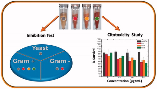
Abstract
We synthesized silver nanoparticles using Rumex hymenosepalus root extract (Rh). Nanoparticles were subjected to a purification process and final product is a composite of Rh and silver nanoparticles (AgNPsC). Transmission electron microscopy (TEM), high-resolution transmission electron microscopy (HRTEM), X-ray diffraction (XRD), and X-ray photoelectron spectroscopy (XPS) were used to perform a microstructure study. Additionally, two fractions (RhA and RhB) were obtained from the original extract by filtration with tetrahydrofuran (THF); both fractions were analyzed using UV-Vis spectroscopy, Fourier transform infrared spectroscopy (FT-IR), and 2,2-diphenyl-1-picrylhydrazyl (DPPH); total polyphenol content was also determined. Separate inhibition tests for AgNPsC and RhA and RhB were applied to Gram-positive bacteria, Gram-negative bacteria, and yeast (Candida albicans) using the well diffusion method. Extract fractions were found to have inhibitory effects only over Gram-positive bacteria, and silver nanoparticles showed inhibitory effects over all the evaluated microorganisms. Cytotoxicity was evaluated using the tetrazolium dye (MTT) assay in mononuclear peripheral blood cells. In addition, we assessment AgNPsC in THP-1 monocyte cell line, using the cell viability estimation by trypan blue dye exclusion test (TB) and Live/Dead (LD) cell viability assays by confocal microscopy.
Graphical Abstract
Acknowledgements
The authors wish to thank Dr. Lorena Armenta, from the University of Sonora, for supporting our XRD experiments and for Ms. Kareen Krizzan Encinas Soto of Departamento de Ingenierıa Quımica y Metalurgia, Universidad de Sonora, for access and the use of AAS. The authors also thank the laboratory of transmission electron microscopy from the Physics Department of the University of Sonora for providing TEM micrographs and laboratory of biomaterials from the Physics Department of the University of Sonora for providing the confocal microscopy. This research study was partially funded by Consejo Nacional de Ciencia y Tecnología (Conacyt–Mexico) through projects PROINNOVA 221252 and 221258, project PRODEP and project DSA/103.5/14/10945.
Disclosure statement
The authors report no conflicts of interest in this work.