
 ?Mathematical formulae have been encoded as MathML and are displayed in this HTML version using MathJax in order to improve their display. Uncheck the box to turn MathJax off. This feature requires Javascript. Click on a formula to zoom.
ABSTRACT
Background: Exposure to cold air is associated with increased morbidity and mortality in the general population. It is difficult to study the effects of whole-body exposure to cold air under controlled conditions in real life. Objectives: The aim of this study was to (1) explore and describe the experience of symptoms in humans during experimental and controlled exposures to cold air, by using controlled environmental chamber exposures and qualitative methodology, and to (2) categorise the symptoms. Method: The study used a randomised, double blind design, in which 34 subjects undertook rest and moderate-intensity exercise in an environmental chamber set to two or three different temperatures (0, −10, and −17°C) on separate occasions. During the chamber exposures, subjects were interviewed. Qualitative content analysis was selected as the method of analysis. Findings: Subjects reported 50 distinct symptoms during the exposures. The symptoms were grouped into ten sub-categories and two major categories; airway versus whole-body symptoms. Conclusion: We have identified a broad range of symptoms in humans undertaking rest and moderate-intensity exercise at sub-zero temperatures. The symptoms and their categories may well be used to more extensively and quantitatively map cold-induced morbidity.
Introduction
For the 4 million people who live in the Arctic region [Citation1], engaging in outdoor activities leads inevitably to regular cold air inhalation during the winter months. Amongst the general population, cold air exposure is associated with increased morbidity and mortality [Citation2], especially sensitive are the elderly and those with cardiopulmonary diseases [Citation3–Citation6]. In a Finnish survey, 26% of men and 31% of women reported that they experience cold-related airway symptoms [Citation7]. In otherwise healthy respondents, respiratory mucus production typically occured at −7 °C whereas onset of cough, wheezing and dyspnea was reported at −18°C or colder [Citation7]. A higher prevalence of cold-induced symtptoms in patients with allergic rhinoconjunctivitis and asthma [Citation7] may lead to avoidance of outdoor activities during cold spells [Citation8,Citation9]. In patients with COPD, cold outdoor temperatures may aggravate respiratory symptoms, increase rescue inhaler use and impair lung function [Citation10].
Winter endurance athletes, such as cross-country skiers, are repeatedly exposed to cold air during prolonged training sessions and competitions, and show an increased prevalence of airway symptoms, bronchial hyperreactivity and asthma [Citation11]. Respiratory symptoms are also very common among children participating in physical activity in cold environments [Citation12].
Environmental chambers aim to imitate real climatic conditions. Experimentally controlling environmental exposures, accounting for the effects of facial cooling, whole-body exposure and direct cold air inhalation is a prerequisite to further explore pathophysiological mechanisms responsible for environmentally induced morbidity. Whole-body exposure to sub-zero temperatures in a climatic chamber has been shown to induce proximal airway obstruction in healthy participants as well as patients with obstructive lung disease [Citation13–Citation21].
Symptoms may function as early warnings for subsequent morbidity. In epidemiological surveys, symptoms have been defined a priori, and only a few studies have investigated the emergence of airway symptoms during experimental exposure to cold air. Lower airway symptoms have usually been limited to shortness of breath, wheezing, prolonged cough, phlegm production, and chest pain [Citation9] whereas upper airway responses have been defined as rhinorrhea, congestion, and sneezing [Citation22–Citation25]. To the best of our knowledge, no qualitative studies have identified symptoms that arise during cold air exposure.
The purpose of this study was to (1) explore and describe the experience of symptoms in humans during experimental exposure to cold air, by using controlled environmental chamber exposures and qualitative methodology, and to (2) categorise the symptoms. We expected to detect a large number of distinct symptoms.
Methods
Environmental chamber
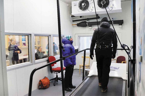
A cold air environment was simulated using the 21 m2 (75 m3) environmental chamber located at the Swedish Winter Sport Research Centre, Mid Sweden University, Östersund. The chamber is equipped with a treadmill (RL 2700E, Rodby Innovation, Vänge, Sweden), 1 m wide and 2.7 m long, outfitted with a safety harness system (). Frost-resistant windows permit visual contact and communication between personnel in- and outside of the chamber. The chamber utilises two systems to control ambient conditions; a temperature controller (TC; Inuwell AB, Östersund, Sweden) and a hypoxic compressor supplying 1500 L/min dehumidified air to the chamber (HC; K2 1500, Hypoxico, New York, USA). Room air composition was set to 20.9% O2 for all trials. For each trial, the desired set point temperature was inputted into the TC system. Chamber temperature was displayed by the TC system as a mean value from four sensors located at opposite ends of the side walls; front left (1.6 m), front right (1.6 m), back left (1.6 m) and back right (1.3 m). Relative humidity (rh) inside the chamber was detected by a humidity sensor located adjacent to the front right temperature sensor.
Figure 1. The environmental exposure chamber. Photo by Sara Rönnberg/Region Jämtland Härjedalen
Absolute humidity (g/m3) in the chamber was calculated based on the relative humidity (rh) and temperature (T) in °C, according to
Chamber performance together with surrounding laboratory and outdoor temperature is presented in .
Table 1. The target and observed milieu in the environmental chamber, surrounding laboratory, and outdoors at Swedish Winter Sport Research Centre, Mid Sweden University, Östersund, Sweden. Data presented as mean (SD)
Design
The study used a randomised, double blind design, in which 20 subjects were exposed to three different air temperatures during three separate testing sessions, and 14 subjects were exposed to two different air temperatures on separate occasions. The exposures were performed in a randomised order and took place in the environmental chamber.
Ethical approval was obtained from the Regional Ethical Review Board, Umeå (DNR 2015–245-31M).
Subjects
In order to obtain extensive descriptions as well as capture similarities and differences [Citation26], 34 adult volunteers were recruited using local advertising into four participant groups.
Group 1 included 13 healthy subjects (8 men and 5 women), without allergy and lung disease, who did not take regular medication and had never been regular smokers. Group 2 included seven subjects (6 men and 1 women) with allergic rhinoconjunctivitis (typical allergic eyes/nose symptoms as well as positive skin prick test for at least one airborne allergen). The subjects had never smoked regularly and had not neither symptoms of nor physician-diagnosed lung disease. Group 3 included 11 subjects (4 men and 7 women) with physician-diagnosed asthma. All except one subject had used daily inhaled corticosteroids for the preceding 3 months. One subject used only a beta-2-agonist approx. two times per week during exercise. The subjects with asthma had a mean (SD) forced expiratory volume in one second (FEV1) of 3.63 (0.85) L, 91 (11) % of predicted values and mean (SD) FEV1/forced vital capacity (FVC) of 0.78 (0.06) post-bronchodilatation. Group 4 included three subjects (1 man, 2 women) with chronic obstructive pulmonary disease (COPD), with > 10 packyears ((number of cigarettes smoked per day/20) x number of years smoked) of smoking and daily inhaled pharmacotherapy. Two subjects had COPD stage-2 category B and one had stage-3 category A [Citation27].
Procedure
After inclusion, all subjects underwent exercise testing and therafter 2 or 3 cold air exposures, all on different test occasions. At visit 1, the subjects were informed about the baseline exercise test, the exposures in the chamber, and that the exposures would be ca 0° C, −10°C, or −20°C (groups 1 and 2 only), and that they should bring appropriate clothing for the exposures.
Baseline exercise test
Prior to first exposure, subjects performed a ramped maximal exercise test to volitional exhaustion on a motorised treadmill (Rodby Innovation, Vänge, Sweden). The test for groups 1–3 followed the Bruce treadmill protocol [Citation28], with the total test duration used to estimate subjects´ maximal oxygen consumption (VO2max) according to previously described methods [Citation28,Citation29]. For group 4, the test was conducted in line with the ramped protocol developed for COPD patients described by Cooper et al. (2010). VO2max was then estimated by entering the final speed and gradient in to ACSM walking equation [Citation30]. Heart rate was monitored continuously throughout the test (Polar S610, Polar Electro, Kempele, Finland). All except two subjects (both in the COPD group) achieved >90% of age-predicted maximum heart rate (HRmax) during the test; a common predetermined end-point for incremental exercise testing (estimated using the formula: age-predicted HRmax = 208–0.7*age; Tanaka et al. [Citation31]). Subject characteristics and test results are reported for all groups in .
Table 2. Subject characteristics and exercise test results. Data are presented as mean (SD), except otherwise stated
Exposures and interviews
Groups 1 and 2 were exposed to target temperatures 0 oC, −10 oC, and −20°C in the environmental chamber for 60 min on three separate occasions. The lowest target temperature of −20°C was below the lower operating limit for the chamber with two occupants, which emerged at approx. −17°C. An intermediate analysis revealed no unique symptoms at the lowest setting (−17°C), and this exposure was dropped for group 3 and 4. Each exposure consisted of alternating 15-minute periods of standing resting and walking on a treadmill at a speed to elict 50% VO2max. The treadmill gradient was set to 6% for groups 1–3 and 1% for group 4. Treadmill speed was adjusted to achieve 50% VO2max solving the ACSM walking equation for desired VO2 at a fixed gradient: VO2 (mL·kg−1·min−1) = 0.1·S + 1.8·S·G + 3.5 where speed (S) is in m·min−1 and gradient (G) in percent [Citation31].
During exposure periods, subjects were interviewed by the authors RS or CK throughout the rest and exercise period of each exposure. All subjects were given the opportunity to talk as long as they wished. After each interview, subjects were asked if she/he had anything more to add. We used focused open interviews in line with recommendations of Tjora [Citation32,Citation33] to use short interviews: (1) if the theme is highly limited, (2) if one believes that trust is created relatively quickly, and (3) when the topics addressed are not very sensitive or difficult for the participants.
According to recommendations for focused interviews [Citation32,Citation33], we used an interview guide to maintain structure and allow subjects to reflect on their experience of symptoms during the exposure. The interview guide included questions concerning experience of symptoms, how symptoms were perceived, and symptom intensity using the Borg CR10 scale [Citation34]. The Borg CR10 scale is a category-ratio (CR) scale anchored at the number 10, which represents extreme intensities. It is a general intensity scale for most subjective magnitudes that with special anchors can be used to measure exertion and pain [Citation34]. Follow-up questions were used: “Earlier you experienced (symptom), how does it feel now?”. Interviews were conducted individually, and the researcher was inside the chamber with the subject for the duration of the exposure.
Qualitative analysis
Qualitative content analysis was selected to systematically and methodically identify and categorise [Citation33] the manner in which various cold-related symptoms were expressed.
Subject interviews were transcribed verbatim including information about symptoms, time of symptom onset, and the timing of symptom reports during the exposure period. Each interview was anonymised prior to analysis. The first step of the analysis was to read through each interview as a whole in order to get a sense of the material. In the next step, a time axis was established for each exposure based on the elements of the exposure (rest 1–15 min, work 1–15 min, rest 2–15 min and work 2–15 min), where each mention of cold-related symptoms was mapped chronologically. The same subject may have stated the same symptoms several times. The analysis is based on the number of times the symptoms were stated [Citation33,Citation35] as well as the proportion of subjects who experienced each symptom.
To improve credibility of the analysis [Citation36–Citation38], RS and CK discussed and analysed the material through parallel processes, together and individually until consensus was reached [Citation39]. The symptoms that were judged to be related were placed into sub-categories with a description of the symptom. These sub-categories were then aggregated into major categories.
Results
The 34 study subjects reported 50 distinct symptoms during the exposures. These symptoms were grouped into ten sub-categories and two major categories.
Frequency of distinct symptoms in each study group
Symptoms occurred during the first rest period and were reported throughout the whole exposure period. A total of 40 distinct symptoms was reported by the healthy (group 1) as well as the asthmatic subjects (group 3). Subjects with allergic rhinoconjunctivitis (group 2) reported 43 distinct symptoms and the three subjects with COPD reported 15 distinct symptoms. A description of the symptoms, and their frequency of occurrence is depicted in .
Table 3. Symptoms reported during exposure to 0, −10 and −17°C in an environmental chamber stratified by subject group. Data are presented as the frequency (n) that each symptom was reported during the exposures and the proportion (%) of symptoms arising in a given sub-category from each group
The five most common symptoms reported by the highest proportion of subjects in each group are presented in .
Table 4. Five most common symptoms by proportion of subjects in an experimental exposure chamber for 1 h at intermittent rest and moderate exercise. Data presented as number of subjects (n)
Sub-categories and major categories
The symptoms that emerged in the analysis were judged to belong to 10 different sub-categories: (1) “mucus in the nose”, (2) “irritation in the nose”, (3) “irritation of the mouth and throat”, (4) “irritation in the chest”, (5) “cold in the airways”, (6) “breathlessness”, (7) “cold in the face”, (8) “cold in the extremities”, (9) “shivering” and (10) “increased body temperature”. Their relative occurrence is reported in bold numbers in .
The ten sub-categories were grouped into two major categories. Sub-categories 1–6 were judged to belong to the major category of symptoms of upper and lower airways, and sub-categories 7–10 were judged to belong to the major category of general whole-body sensations.
Discussion
We have identified 50 distinct symptoms in humans at rest and during moderate exercise that arise during controlled experimental exposures to temperatures between 0° and −17°C. These symptoms were grouped into 10 sub-categories and two major categories; airway versus whole-body symptoms.
Our qualitative interviews revealed numerous symptoms of cold air exposure, many more than those commonly used in epidemiological surveys on cold air associated morbidity. Overall, symptoms arising from the upper airways were frequent in healthy subjects as well as in subjects with allergic rhinoconjunctivitis or obstructive lung disease. The most common symptom, runny nose, has regularly been associated with exposure to cold air [Citation23,Citation24,Citation40].
Other common airway symptoms detected in the present study were symptoms belonging to the sub-category “irritation in the nose”, such as burning and stinging sensation, and in the sub-category of “irritation in the mouth and throat”, the symptoms most commonly reported such as dryness of the mouth, pharynx and throat, and increased mucus production had generally not been reported in other epidemiological surveys.
The most common whole-body symptoms arose within the sub-category of “cold in the face”, where cold at the tip of the nose and cold around the mouth were the most commonly described symptoms.
Cold air exposures may induce cold extremities, also detected in our experimental setup. Cold in the fingers, legs and a general cold sensation in the body were the most frequent symptoms in the sub-categories “cold in the extremities” and “shivering”, respectively. Pienimäki et al. [Citation41] reported that as ambient temperature decreases more physical symptoms are reported, with 90% of participants reporting feeling cold in the extremities at -23°C. Piedrahita et al. [Citation42] and Dovrat and Katz Leurer [Citation43] showed similar results in participants who worked in a cold indoor environment, where physical symptoms were most prominent, mostly in the fingers but also in the musculoskeletal system.
Symptoms from the lower airways were limited to subjects with asthma or COPD. This is in line with epidemiological studies showing that cold-related symptoms from lower airways are more often reported by subjects with obstructive lung disease [Citation7,Citation9,Citation11,Citation44].
In this study, there were few subjects with COPD which has to be taken into consideration. Among the subjects with COPD, cough was the most commonly reported symptom followed by difficulty breathing and constriction in the airways. These symptoms have also been detected in previous epidemiological surveys of patients with chronic bronchitis and COPD [Citation7,Citation10].
Although we did not perform any formal statistical comparisons, our impression is that some differences in symptoms arose between groups. In the healthy group, the sub-categories “mucus in the nose” and “cold in the face” appear to be more common compared to other groups. Among subjects with allergic rhinoconjunctivits, the sub-category “irritation in the nose” occurred frequently. Emergence of lower airway symptoms was limited to subjects with asthma or COPD, even though these subjects were only exposed to 0 and −10°C. We did not observe any obvious gender differences.
In these qualitative interviews, we used systematic and quantitative description of the manifest content of communication [Citation33]. The symptoms described by the study subjects were presented in 10 sub-categories and two major categories based on the symptoms and their anatomical origin. Some of the symptoms may predominantly arise as a consequence of cold air inhalation, such as” irritation in the chest” and difficulty breathing whereas other symptoms may have an association with the physical activity, such as “breathlessness”, shortness of breath, and increased breathing.
Limitations and strengths of the study
The experimental set-up may have some limitations to internal and external validity. As clothing was not standardised between or within subjects, poor or inappropriate clothing may have influenced the perception of cold air. The cooling fans in the roof of the environmental chamber contributed to a high noise level and several subjects felt that the wind swirled in the chamber, which was perceived as artificial since the wind does not usually spin in the same way when one is outdoors. Despite this, subjects perceived the environmental chamber exposures as realistic in comparison to outdoor conditions, which can be seen as a strength of the study. Another strength of the study was that the environmental chamber had a “sterile” appearance, removing visual cues, such as frost or ice, that could be used to evaluate ambient temperature.
The symptoms detected in the present study are a consequence of low to moderate intensity exercise in cold air. We believe that the moderate exercise intensity of 50% VO2 max used in the present study may have been too low to trigger lower airway symptoms in subjects without respiratory disease at these temperatures. The results therefore highlight the need for further studies in sub-zero temperatures with higher workloads.
The study was neither designed to nor had the sample size/distribution to evaluate the effect of age on symptoms arising during cold air exposure.
Conclusion
We have identified a broad range of symptoms that arise during rest and moderate exercise in sub-zero temperatures, in both healthy participants and patients with respiratory disease. These symptoms were grouped into 10 sub-categories and two major categories; airway versus whole-body symptoms. These symptoms and their sub-categories may be used to more extensively and quantitatively map cold-induced morbidity.
Acknowledgments
We are grateful to all the study participants as well as the students and test leaders at the Swedish Winter Sports Research Centre who assisted with data collection. The study was supported by grants from Region Jämtland Härjedalen and Gunhild and Assar Karlssons Donationsfond.
Disclosure statement
No potential conflict of interest was reported by the authors.
Related Research Data
References
- Larsen JN, Fondahl G, editors. Arctic human development report: regional processes and global linkages. TemaNord, Nordisk Ministerråd; Denmark: Rosendahls-Schultz grafisk; 2015.
- Rocklöv J, Forsberg B. The effect of temperature on mortality in stockholm 1998–2003: a study of lag structures and heatwave effects. Scand J Public Health. 2008 Jul;36(5):516–8. PubMed PMID: 18567653.
- The Eurowinter Group. Cold exposure and winter mortality from ischaemic heart disease, cerebrovascular disease, respiratory disease, and all causes in warm and cold regions of Europe. Lancet. 1997 May;349(9062):1341–1346. PubMed PMID: 9149695.
- Analitis A, Katsouyanni K, Biggeri A, et al. Effects of cold weather on mortality: results from 15 European cities within the phewe project. Am J Epidemiol. 2008 Dec 15;168(12):1397–1408. Pub Med PMID: 18952849.
- Hajat S, Kovats RS, Lachowycz K. Heat-related and cold-related deaths in England and Wales: who is at risk? Occup Environ Med. 2007 Feb;64(2):93–100. PubMed PMID: 16990293. PMCID: PMC2078436.
- Schwartz J. Who is sensitive to extremes of temperature?: a case-only analysis. Epidemiology. 2005 Jan;16(1):67–72. PubMed PMID: 15613947.
- Harju T, Mäkinen T, Näyhä S, et al. Cold-related respiratory symptoms in the general population. Clin Respir J. 2010 Jul;4(3):176–185. PubMed PMID:20565497.
- Millqvist E, Bengtsson U, Bake B. Occurrence of breathing problems induced by cold climate in asthmatics–a questionnaire survey. Eur J Resp Dis. 1987 Nov;71:444–449. PubMed PMID:3443165.
- Hyrkäs H, Jaakkola MS, Ikäheimo TM, et al. Asthma and allergic rhinitis increase respiratory symptoms in cold weather among young adults. Respir Med. 2014 Jan;108(1):63–70. PubMed PMID: 24239316.
- McCormack M, Paulin LM, Gummerson CE, et al. Colder temperature is associated with increased COPD morbidity. Eur Respir J. 2017 Jun 29;49(6). DOI:10.1183/13993003.01501-2016. PubMed PMID: 28663313. PMCID: PMC5817981.
- Carlsen KH, Anderson SD, Bjermer L, et al. Exercise-induced asthma, respiratory and allergic disorders in elite athletes: epidemiology, mechanisms and diagnosis: part i of the report from the joint task force of the European Respiratory Society (ers) and the European Academy of Allergy and Clinical Immunology (EAACI) in cooperation with Ga2len. Allergy. 2008 Apr;63(4):387–403. PubMed PMID: 18315727.
- Rasi H, Kuivila H, Pölkki T, et al. A descriptieve quantitative study of 7- and 8-year-old children´s outdoor recreation, cold exposure and symptoms in winter in Northern Finland. Int J Circumpolar Health. 2017;76(1):1298883. PubMed PMID: 28346080. PMCID: PMC5405444.
- Pekkarinen H, Tukiainen H, Litmanen H, et al. Effect of submaximal exercise at low temperatures on pulmonary function in healthy young men. Eur J Appl Physiol Occup Physiol. 1989;58(8):821–825. PubMed PMID: 2767062.
- Chapman KR, Allen LJ, Romet TT. Pulmonary function in normal subjects following exercise at cold ambient temperatures. Eur J Appl Physiol Occup Physiol. 1990;60(3):228–232. PubMed PMID: 2347327.
- Koskela H, Tukiainen H, Kononoff A, et al. Effect of whole-body exposure to cold and wind on lung function in asthmatic patients. Chest. 1994 Jun;105(6):1728–1731. PubMed PMID: 8205867.
- Koskela H, Tukiainen H. Facial cooling, but not nasal breathing of cold air, induces bronchoconstriction: a study in asthmatic and healthy subjects. Eur Respir J. 1995 Dec;8(12):2088–2093. PubMed PMID: 8666105.
- Koskela HO, Koskela AK, Tukiainen HO. Bronchoconstriction due to cold weather in COPD. The roles of direct airway effects and cutaneous reflex mechanisms. Chest. 1996 Sep;110(3):632–636. PubMed PMID: 8797403.
- Koskela H, Pihlajamäki J, Pekkarinen H, et al. Effect of cold air on exercise capacity in copd: increase or decrease? Chest. 1998 Jun;113(6):1560–1565. PubMed PMID: 9631794.
- Therminarias A, Oddou MF, Favre-Juvin A, et al. Bronchial obstruction and exhaled nitric oxide response during exercise in cold air. Eur Respir J. 1998 Nov;12(5):1040–1045. PubMed PMID: 9863994.
- Sandsund M, Sue-Chu M, Helgerud J, et al. Effect of cold exposure (−15°C) and salbutamol treatment on physical performance in elite nonasthmatic cross-country skiers. Eur J Appl Physiol Occup Physiol. 1998 Mar;77(4):297–304. PubMed PMID: 9562357.
- Kennedy MD, Faulhaber M. Respiratory function and symptoms post cold air. exercise in female high and low ventilation sport athletes. Allergy Asthma Immunol Res. 2018 Jun;10(1):43–51. PubMed PMID: 29178677. PMCID: PMC5705483.
- Silvers WS. The skier’s nose: a model of cold-induced rhinorrhea. Ann Allergy. 1991 Jul;67(1):32–36. PubMed PMID: 1859038.
- Van Gerven L, Boeckxstaens G, Jorissen M, et al. Short-time cold dry air exposure: a useful diagnostic tool for nasal hyperresponsiveness. Laryngoscope. 2012 Dec;122(12):2615–2620. PubMed PMID: 22865676.
- Kim YH, Jang TY. Usefulness of the subjective cold hyperresponsiveness scale as evaluated by cold dry air provocation. Am J Rhinol Allergy. 2012 Jan-Feb;26(1):45–48. PubMed PMID: 22391082.
- Kim T, Jang YH. Subjective cold hyper-responsiveness grade reflects age- and duration-related increase of nonspecific nasal hyperreactivity. Auris Nasus Larynx. 2013 Apr;40(2):184–188. PubMed PMID: 22938731.
- Sandelowski M. Whatever happened to qualitative description? Res Nurs Health. 2000 Aug;23(4):334–340. PubMed PMID: 10940958.
- Vogelmeier CF, Criner GJ, Marinez FJ, et al. Global strategy for the diagnosis, management, and prevention of chronic obstructive lung desiease 2017 report. GOLD executive summary. Am J Respir Crit Care Med. 2017 Mar 1;195(5):557–582. PubMed PMID: 28128970.
- Bruce R, Kusumi F, Hosmer D. Maximal oxygen intake and nomographic assessment of functional aerobic impairment in cardiovascular disease. Am Hear J. 1973 Apr;85(4):546–562. PubMed PMID: 4632004.
- Heyward V. Advanced fitness assessment and exercise prescription. 6th ed. Champaign, IL: Human Kinetics; 2010.
- Cooper CB, Abrazado M, Legg D, et al. Development and implementation of treadmill exercise testing protocols in COPD. Int J Chron Obstruct Pulmon Dis. 2010 Oct 12;5:375–385. PubMed PMID: 21103404. PMCID: PMC2981152.
- Tanaka H, Monahan KD, Seals DR. Age-predicted maximal heart rate revisited. J Am Coll Card. 2001 Jan;37(1):153–156. PubMed PMID: 11153730.
- Tjora A. Från nyfikenhet till systematisk kunskap – kvalitativ forskning i praktiken. 1st ed. Lund: Studentlitteratur; 2012.
- Bryman A. Social research methods. 5th ed. Oxford, England: OUP Oxford; 2015.
- Borg E, Kaijser L. A comparison between three rating scales for perceived exertion and tvo different work tests. Scand J Med Sci Sports. 2006 Feb;16(1):57–69. PubMed PMID: 16430682.
- Hsieh HF, Shannon SE. Three approaches to qualitative content analysis. Qual Health Res. 2005 Nov;15(9):1277–1288. PubMed PMID: 16204405.
- Patton MQ. Qualitative research & evaluation methods. 3rd ed. London, UK: SAGE; 2002.
- Cavanagh S. Content analysis: concept, methods and applications. Nurse Res. 1997;4(3):5–16.
- Polit DF, Beck CT. Nursing research. 9th ed. Australia: Lippincott Williams & Wilkins; 2012.
- Svensson P-G, Starrin B. Kvalitativa studier i teori och praktik. Lund: Studentlitteratur; 1996.
- Hassi J, Rytkönen M, Kotaniemi J, et al. Impacts of cold climate on human heat balance, performance and health in circumpolar areas. Int J Circumpolar Health. 2005 Dec;64(5):459–467. PubMed PMID: 16440608.
- Pienimäki T, Karppinen J, Rintamäki H, et al. Prevalence of cold-related musculoskeletal pain according to self-reported threshold temperature among the Finnish adult population. Eur J Pain. 2014 Feb;18(2):288–298. PubMed PMID: 23881586.
- Piedrahita H, Oksa J, Malm C, et al. Health problems related to working in extreme cold conditions indoors. Int J Circumpolar Health. 2008 Jun;67(2–3):279–287. PubMed PMID: 18767348.
- Dovrat E, Katz-Leurer M. Cold exposure and low back pain in store workers in Israel. Am J Ind Med. 2007 Aug;50(8):626–631. PubMed PMID: 17595006.
- Sue-Chu M. Winter sports athletes: long-term effects of cold air exposure. Br J Sports Med. 2012 May;46(6):397–401. PubMed PMID: 22267570.