
 ?Mathematical formulae have been encoded as MathML and are displayed in this HTML version using MathJax in order to improve their display. Uncheck the box to turn MathJax off. This feature requires Javascript. Click on a formula to zoom.
Abstract
The total phenol content, antioxidant and antidiabetic activity of Myrothamnus flabellifolius (MF) were investigated. The study revealed 70% ethanol/water (EW70), 70% methanol/water (MW70) and methanol (Me) extracts to contain the highest phenol content, 350 mg GAE/g, 300 mg GAE/g and 250 mg GAE/g, respectively (GAE/g—Gallic Acid Equivalent per gram). MW70 exhibited the highest radical [diphenyl picryl hydrazine (DPPH) and 2, 2-Azobis-3-ethyl benzthiazoline-6-sulphonic acid (ABTS+)] scavenging power. About 70% ethanol/water exhibited the highest reducing power, hydrogen peroxide and nitric oxide scavenging activities. The results of antidiabetic activity showed EW70 to possess the highest α-amylase inhibitory effect. The α-glucosidase activity was inhibited by EW70, n-Butanol fraction and ethyl acetate fraction, respectively. The results of the study suggest MF as a beneficial natural medicine.
Public Interest Statement
Myrothamnus flabellifolius is a medicinal plant found to be growing in southern part of Africa including Botswana. The plant is well known for its medicinal purposes as it has been used in traditional medicine for management of various ailments such as stroke, common colds, microbial infections and kidney problems. For medicinal purposes, a decoction of any part of the plant can be boiled and orally taken as tea for relief of different ailments. Hence, the aim of this study was to elucidate possible antidiabetic effects of the plant extract. The antioxidant properties of the plant were investigated for the plant’s preventive and curative ability on oxidative stress linked ailments such as diabetes, metabolic syndrome and other degenerative diseases.
Competing Interests
The authors declare no competing interest.
1. Introduction
Myrothamnus flabellifolius (Myrothamnaceae) is one of the wild African plants that are used for medicinal purposes. It is used as treatment for a wide range of ailments including asthma, backaches, kidney problems and microbial infections (Molefe-Khamanga, Mookets, & Kensley, Citation2012). Setshogo and Mbereki (Citation2011) reported M. flabellifolius as a medicinal plant for the cure of stroke and shingles. In Botswana, the local people ground any part of the shoot to powder and prepare it for drinking as tea (Setshogo & Mbereki, Citation2011). In Southern Zimbabwe, the leaves and twigs of the plant are boiled and drank as a remedy for colds and other discomforts of the chest. Maroyi (Citation2013), Motlhanka and Mathapa (Citation2011), interestingly confirmed the medicinal value of M. flabellifolius by pointing out its use in the treatment of diabetes, hypertension and stroke.
Medicinal plants like M. flabellifolius contain compounds with potent antioxidant activity (Maroyi, Citation2013). Some studies have shown the plant to possess compounds that have good biological activities such as saponins, flavanoids, polyphenols, gallo tannins, terpenoids and many others (Atawodi, Citation2005; Molefe-Khamanga et al., Citation2012; Viljoen et al., Citation2000).
Despite the traditional claims on the antidiabetic and other important medicinal properties, there are inadequate scientific studies to back the claims with scientific evidence. The aim of this study was to evaluate the in vitro antioxidant and antidiabetic potential of various extracts from M. flabellifolius.
2. Results and discussion
2.1. Total phenol content
Phenols have been reported to possess inhibitory effects on starch digestive enzymes (Ali Asgar, Citation2013). MF extracts contain phenolic compounds (Table ). All the tested extracts contained phenolic compounds and a higher content of phenols was observed in EW70 extract 350 mg GAE/g, followed by MW70 and Me, 300 mg GAE/g and 250 mg GAE/g, respectively. Chloroform (Ch) and hexane (Hex) are with the least phenol content 150 mg GAE/g and 45 mg GAE/g, respectively.
Table 1. Total phenol content of the Myrothamnus flabellifolius extracts
2.2. Free radical scavenging activity
The free radical scavenging activity of MF was tested on the TLC-DPPH assay, DPPH spectrophotometric assay and ABTS assay. On the TLC-DPPH assay, the TLC plate was observed in light as in Figure . The different concentrations of M. flabellifolius extracts, were spotted on the TLC-sheet before being sprayed with DPPH reagent after drying for 1 h. The spots producing yellow coloration against the purple background were considered as containing antioxidants (Wang, Yue, Tang, & Sun, Citation2012). All the MF extracts texted exhibited potent antioxidants, with hex giving the least depicted by a fainter yellow coloration. The bleaching of DPPH absorption occurs when the odd electron of the radical is paired; thus it is a representative of the capacity of antioxidant compounds to scavenge free radicals (Motlhanka & Mathapa, Citation2011). ABTS method is based on the ability of antioxidant molecules to quench the long-lived ABTS radical cation, Pellegrini, Proteggente, Pannala, Yang, & Rice-Evans, Citation1999). As seen in Figure and Table . EW70 exhibited high antioxidant activity both on DPPH-70%] and ABTS assay [25.22; IC50 (μg/ml)]. It was followed by MW70 [DPPH-55%, ABTS-30.21; IC50 (μg/ml], Me [DPPH-44%, ABTS-50.13; IC50 (μg/ml)], Hex [DPPH-17.33%, ABTS-77.21; IC50 (μg/ml)] and Ch [DPPH-16.33%, ABTS-55.34].
Figure 1. TLC-DPPH assay showing the anti-oxidant activities of different concentrations of MF spotted on a TLC sheet.
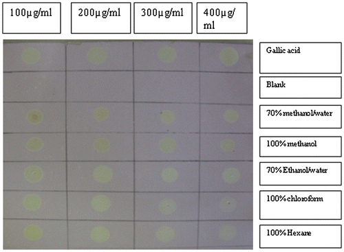
Figure 2. Comparative DPPH scavenging activity of Gallic acid and Myrothamnus flabellifolius.
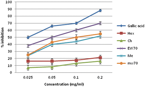
Table 2. In vitro ABTS free radical scavenging activity of various extracts of Myrothamnus flabellifolius
2.3. Reducing power
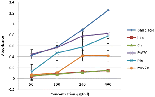
Oxidised form of iron (Fe3+) in ferric chloride is converted to ferrous (Fe2+) hence indicating the presence of an antioxidant compound responsible of donating an electron to the oxidised form iron resulting in the more stable reduced form (Moein, Moein, & Ahmadizadeh, Citation2008). In the present study, Figure shows hexane and chloroform have the lowest reducing power in the highest concentration among other extracts 0.149 and 0.153 Abs, respectively. The highest reducing power was exhibited by EW70 at 0.825 Abs, followed by methanol and MW70 at 0.781 and 0.422 Abs, respectively. The reducing power of all the extracts increased with increasing concentration and were all compared to gallic acid with the highest reducing power of 1.253 Abs.
Figure 3. Comparative reducing power of Gallic acid and Myrothamnus flabellifolius leaf.
2.4. Hydrogen peroxide scavenging activity
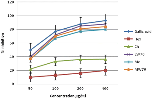
H2O2 is rapidly decomposed in to oxygen and water and this may produce hydroxyl radical (OH) that can initiate lipid peroxidation and cause DNA damage (Alam & Bristi, Citation2013). The ability of MF to scavenge H2O2 was estimated in different extracts. In Figure , Hex and Ch exhibited the lowest scavenging ability, 35.0 and 36.5%, respectively. EW70 exhibited 88% scavenging ability and found to be the highest among other extracts. Me and MW70 both exhibited 84%. The plant thus can scavenge the H2O2 with increasing concentrations in a similar way to gallic acid standard.
Figure 4. Comparative hydrogen peroxide scavenging activity of Gallic acid Myrothamnus flabellifolius extract.
2.5. Nitric oxide
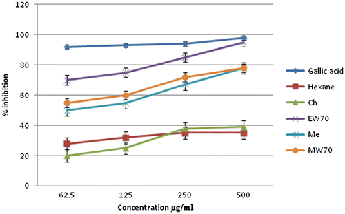
Nitric oxide (NO) is a free radical generated by endothelial cells, macrophages, neurons etc … and involved in the regulation of various physiological processes. Excess NO is associated with various diseases as it can react with oxygen to yield nitrite and peroxy nitrite acting as free radicals (Bala, Manigundan, Usha, & Priya, Citation2014; Lata & Ahuja, Citation2000). Cancer, inflammation and other pathological conditions can emerge as a result of nitric oxide (Bala et al., Citation2014). The various MF extracts reduced the generation of nitric oxide from sodium nitroprusside in buffered saline. As depicted in Figure EW70 show the most effective inhibition (95%) on the highest concentration 500 μg/ml. It is followed by MW70 (78%), Me (70%) Ch (45%). The lowest recorded nitric oxide inhibition was Hex at 35%.
Figure 5. Comparative nitric oxide scavenging activity of Gallic acid Myrothamnus flabellifolius extract.
2.6. Phytochemical screening
MF phytochemical screens in Table show various components in the various extracts. Flavanoids and tannins were present in all extracts except chloroform and hexane; Terpenoids are present in all extracts except in MW70 and Hex. Amino acids, proteins, fatty acid quinines and oxalate were all absent. Phenols show presence only in MW70 and EW70. Triterpenoids are present in Ch, EW70 and hexane extracts. The saponins are present in the EW70 and MW70 only. The presence of phenols, terpenoids, flavanoids, triterpenoids and saponins is associated with medicinal values such as the anti-inflammatory and antidiabetic (Mazimba, Wale, Kwape, Mihingo, & Kokengo, Citation2015).
Table 3. Phytochemical screening of Myrothamnus flabellifolius extracts
2.7. Antidiabetic activity
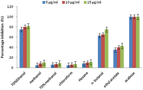
Different extracts of MF were investigated for antidiabetic activity. α-glucosidase and α-amylase assays were employed in the study. In both studies, Acabose a drug used in the treatment of diabetes and mainly targeting enzyme inhibition was used as reference standard. In Figure , EW70 and BuF show the highest α-glucosidase inhibitory effect 15 μg/ml-82% and 15 μg/ml-75%, respectively. They were followed by EtF, Ch, Me and Hex, respectively. The inhibition effect of all the extracts increased with increasing concentration. In Figure , EW70 and BuF were the most effective in alpha-amylase inhibition, 15 μg/ml-83%, 15 μg/ml-76%, respectively, followed by EtF, Ch, MW70, Me and Hex with the lowest inhibition of 15 μg/ml-11%. For all the extract the inhibitory effect was concentration dependant, the higher the concentration the higher the inhibition.
Figure 6. Percentage inhibition of Myrothamnus flabellifolius extracts on α-amylase. Each value is mean ± SEM of three trials.
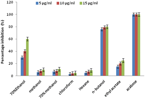
Figure 7. Percentage inhibition of Myrothamnus flabellifolius extracts on α-Glucosidase. Each value is mean ± SEM of three trials.
3. Experimental
3.1. Plant material and extraction procedure
The plant was collected in Ranaka Hills. The plant authentication was performed by Mr M. Muzila (Voucher no 008), University of Botswana Herbarium. The plant was then washed with distilled water and sun dried. The dried plant material was crushed with a laboratory grinder to obtain 10 kg of powder. The powder was soaked and extracted successively at room temperature in the following solvent system: 100% hexane; 100% chloroform; 100% methanol and 70% methanol/water and a separate 10 kg powder were extracted separately with 70% ethanol/water. The extracts were evaporated in a rota vapour. The 70% methanol/water and 70% ethanol/water extracts were freeze dried. Yields obtained were as follows: 100% hexane (14% yield) 100% chloroform (32% yield), 100% methanol (40% yield) 70% methanol/water (40 yield) 70% Ethanol/water (48% yield). The extracts were kept in a refrigerator until required.
3.2. Total phenol content
The total phenol content (TPC) was determined by the method described by Stoilova, Krastanov, Stoyanova, Denev, and Gargova (Citation2007), using Folin-Ciocalteu reagent. About 1 ml of extract or standard solutions (Gallic acid) (0–500 mg/l) was added to a mixture of 10-ml deionised water and 1.0 ml of Folin–Ciocalteu phenol reagent. After 5 min, 2.0 ml of 20% sodium carbonate was added to the mixture. After 1 h of incubation at room temperature in darkness the absorbance was measured at 750 nm. The TPC was calculated from the linear regression equation of the standard curve, from this equation, the concentration of gallic acid was determined for each extract and converted to mg of gallic acid equivalents/g of dry extract (mg GAE/g).
3.3. Reducing power
The reducing power of the extracts of M. flabellifolius was determined according to the method of described by Nandhakumar and Indumathi (Citation2013). About 1 mL of extract was added with 2.5 mL of phosphate buffer and 2.5 mL of 1% potassium ferricyanide. The reaction mixture was incubated for 20 min at 50°C, and, after that, 2.5 mL of 10% TCA was added and centrifuged. The supernatant was mixed with 2.5 mL of distilled water and 0.5 mL of FeCl3, and the absorbance was read at 700 nm. The assay was carried out in triplicate, and the results are expressed as mean ± standard error (SE). Increase in absorbance of sample with concentrations indicates high reducing potential of the samples.
3.4. Hydrogen peroxide scavenging activity
The scavenging activity of extract towards hydrogen peroxide radicals was determined by the modified method described by Ngonda (Citation2013). Solution of hydrogen peroxide (40 mM) was prepared in phosphate buffer pH 7.4 and its concentration was determined by measuring the absorbance at 560 nm using UV spectrophotometer. About 15.6–250 μg of the extract were added to hydrogen peroxide solution and absorbance measured at 560 nm using UV spectrophotometer against a blank solution containing phosphate buffer without hydrogen peroxide. The percentage of hydrogen peroxide scavenging by the extract and standard compound was calculated using the given formula:
3.5. Nitric oxide
Nitric oxide was performed according the method described by Rana et al. (Citation2010). Sodium nitroprusside 5 mM was prepared in phosphate buffer pH 7.4. To 1 ml of various concentrations of test extracts, 0.3 ml of sodium nitroprusside was added. The test tubes were incubated at 25°C for 5 h after which, 0.5 ml of Griess reagent was added. The absorbance of the chromophore was read at 546 nm. The experiment was performed in triplicate.
3.6. Phytochemical screening
Phytochemical screening was performed according to the methods described by (Mazimba et al., Citation2015).
3.7. In vitro antidiabetic activity
3.7.1. α-Glucosidase inhibition assay
MF α-glucosidase inhibition assay was carried as described by Ju Jeong et al. (Citation2013). α-glucosidase (50 μl, 0.5 U/ml) and 0.2 M K3PO4 buffer (pH 6.8, 5 μl) were mixed with 50 μl of the test sample pre-incubation at 37°C for 3 mM PNGP (50 μl) was added and was stopped by the addition of 750 μl of 0.1 M Na2CO3 The 4-nitrophenol absorption was measured was measured at 405 mM using a spectrophotometer. A solution without sample substrate was used as a control and a solution without sample was used as blank. The antidiabetic drug Acarbose was also assayed as a reference standard. The percentage inhibition of α-glucosidase was calculated as
3.7.2. α-Amylase inhibitory activity
α-Amylase inhibitory activity was tested by the method described by Ali, Houghton, and Soumyanath (Citation2006) with little modification. A total of 500 μl of test samples and standard drug (5–15 μg/ml) were added to 500 μl of 0.20 mM phosphate buffer pH (6.9) containing α-amylase (0.5 mg/ml) solution and were incubated at 25°C for 10 min. The reaction mixture was then incubated at 25°C for 10 min. About 500 μl of starch solution in 0.02 M sodium phosphate was added to each tube and the reaction mixture incubated at 25°C for 10 min. The reaction mixture was stopped with 1.0 ml of 3, 5 dinitrosalicylic colour reagent; the test tubes were then incubated in a boiling water bath for 5 min, cooled to room temperature. The reaction mixture was then diluted after adding 10-ml distilled water and absorbance was measured at 540 nm. Control represent 100% enzyme activity and were conducted in a similar way by replacing extract with vehicle. The percentage inhibition of α-amylase was calculated as
3.8. Statistical analysis
The experimental assays were carried out in triplicates, and the results are expressed as mean (n = 3) ± standard error (SE).
4. Conclusion
The present study is the first study of its nature in Botswana to assess the phenol content, antioxidant and antidiabetic activity of MF. The antioxidants and antidiabetic effect of the plant coincides with important compounds of Biological importance depicted by the phytochemical screening and phenol content. The study confirms the medicinal use of MF as a traditional medicine in the treatment of diabetes and possibly other ailments linked to oxidative stress, hence the plant applicability as a natural medicine. More studies in in vivo systems and on the individual components are needed to further confirm the medicinal properties of this plant.
Acknowledgements
The authors are thankful to Forest conservation Botswana (FCB) and Office of research and development (ORD) of University of Botswana for providing the funding of the study.
Additional information
Funding
Notes on contributors
Tebogo E. Kwape
Tebogo E. Kwape has completed PhD and his current research interests are on diabetes, antioxidants and antihypertensive properties of medicinal plants. The pharmacological aspects focussing on the possible mechanisms of action and histopathology of target organs like pancreas and liver.
References
- Ali Asgar, M. A. (2013). Anti-diabetic potential of phenolic compounds: A review. International Journal of Food Properties, 16, 91–103.10.1080/10942912.2011.595864
- Ali, H., Houghton, P. J., & Soumyanath, A. (2006). α-Amylase inhibitory activity of some Malaysian plants used to treat diabetes; with particular reference to Phyllanthus amarus. Journal of Ethnopharmacology, 107, 449–455.10.1016/j.jep.2006.04.004
- Atawodi, S. E. (2005). Antioxidant potential of African medicinal plants. African Journal of Biotechnology, 4, 128–133.
- Alam, M. N., & Bristi, J. N. (2013). Review on in vivo and in vitro methods evaluation of antioxidant activity. Saudi Pharmaceutical Journal, 21, 143–152.
- Bala, I. M., Manigundan, K., Usha, J., & Priya, S. (2014). Antioxidant and anti-proliferative activity of methanolic leaf extract of Eupatorium glandulosum. International Journal of Advanced Research, 2, 717–723.
- Ju Jeong, H., Kim, J., Kyung Hyun, T. K., Yang, J., Kang, H., Cho, J., … Jo Kim, M. (2013). In vitro antioxidant and antidiabetic activities of Rehmannia glutinosa tuberous root extracts. ScienceAsia, 39, 605–609.10.2306/scienceasia1513-1874.2013.39.605
- Lata, H., & Ahuja, G. K. (2000). Role of free radicals in health and disease. Indian Journal of Physiology and Allied Sciences, 57, 124.
- Maroyi, A. (2013). Traditional use of medicinal plants in south-central Zimbabwe. Journal of Ethnobiology and Ethnomedicine, 9, 1–18.
- Mazimba, O., Wale, K., Kwape, T. E., Mihingo, S. O., & Kokengo, B. M. (2015). Cinnammon verum: Ethylacetate and methanol extracts antioxidants and microbial activity. Journal of Medicinal Plants Studies, 3, 28–32.
- Moein, M. R., Moein, S., & Ahmadizadeh, S. (2008). Radical scavenging and reducing power of salvia mirzayanii subfractions. Molecules, 13, 2804–2813.10.3390/molecules13112804
- Molefe-Khamanga, D. M., Mookets, I. N.A., Matsabisa, M. G., & Kensley, R. M. (2012). Qualitative phytochemical studies of solvent extracts from Myrothamnus flabellifolius. International Journal of Medicinal Plant Research, 1, 1–5.
- Motlhanka, D. M. T., & Mathapa, G. (2011). Antioxidant activities of crude extracts from medicinal plants used by diabetic patients in Eastern Botswana. Journal of Medicinal Plants Research, 6, 5460–5463.
- Nandhakumar, E., & Indumathi, P. (2013). In vitro antioxidant activities of methanol and aqueous extract of annona squamosa (L) fruit pulp. Journal of Acupuncture and Meridian Studies, 6, 142–148.10.1016/j.jams.2012.09.002
- Ngonda, F. (2013). In vitro anti-oxidant activity and free radical scavenging potential of roots of malawian trichodesma zeylanicumm (burm. f.). Asian Journal of Biomedical and Pharmaceutical Sciences, 3, 21–25.
- Pellegrini, N., Proteggente, A., Pannala, A., Yang, M., & Rice-Evans, C. (1999). Antioxidant activity applying an improved ABTS radical cation decolorization asszy. Free Rad Biol Med, 26, 1231–1237.
- Rana, M. G., Katbamna, R. V., Padhya, A. A., Dudhrejiya, A. D., Jivani, N. P., & Sheth, N. R. (2010). In vitro antioxidant and free radical scavenging studies of alcoholic extract of medicago sativa l. Journal of Plant Biology, 1, 15–22.
- Setshogo, M. P., & Mbereki, C. M. (2011). Floristic Diversity and uses of medicinal plants sold by street vendors in Gaborone, Botswana. The African Journal of Plant Science and Biotechnology, 5, 69–74.
- Stoilova, I., Krastanov, A., Stoyanova, A., Denev, P., & Gargova, S. (2007). Antioxidant activity of a ginger extract (Zingiber officinale). Food Chemistry, 102, 764–770.10.1016/j.foodchem.2006.06.023
- Viljoen, A., Klepser, M. E., Ernst, E. J., Keele, D., Roling, E., van Vuuren, S., Demirci, B., … van Wyk, B. E. (2000). The composition and antimicrobial activity of the essential oil of the resurrection plant Myrothamnus flabellifolius. South African Journal of Botany, 68, 100–105.
- Wang, J., Yue, Y., Tang, F., & Sun, J. (2012). TLC screening for antioxidant activity of extracts from fifteen bamboo species and identification of antioxidant flavone glycosides from leaves of bambusa. textilis McClure. Molecules, 17, 12297–12311.10.3390/molecules171012297