
Abstract
This study reports a unique 10 years follow-up case of a patient who underwent a free fillet of sole flap for left leg stump coverage and free dorsalis pedis flap for soft tissue reconstruction of contralateral popliteal fossa following severe bilateral lower leg injury.
Keywords:
Introduction
Clinical advances in plastic surgery provide the means to reconstruct lower leg complex injuries. However, post-traumatic amputation is still sometimes mandatory, especially following Gustilo grade IIIB or IIIC open fractures,[Citation1,Citation2] and surgical planning needs to consider limb length preservation, durable soft-tissue coverage and maintenance of viable joints.
Several reconstructive options for soft tissue coverage have been already suggested including skin graft, tissue expansion and free flaps. Skin grafts provide insufficient tissue coverage resulting in easy skin ulcerations, while tissue expansion is often considered as a too long and discomfortable procedure. Furthermore, both procedures do not provide innervated tissues with consequent poor sensation and prosthesis compliance. Free flaps are a valuable option to improve stump durability and gait mechanics due to length preservation, even if an additional donor site is required.
Russell et al. [Citation3] and Frykman and Jobe [Citation4] described the use of undamaged ‘spare parts’ for reconstruction of a below-knee amputation in an emergency setting. The fillet foot flap [Citation5–9] is the extension of this concept that includes harvesting the entire soft-tissue envelope of the foot with both the dorsalis pedis and posterior tibial arteries as dual pedicles.
We report a 10 years follow-up of a patient who underwent a free fillet of sole foot flap for left leg stump coverage and free dorsalis pedis flap for soft tissue reconstruction of contralateral popliteal fossa following severe bilateral lower leg injury.
Case report
A 35-year-old man was a victim of a high-energy accident by his mowing machine. His injuries consisted of a Gustilo IIIc left tibial and fibula fracture and circumferential degloving of the right thigh associated with deep soft tissue loss at the popliteal fossa. X-ray showed pelvis, left distal and right proximal femoral fractures. Furthermore, a bilateral lower limb angiogram showed a partial obliteration of the left anterior tibial and peroneal arteries while it demonstrated patent vessels on the right side.
At the admission, initial management by the orthopaedic surgeons consisted of cleaning, surgical debridement, fasciotomies of the left leg compartments and application of an external fixator to stabilize the tibial fracture. The left leg wound became grossly infected and remittent fever was observed four days after injury. Surface bacteriology swabs were positive for methicillin-resistant.
Staphylococcus aureus and Pseudomonas. Appropriate antibiotic therapy was commenced. Due to the progressive worsening of left leg wound and medical clinical condition, the patient was transferred to the department of Plastic Surgery for further management 20 days after the injury. He underwent serial surgical debridements to remove the infected and necrotic tissue. The extensive soft tissue loss involved the anterior, lateral and superficial posterior muscle compartments of the left lower limb with exposure of the tibia and fibula ( and ). The dorsal and plantar surfaces of the foot were uninjured.
Figure 1. Injury three weeks after admission showing extensive soft-tissue and muscle loss in the left lower extremity.
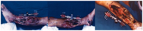
Figure 2. Injury three weeks after admission showing circumferential degloving of the right thigh with deep soft tissue injury at the popliteal fossa.
Thirty days post-injury following hemodynamic stabilization of the patient, a below-the-knee amputation was performed 12 cm below the tibial tuberosity and a free fillet of sole flap was fashioned. The neurovascular bundle was dissected through dense scar tissue, so it was impossible to harvest pedicled flap. The posterior tibial artery and both venae comitantes were anastomosed in an end-to-side fashion to the popliteal vessels while neurorrhaphy was performed between the tibial nerve and a branch of the sciatic nerve.
At the same time, a free dorsalis pedis fillet flap from the same limb was harvested to reconstruct the wide defect of the right popliteal fossa, using popliteal vessels as recipients. The length of the vascular pedicle was 12 cm. In order to avoid twisting and kinking of the vessels, the pedicle entered the recipient vessels at a gentle angle. The circumferential degloved wound of the right thigh was covered by split-thickness skin graft.
Preoperative sensitivity assessment by the use of cotton wool, pinprick test and PSSDTM [Citation10] demonstrated normal sensation of two skin areas on the left foot (the plantar medial and lateral surface of the heel) compared to the uninjured side (). Postoperatively neurological examination was obtained at six, 12 and 24 months. At six months the patient could localize pressure but was not able to feel cotton wool and a pinprick test (). There was a clear improvement in cutaneous pressure sensation thresholds and in a pinprick test over the time (). At 24 months he could recognize cotton wool, but a pinprick was felt as touch. The patient was discharged on 21st postoperative day and was then transferred to the rehabilitation centre where he was fitted with a below knee prosthesis. At one-year follow-up, the patient regained a stable and painless gait. At 10 years review, the patient was fully ambulating without any limitation of activities and did not have problem of wound stump breakdown over the time ().
Figure 3. Anterior and posterior postoperative view at 12-month follow-up.
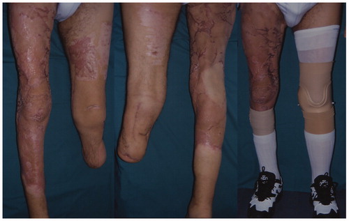
Figure 4. At 10-year follow-up a stable wound closure can be observed following left knee amputation by the use of free fillet of sole of foot flap.
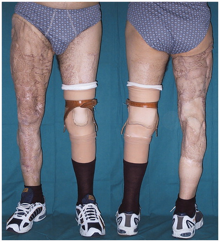
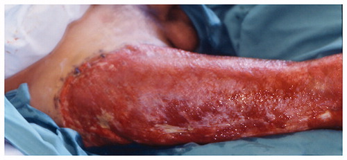
Figure 5. No skin retraction was observed following a free dorsalis pedis fillet flap coverage for the wide defect of the right popliteal fossa.
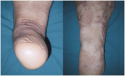
Table 1. Preoperative assessment of the cutaneous pressure sensation thresholds for the left and right heel in g/mm2.
Table 2. Cutaneous pressure sensation thresholds for the medial and lateral left heel in g/mm2 at six, 12 and 24 months of follow-up.
Discussion
The decision to proceed to lower limb amputation following traumatic injuries is never easy taking into account the significant cost to both the patient and the community. The patient experiences physical pain and emotional distress during the recovery from surgery and the rehabilitation phase. The patient faces loss of physical capabilities, the need for occupational re-training and the changes in his lifestyle. Previous report claimed that the limb amputation is more expensive than limb salvage.[Citation11] Consequently, the amputation choice needs to be carefully evaluated.
Obtaining suitable coverage to salvage a below-the-knee amputation has been performed by the use of a variety of reconstruction techniques. Among those, many authors have reported a long-term success using the pedicled or free fillet flap.[Citation5–9] The fillet flaps are axial pattern flaps that can be harvested as composite tissue transfers for reconstruction of complex defects.[Citation5] The fillet of sole flap can be raised on the posterior/anterior tibial neurovascular bundle. Whether it is used as a pedicled flap or free tissue transfer, it has several advantages. The thick plantar skin is ideal for weight bearing and can cover a relatively large area. Moreover, the septae between the skin and the plantar fascia prevent shearing forces transmitted to the soft tissue when the patient is walking with the prosthesis. In addition to the above aforementioned, the sole of the foot is preferred to the dorsalis pedis flap since it carries plantar innervated skin that improves prosthesis compliance by providing proprioceptive feedback and reduces prosthesis-related complications such as ulcers and painful neuromas.[Citation6] The dorsalis pedis fasciocutaneous flap is also useful for tissue transfer because of reliability and the long and thick vascular pedicle.[Citation9]
In our case, due to bilateral extended soft tissue loss and extensive necrotic muscle with bone exposure on the left side, an amputation was indicated. A reconstruction with free fillet of sole flap and a free dorsalis pedis flap was respectively performed to cover the left leg stump and the soft tissue defect on the right popliteal fossa. A pedicled flap was not harvested because of the partial obliteration of the anterior tibial and peroneal arteries and because of the long posterior tibial pedicle. The dissection of the lengthy neurovascular tibial bundle was judged to be too difficult in the chronically inflamed wound because of the fragility and adhesions to the surrounding tissue with pedicle redundancy such as pedicle kinking during flap inset and compression of the twisted pedicle in the stump pocket might compromise the final outcome. Cheng and colleagues were the first to describe the transfer of a free sole flap to the contralateral lower limb.[Citation7] The soft tissue defect on the right side was covered with a free dorsalis pedis flap in order to avoid skin retraction inducing knee joint complications in the active range of motion as generally observed after skin graft reconstruction. Intervention was delayed in this patient because he was too unstable at the time of injury and despite fasciotomies, the muscles of the lower leg infarcted while those above the knee and distal to the ankle remained viable. Whenever the injury involves wide zones, it is often unclear which tissue is devitalized and how much initial debridement needs to be carried out in order to decide for amputation or reconstruction. Reinnervation of large nerve fibers resulted in a marked improvement in cutaneous pressure threshold and the patient could detect cotton wool over the sole 24 months after surgery.
A double free fillet foot flap from the same limb was performed only by Pribaz et al. [Citation12] in a case of an emergency leg and hand reconstruction. To the best of our knowledge, our case report is the first description of a ‘double free flap’ for bilateral lower leg trauma. The 10-year follow-up demonstrated that the fillet free flap for reconstruction of complex bilateral lower extremity defects after high energy trauma is a valid alternative for achieving stable wound closure and maximising length preservation which facilitates gait rehabilitation without the common complications associated with the weight-bearing portion.
Disclosure statement
The authors declare that they have no conflict of interest.
References
- Gustilo RB, Mendoza RM, Williams DN. Problems in the management of type III (severe) open fractures: a new classification of type III open fractures. J Trauma. 1984;24:742–746.
- Puno RM, Grossfeld SL, Henry SL, et al. Functional outcome of patients with salvageable limbs with grades IIIB and IIIC open fractures of the tibia. Microsurgery. 1996;17:167–173.
- Russell RC, Vitale V, Zook EC. Extremity reconstruction using the “fillet of sole flap.” Ann Plast Surg. 1986;17:65–72.
- Frykman GK, Jobe CM. Amputation salvage with microvascular free flap from the amputated extremity. J Trauma. 1987;27:326–329.
- Küntscher MV, Erdmann D, Homann HH, et al. The concept of fillet flaps: classification, indications, and analysis of their clinical value. Plast Reconstr Surg. 2001;108:885–896.
- Ghali S, Harris PA, Khan U, et al. Leg length preservation with pedicled fillet of foot flaps after traumatic amputations. Plast Reconstr Surg. 2005;115:498–505.
- Cheng YC, Wei FC, Wang JW, et al. Reconstruction of below-knee stump using the salvaged foot fillet flap. Plast Reconstr Surg. 1995;96:731–738.
- Tran NV, Evans GR, Kroll SS, et al. Free fillet extremity flap: indications and options for reconstruction. Plast Reconstr Surg. 2000;105:99–104.
- Hidalgo DA, Shaw WW. Dorsalis pedis and “foot fillet” free flaps. In: Microsurgery in trauma. Mount Kisco (NY): Futura; 1987.
- Dellon AL. Somatosensory testing and rehabilitation. Baltimore: Institute for Peripheral Nerve Surgery; 2000.
- Chung KC, Saddawi-Konefka D, Haase SC, et al. A cost–utility analysis of amputation versus salvage for Gustilo Type IIIB and IIIC open tibial fractures. Plast Reconstr Surg. 2009;124:1965–1973.
- Pribaz JJ, Morris DJ, Barrall D, et al. Double fillet of foot free flaps for emergency leg and hand coverage with ultimate great toe to thumb transfer. Plast Reconstr Surg. 1993;91:1151–1153.