Abstract
The association of malignancy with nephrotic syndrome and renal histopathologic abnormalities is well documented. Paraneoplastic proteinuria caused by membranous glomerulonephritis usually is made simultaneously with the diagnosis of a malignant tumor, or the two conditions are diagnosed within a year of each other. We reported a patient who presented with nephrotic syndrome initially. Incidentally, in kidney specimens, pathologic findings showed perirenal fatty tissue with malignancy tumor emboli in lymphatics. Thereafter, gastric adenocarcinoma was diagnosed by gastrointestinal panendoscopy with gastric biopsy under impression of malignancy associated with glomerulonephritis. Patient died of complications of malignancy-related disseminated intravascular coagulation without chemotherapy after confirming diagnosis was made three months later.
INTRODUCTION
Significant proteinuria has become a recognized systemic manifestation of a wide variety of neoplastic disease. The first clinicopathologic study to convincingly present an association between neoplasia and nephrotic syndrome was published in 1966 by Lee et al. Citation[[1]]. In the other report, linking nephrotic syndrome with underlying cancer compiled by Eagen et al. Citation[[2]], 67 of 171 patients with malignancy had a carcinoma. Forty-eight of these 67 had a renal biopsy performed; 33 had membranous nephropathy. There seems to be an association between membranous glomerulonephritis and digestive tumors. The diagnosis of symptomatic proteinuria caused by membranous glomerulonephritis usually is made simultaneously with the diagnosis of a malignant tumor, or the two conditions are diagnosed within a year of each other. Most authors are willing to impute a causal relationship between the cancer and membranous glomerulonephritis only when this sequence is observed but not when a period of several years, with intervening treatment and complications, separate them Citation[3-4].
We described a patient who presented with nephrotic syndrome and incidentally, in kidney specimens, pathologic findings of perirenal fatty tissue with malignancy tumor emboli in lymphatics. Thereafter, he was diagnosed to have gastric cancer by gastrointestinal panendoscopy with gastric biopsy under impression of malignancy associated with glomerulonephritis. Simultaneously, malignancy associated with membranous glomerulonephritis was documented by renal and gastric biopsy. To our knowledge this is the first reported instance of such an association. Patient died of complications of malignancy-related disseminated intravascular coagulation three months later.
CASE REPORT
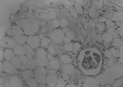
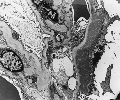
A 52-year-old male was admitted to the hospital because of progressive leg edema and abdominal distension for 3 months. He was referred from his local physician under impression of nephrotic syndrome. On examination the patient was clear consciousness and general anasarca. The body temperature was 37.4°C, pulse rate 84/min and respiratory rate 20/min. The blood pressure was 116/70 mmHg. The thyroid gland was firm and no bruit without enlargement. There was no supraclavicular and axillary lymphadenopathy. The jugular venous pressure was not elevated. The both lung fields were clear. The heart sounds were normal and abdominal examination showed distended abdomen with shifting dullness. There was moderate pitting edema over the both legs and no clubbing finger. There was no past history of diabetes mellitus and other systemic disease. Laboratory studies revealed hemoglobin 11.5 gm/dL, hematocrit 33.3%, platelete 338 × 103, white blood count 9100 with segment 89%, monocyte 3%, lymphocyte 8%; blood urea nitrogen 14 mg/dL, serum creatinine 1.3 mg/dL, uric acid 6.2 mg/dL, serum albumin 2.4 gm/dL, total serum protein 5.4 gm/dL, serum cholesterol 439 mg/dL, serum sodium 141 mmol/L, serum potassium 5.3 mmol/L. A urinalysis revealed 4+ proteinuria, 2 to 4 red blood cells/hpf. A 24-hour urine creatinine clearance and protein excretion was 82 mL/min and 9.3 gm, respectively. Immunoglobulin level was IgG 690 mg/dL, IgA 298 mg/dL, IgM 39.7 mg/dL, IgE < 28.7 mg/dL. Hepatitis B antigen and anti-HCV antibody were negative. Anti-nuclear antibody was negative with normal complement level. Kidney sonography showed renal parenchymal disease. Chest roentgenogram was normal. Taken together, these data suggest a diagnosis of nephrotic syndrome. A percutaneous renal biopsy was performed and histopathology showed membranous glomerulonephritis and light microscopy with hematoxylin and eosin stain revealed perirenal fatty tissue had malignant tumor emboli in lymphatics (). Immunofluorescent studies showed glomeruli with 4 + IgG and 4 + C3granular deposition in loop pattern and in tubular basement membrane. Electron microscopy revealed subepithelial electron dense deposits (). Gastrointestinal panendoscopy was arranged and showed a cardiac polypoid mass lesion in the stomach after we got the report of kidney biopsy.
Figure 1. Light microscopy of the kidney biopsy specimen shows perirenal fatty tissue has malignant tumor emboli in lymphatics, HE stain (×200).

Figure 2. Electron micrography of a glomerulus revealing subepithelial electron dense deposits (×4000).

Histopathology of stomach biopsy demonstrated poorly differentiated adenocarcinoma. Abdominal computer tomography (CT) scan showed omental cake with mesenteric traction and increased density in mesentery; there was thick wall of stomach and gastric malignancy with carcinomatosis was suspected. There were adenocarcinoma cells in ascites cytology study. Tumor marker data showing that CEA was 4.9, CA-125 was 426, CA 19.9 was 95.1. General anasarca and ascites had good response to albumin and furosemide therapy although he refused chemotherapy. He was discharged with edema-free status and was on symptomatic therapies without chemotherapy.
Three months later, he was readmitted because of intermittent fever with ecchymosis and subconjuntival hemorrhage of left eye. Laboratory studies showed a picture of disseminated intravascular coagulation (DIC). He died of sudden loss of consciousness with refractory shock and respiratory failure, which contributed to complications of malignancy-related DIC and possible central nervous hemorrhage later.
DISCUSSION
The association of malignancy with nephrotic syndrome and renal histopathologic abnormalities is well documented. The incidence of cancer reported in patients with membranous nephropathy has be variable, from 1.4% to 10.9% Citation[1-5]. A variety of malignancies associated with membranous nephropathy have been reported in literature; the majority have been carcinomas, the most frequent being bronchogenic carcinoma, adenocarcinoma of the stomach and colon Citation[6-7]. Further, if there has been an association of nephrotic syndrome, membranous glomerulonephritis, and malignancy, the discovery of the tumor presented simultaneously with the diagnosis of the glomerulonephritis or within 14 months following the renal diagnosis Citation[[1]]. In one previous study, malignancy will become detectable within a relatively short period after the diagnosis of membranous glomerulonephritis, and the malignancy rate is five times greater than the incidence in a baseline population. This risk is also highest in the elderly Citation[4-5]. This patient presented with nephrotic syndrome, and a renal biopsy revealed membranous glomerulonephritis and tumor emboli in lymphatic vessel. Thus, we searched for primary tumor and gastric adenocarcinoma was found. In addition, ascites investigation demonstrated adenocarcinoma cell and malignant ascites was documented. These findings suggest a disseminated metastasis of gastric cancer should be associated with membranous glomerulonephritis. This is the first reported case of nephrotic syndrome presenting as initially linking malignancy with perirenal fat lymphatic tumor emboli.
The diagnosis of malignancy-associated nephrotic syndrome caused by membranous glomerulonephritis usually is made simultaneously with the diagnosis of a malignant tumor, or the two conditions are diagnosed within a year of each other Citation[[2]], Citation[[5]], Citation[[7]]. The presence of a gastric adenocarcinoma in our patient with nephrotic syndrome suggests a pathogenic link with membranous glomerulonephritis, especially lymphatic vessel with tumor emboli of perirenal fatty tissue is detected, although this is difficult to prove. The pathogenesis, however, remains unclear. Using indirect immunofluorescence techniques, carcinoembryonic antigen (CEA) and its corresponding antibody has been demonstrated in the same glomerular distribution as the patient's own IgG in case of gastric adenocarcinoma Citation[8-9]. Lymphatic tumor emboli in perirenal fatty tissue may be implicated the pathogenesis of immune-complex-mediated mechanism because of producing antibody in lymphatic system.
Mean survival of cancer-associated nephrotic syndrome after recognition of the tumor was only 3 months; most patients died of their malignancy Citation[[2]]. In our patient, he refused chemotherapy and died of cancer-related DIC with possible central nervous hemorrhage, which presented with sudden loss of consciousness. The pathogenesis of DIC secondary to malignancy is not fully understood. The etiology of death is underlying malignancy and survival time is 3 months which consistent with previous report.
From our patient patient's experiences, as Ronco recommends Citation[[10]] that patients over the age of 50 with membranous glomerulopathy, in addition to a thorough history of the patient with a search for personal and hereditary cancer risk factors, physical examination, and standard biologic tests, one should undertake basic routine cancer screening procedures, including a chest radiography and a flexible colonoscopy. Further investigation, including flexible bronchoscopy or gastroscopy and CT scan, might be in order after first-line evaluation.
In summary, we report a patient presenting nephrotic syndrome diagnosing as membranous glomerulonephritis by kidney biopsy. Simultaneously, gastric cancer was suggested by perirenal fatty tissue with malignancy tumor emboli in lymphatics and confirmed by gastric biopsy as well as malignant ascites. Carcinoma-associated membranous glomerulonephritis is implicated as pathogenesis of nephrotic syndrome.
REFERENCES
- Lee J R, Yamauchi H, Hopper J, Jr. The association of cancer and the nephrotic syndrome. Ann Intern Med 1966; 64: 41–51
- Eagen J W, Lewis E J. Glomerulopathies of neoplasia. Kidney Int 1977; 11: 297–306
- Norris S H. Paraneoplastic glomerulopathies. Semin in Nephrol 1993; 13: 258–272
- Brueggemeyer C D, Ramirez G. Membranous nephropathy: a concern for malignancy. Am J Kid Dis 1987; 1: 23–26
- Burstein D M, Korbet S M, Schwarz M M. Membranous glomerulonephritis and malignancy. Am J Kid Dis 1993; 22: 5–10
- Alpers C E, Cortran R S. Neoplasia and glomerular injury. Kidney Int 1986; 30: 465–473
- Pai P, Bone J M, McDicken I, Bell G M. Solid tumor and glomerulopathy. Q J Med 1996; 89: 361–367
- Costanza M E, Oinn V, Schwartz R E. Nathanson: Carcinoembryonic antigen-antibody complexes in a patient with colonic carcinoma and nephrotic syndrome. N Eng J Med 1973; 289: 520–522
- Wakashin M, Wakashin Y, Iesato K, et al. Association of gastric cancer and nephrotic syndrome: An immunologic study in three patients. Gastroenterology 1980; 78: 749–756
- Ronco P M. Paraneoplastic glomerulopathies: New insights into an old entity. Kidney Int 1999; 56: 355–377