Abstract
Endothelial factor VIII staining in renal microcirculation was performed in eight nephrotic patients associated with mesangial proliferation (MesP) and six nephrotic patients associated with focal segmental glomerulosclerosis (FSGS). The result in MesP revealed a greater staining for glomerular endothelial factor VIII (35 ± 15%) and for postglomerular capillary endothelial factor VIII (65 ± 21%) than that observed in FSGS, which revealed a 11 ± 8% staining for glomerular endothelial factor VIII and 19 ± 15% staining for postglomerular capillary endothelial factor VIII. This finding implies that there is a greater loss of endothelial cell in renal microcirculation in FSGS. Such a finding correlates with the intrarenal hemodynamics which illustrated (Futrakul, P.; Sitprija, V.; Yenrusi, S. Glomerular endothelial dysfunction determines disease progression: a hypothesis. Am. J. Nephrol. 1997, 17, 533–540.) a mild reduction in renal plasma flow (535 ± 106 mL/min/1.73 m2, normal 600 mL/min/1.73 m2) and in peritubular capillary flow (422 ± 80 mL/min/1.73 m2, normal 480 mL/min/1.73 m2) in MesP and (Futrakul, P. Coagulation in glomerulonephritis and nephrotic symdrome: Its therapeutic intervention. In Asian Manual of Nephrology, Takeuchi, T.; Sugino, N.; Ota, K., Eds.; SEAMIC Publication, Tokyo, 1981; pp. 89–95.) a greater reduction in renal plasma flow (108 ± 50 mL/min/1.73 m2) and in peritubular capillary flow (87 ± 42 mL/min/1.78 m2) in FSGS. Therefore the study has emphasized both the structural and functional defects of endothelium in renal microcirculation in particular in FSGS.
Introduction
The function of glomerular endothelial cell normally expresses anticoagulant activity and vasodilating property by which it allows a free flowing of noncoagulant blood.Citation[[1]] However, in nephrotic syndrome, the function of glomerular endothelial cell is altered and expresses procoagulant activity namely the shortened platelet half-life, blood hypercoagulability, and elevated level of fibrin degradation product in serum and urineCitation[[2]], Citation[[3]] in conjunction with the defect in releasing vasodilator which is reflected by the reduction in renal plasma flow encountered in both mild form such as mesangial proliferative, steroid resistant nephrosis (MesP-NS),Citation[[4]] and in severe form namely focal segmental glomerulosclerosis (FSGS).Citation[[5]]
In respect to the reduction in renal plasma flow (RPF) observed in severe form of nephrosis such as FSGS, it is of notion that the reduction in RPF has been sustained and become progressive as the disease severity progresses.Citation[[1]] This remark renders a suggestive view that the endothelial structure as well as its function is likely to be defective.
Subjects and Methods
We studied endothelial factor VIII by mean of immunohistochemical staining using specific antibody to factor VIII in kidney biopsied specimens obtained from eight idiopathic nephrotic patients associated with MesP and six idiopathic nephrotic patients associated with FSGS. In addition, intrarenal hemodynamic study was performed in these nephrotic patients using a simultaneous determination of RPF by mean of 131I-labeled orthoiodohippuric acid (hippuran) and of glomerular filtration rate (GFR) by mean of 99mTc-labeled diethylene triamine pentaacetic acid (DTPA) and of peritubular capillary flow (PTCF) which was determined by the substraction of GFR from RPF in accordance with the previously described method.Citation[[5]]
Results
In MesP-NS, the positive stainings for endothelial factor VIII in glomerular microcirculation and in postglomerular capillary (peritubular capillary) microcirculation were 35 ± 15% and 65 ± 21% respectively (). In contrast, the positive stainings in FSGS-NS for endothelial factor VIII in glomerular microcirculation and in postglomerular capillary microcirculation were 11 ± 8% and 19 ± 15% respectively ( and ). The intrarenal hemodynamic study revealed thatCitation[[1]] in MesP-NS, the RPF, and PTCF were 535 ± 106 mL/min/1.73 m2 and 422 ± 80 mL/min/1.73 m2 respectively andCitation[[2]] in FSGS-NS, the RPF, and PTCF were 108 ± 50 mL/min/1.73 m2 and 87 ± 42 mL/min/1.73 m2 respectively.
Figure 1. Immunohistochemical staining for endothelial factor VIII of peritubular capillary in MesP-NS demonstrates a rather normal staining.
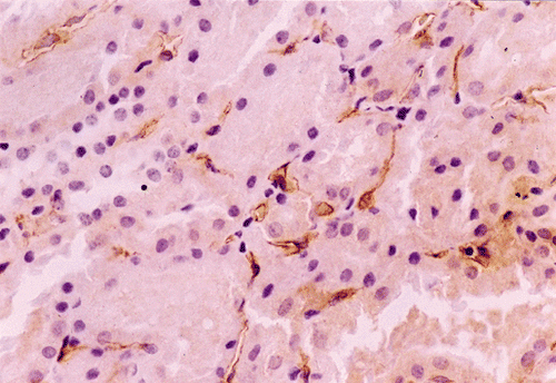
Figure 2. Immunohistochemical staining for endothelial factor VIII of peritubular capillary in FSGS demonstrates a diminished staining.
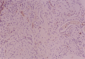
Table 1. Endothelial factor VIII staining and intrarenal hemodynamics in nephrosis
Discussion
The study of endothelial factor VIII staining demonstrates a greater loss of endothelial cell in renal microcirculation in nephrosis associated with FSGS. Such a defect in glomerular and postglomerular endothelial structure in nephrosis correlates with the degree of glomerular endothelial dysfunction determined by intrarenal hemodynamic study in such a way that the degree of endothelial factor VIII staining in renal microcirculation corresponds with the magnitude of reduction in renal plasma flow and peritubular capillary flow.
The significances of glomerular and postglomerular endothelial structural defect and dysfunction have recently been related to the disease severity and renal disease progression. Bohle and associatesCitation[[6]], Citation[[7]] noted an inverse correlation between the postglomerular capillary patency and the development of tubulointerstitial fibrosis. Recently, Yenrudi and associates have also demonstrated that there is an inverse correlation between renal perfusion and a relative area of renal cortical interstitium in idiopathic nephrotic syndrome.Citation[[8]] The preceding correlation between the renal perfusion deficit or the renal microcirculatory defect and the development of tubulointerstitial fibrosis is an interesting phenomenon and it would raise an interesting issue as to what would be the cause-and-effect relationship. In this regard, we have recently demonstrated thatCitation[[1]] the reduction in renal perfusion is inversely proportional to the degree of tubulointerstitial fibrosis andCitation[[2]] the reduction in RPF or PTCF precedes the development of tubulointerstitial fibrosis in severe form of nephrosis.Citation[[9]] Furthermore, a serum from severe nephrosis such as FSGS is able to induce a greater degree of endothelial cytotoxicity in vitro than that of a mild form of nephrosis or MesP.Citation[[10]] This finding is relevant to the spontaneous reduction in renal perfusion as the disease severity progresses, which has been uniquely observed in severe nephrosis.
The preceding information renders a support to the hemodynamically mediated mechanism of renal disease progression and it would offer a new therapeutic strategy of preventing the renal disease progression or the development of tubulointerstitial fibrosis by enhancing the renal perfusion. This can be accomplished by restoring the glomerular endothelial function and correcting the hemodynamic maladjustment commonly observed in severe form of nephrosis with vasodilators and antioxidants.Citation[[11]] Treatment in this regard has been performed with a greater success in not only preventing the progression of renal disease but also improving the renal function following the improvement in renal perfusion.Citation[[11]], Citation[[12]]
References
- Futrakul P., Sitprija V., Yenrusi S. Glomerular endothelial dysfunction determines disease progression: a hypothesis. Am. J. Nephrol. 1997; 17: 533–540
- Futrakul P. Coagulation in glomerulonephritis and nephrotic syndrome: its therapeutic intervention. Asian Manual of Nephrology, T. Takeuchi, N. Sugino, K. Ota. SEAMIC Publication, Tokyo 1981; pp. 89–95
- Stiehm E.R., Kuplic L.S., Nehling D.T. Urinary fibrin split products in human renal disease. J. Lab. Clin. Med. 1971; 77: 843–848
- Futrakul N., Futrakul P., Pohyachinda M., Apaiwong S., Sensirivatana R., Thamaree S., Chairatanarath T. Intrarenal hemodynamic alteration in mesangial proliferative nephrotic syndrome with steroid resistance: effect of vasodilators. Nephron 1994; 66: 366–367
- Futrakul P., Poshyachinda M., Futrakul. N., Chairatanarath T., Sensirivatana R., Thamaree S., Watana D., Kingwatanakul P. Intrarenal hemodynamic alterations and tubular functions in nephrotic syndrome associated with focal segmental glomerulosclerosis (FSGS): a pathogenetic and therapeutic implication. Current Therapy in Nephrology, V.E. Andreucci, A. Dal Canton. Wichtig Editore, Milano 1993; pp. 107–114
- Bohle A., Gise H., Mackensen-Haen S., Stark-Jacob B. The obliteration of the postglomerular capillaries and its influence upon the function of both glomeruli and tubuli, function interpretation of morphologic findings. Klin. Wochr. 1981; 59: 1043
- Bohle A., Mackenson-Haen S., Wehrmann M. Significance of postglomerular capillaries in the pathogensis of chronic renal failure. Kidney Blood Press. Res. 1996; 19: 191–195
- Yenrudi S., Laohapaibul A., Kittidiwit W., Suteparuk S., Futrakul N. A correlation between renal morphology and renal circulation in pediatric nephrotic syndrome. Ren. Fail. 2001; 23: 85–90
- Futrakul N., Yenrudi S., Sensirivatana R., Watana D., Laohapaibul A., Watanapenphaibul K., Kingwatanakul P., Futrakul P., Futrakul S. Peritubular capillary flow determines tubulointerstitial disease in idiopathic nephrotic syndrome. Ren. Fail. 2000; 22: 329–335
- Futrakul N., Panichakul T., Chaisuriya S., Sirisinha S., Futrakul P., Patumraj S. Endothelial cell cytotoxicity induced by nephrotic serum. 2nd Congress of the Federation of Immunologic Societies of Asia Oceania, Bangkok, 2000, S. Sirisinha, S.L. Chaiyaroj, F. Tapchaisri, Monduzzi Editore. pp. 27–31
- Futrakul N., Tosukhowong P., Valyapongpichit Y., Tipprukmas N., Futrakul P., Patumraj S. Oxidative stress and hemodynamic maladjustment in chronic renal disease: a therapeutic implication. Ren. Fail. 2002; 24: 433–445
- Futrakul P., Futrakul N., Sitprija V. Enhanced renal perfusion improves function in severe nephrosis with focal segmental glomerulosclerosis. Nephrology 1995; 1: 51–57