ABSTRACT:
Head trauma and subsequent brain injury is observed frequently in practice; although it is very difficult to estimate the number of cases seen each year. Cats are most often the victims admitted for this clinical reason.
In practice, common causes of head trauma include the following:
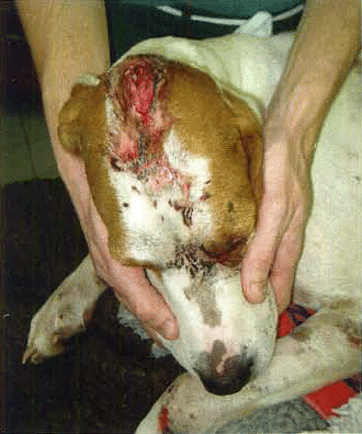
| • | road traffic accident (Figure 1) | ||||
| • | fall from a height | ||||
| • | big dog versus small dog incident | ||||
| • | kicked by a horse. | ||||
Factors such as the height of the fall or the force and velocity of a missile all have an effect on the severity of the injury. Concussion with depressed mentation, facial fractures and ocular injuries are common; other complications include soft tissue damage and proptosis.
Supportive care and first aid treatment should be given whilst secondary assessment is carried out. It is not unusual for multiple injuries to be present in these cases – in particular, thoracic and forelimb damage. This underlines the absolute importance of global assessment.
The aim of treating traumatic brain injury is to minimise or reverse the secondary changes that occur after the initial impact damage. Blood flow and oxygenation to the brain need to be optimal to prevent ischaemia and cell death, while at the same time avoiding sudden increases in intracranial pressure (ICP). Secondary damage occurs in the hours and days following the incident.
There are very few clinical data available regarding head trauma that are specific to dogs and cats. However, the guidelines produced for human head trauma patients can be almost directly transposed to veterinary patients for practical use.
This is supported by research showing that the histological changes (and magnetic resonance imaging changes) in a cat brain were identical to those in humans, (CitationSato et al, 2003).
The frequent assessment and monitoring of mentation status is key to providing the appropriate level of care for these cases.
Assessing outcome
There are a number of coma scales devised to predict whether a prognosis will be good, guarded or grave. The results are dependent on physiological factors that are graded numerically; these numbers are added together to give a final score. As would be expected, there is a degree of similarity between small animal charts and human charts ().
Table 1. Small animal coma scale
One example is the Glasgow Coma Scale, described in medical dramas as the GCS, (). A modified version, the MGCS has been produced for young children to address their inability to communicate verbally. This version has also been used in clinical veterinary practice.
Table 2. Glasgow Coma Scale (GCS)
Mentation status can be classified using the following terms:
| • | alert | ||||
| • | obtunded | ||||
| • | stuporous | ||||
| • | comatose. | ||||
The assessment is broadly based upon the following: verbal response, response to touch, ability to move around and show interest in surroundings. It is critical that an observation of mentation is recorded on admission, otherwise it is impossible to assess whether the patient has deteriorated or improved subsequently.
Protocol within the medical field suggests assessment every 10 minutes for at least the first hour after admission of a head trauma victim. The nursing plan for the individual will depend upon the level of consciousness – the more depressed the patient, the greater the list of concerns.
I consider the following to be the major nursing concerns:
| • | oxygenation and ventilation | ||||
| • | handling and positioning | ||||
| • | analgesia | ||||
| • | nutritional support. | ||||
Oxygenation and ventilation
If the patient is admitted in an unconscious state, it must be intubated. This is the only way to protect and manage the airway; not to do so is suboptimal care. If the patient is intubated, they will have to be monitored continuously, around the clock.
It is recommended that all head trauma patients should be supplemented with oxygen – pulse oximetry can be used to assess status and oxygen therapy provided by the most appropriate route. Blood gas analysis will confirm whether oximetry is accurate, as critically ill patients may not provide optimal conditions for reliable oximetry readings.
Venous jugular blood samples may be useful in these patients to monitor for cerebral ischaemia. Frequent blood pressure monitoring is also recommended to assess changes in blood pressure – hypoxaemia and hypotension have been observed in more than one third of human severe head injury patients.
Historically, hyperventilation was a longstanding protocol for severe head trauma in human patients, but more recently this has been reviewed and the conclusion is that it may not be warranted in all individuals. It is ‘no longer recommended as a first line therapy for intracranial hypertension or as prophylactic therapy following severe traumatic brain injury’ (CitationMarion et al 1995).
Explained simply – aggressive hyperventilation reduces intracranial pressure (ICP) rapidly, which in turn reduces cerebral blood flow, which can result in cerebral ischaemia. Cerebral ischaemia may be ‘the single most important event following severe traumatic brain injury’ (Brain Trauma Foundation, 1995).
Hyperventilation should probably be reserved for those patients showing clinical signs of dangerously high intracranial pressure. ICP and cerebral perfusion pressure are inextricably linked as demonstrated in the following equation, where CPP = cerebral perfusion pressure, MAP = mean arterial pressure and ICP = intracranial pressure:
CPP = MAP – ICP
Respiratory effort – along with pattern and rate – must be observed closely as the nature of the respiratory pattern can often indicate which area of the brain has been damaged.
Handling and positioning
Careful positioning and handling of these patients is vital to prevent sudden changes in blood pressure and subsequent ICP changes, as well as respiratory arrest and aspiration pneumonia.
Handling
Great care must be exercised when taking jugular blood samples, owing to the risk of respiratory arrest, should brain swelling and subsequent herniation be present. This also applies to general lifting and turning of the patient. Stress must be avoided and sedation may be necessary if the patient becomes distressed or restless.
Ketamine and diazepam are two drugs that have been used for this purpose and, although there does not appear to be conclusive evidence, it is thought that ketamine may have a protective effect against cerebral ischaemia, (CitationGremmelt & Braun, 1995).
Consider the environment – can noise or lighting levels be adjusted to reduce these stimuli? Ideally the patient should be housed somewhere that is conducive to easy viewing and monitoring, balanced with a perfectly calm recovery area.
Retching, sneezing and gagging reflexes should also be avoided – one reason to avoid placing a naso-oesophageal feeding tube!
Positioning
Positioning is particularly important if the patient is immobile, in order to prevent aspiration in case of regurgitation, as well as for comfort.
Traditionally, positioning to prevent passive regurgitation and subsequent aspiration involved placing padding beneath the head to elevate it. However, if the patient regurgitated in this position, the only way for food and fluid to go was straight back into the airway. The result of this was the horrendous complication of pneumonia.
Therefore, it is preferable to place padding underneath the shoulder so that any reflux can flow away from the airway and out of the mouth. It is suggested that the head should be elevated to no more than 30° to decrease ICP; so the nursing and veterinary teams together need to decide which is the priority in this instance.
Analgesia
Evaluating analgesic requirements can be problematic in the presence of dull or altered responses where normal pain scoring regimens cannot be used. Therefore, a process of deduction may have to be relied upon – if there are fractures and major soft tissue trauma present, then it is reasonable to assume that pain is present and that it requires treatment.
Decisions to give pain relief, in cases with obvious injury, are straightforward. Consideration of history and injuries sustained, as well as observing physiological signs are reasonable starting points as long as the patient is continuously re-evaluated.
Opioid analgesia would appear to be the obvious choice, as it is well suited to use in acute trauma; but there are concerns regarding opioids and their effects on respiration as they can cause respiratory depression and in an already compensating animal this may create further problems.
Fentanyl is considered the opioid of choice in head trauma because it is thought to preserve cerebral blood flow; with judicious use, it may not necessarily worsen intracranial pressure. Fentanyl also has the advantageous properties of rapid effect as well as short duration to provide opportunity for frequent reassessment. It is also a potent and effective analgesic.
If the patient is already experiencing respiratory depression, opioids are probably best avoided; otherwise, judicious use of an appropriate dose is acceptable. Buprenorphine has, however, been cited as useful in these cases ‘as it does not depress the respiratory system or CNS as much as Fentanyl’ (CitationO'Dwyer 2013).
The presence of pain itself can be responsible for raised ICP. Monitoring the response to analgesia is imperative to decide whether it has had a positive effect or not.
Nutrition
Nutritional support may not be at the top of the list of priorities for the first 48 hours, but some thought must be given to the options available to present a practical plan if the need arises.
The patient may be severely depressed but still able to lick and swallow; it is all too easy to assume that the ability to do so has been lost. A sensible quantity of food can be offered and the ability to eat can be evaluated.
If the animal is unable to eat, some form of nutritional support has to be considered. This raises several difficult questions: placing a naso-oesophageal tube is easy to do; but aspiration pneumonia is a real concern in these patients and, as previously mentioned, it is advisable not to stimulate coughing or sneezing reflexes.
Additionally the usual safety checks, such as checking for coughing performed prior to feeding, cannot be relied upon, because of depression or absence of normal protective reflexes. The placement of another type of feeding tube would necessitate general anaesthesia and this might not be advisable in an unstable patient.
Parenteral nutrition can be considered and, more specifically, peripheral parenteral nutrition could be a valid option for short-term use in this situation. Total parenteral nutrition may not be an option – either because of cost or concerns regarding elevation of ICP following jugular catheter insertion.
Monitoring and observation
Coma-scoring systems devised to evaluate mentation status have already been discussed; but there are several other specific areas to observe when nursing these individuals.
Respiration must be monitored closely for changes, as mentioned previously, as well as:
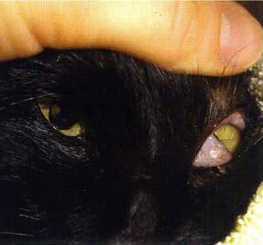
| • | pupillary size and light response (Figure 2) | ||||
| • | posture | ||||
| • | signs of seizure activity. | ||||
Pupil size is used as a prognostic tool – constricted, responsive pupils carry a more favourable outlook than dilated, unresponsive pupils. Other observations include ocular position and movement abnormalities. Anisocoria is frequently observed in these individuals.
Again it is critical that the veterinary nurse monitors and records these observations at the time of admission. If any changes in pupil size or reactivity are observed they must be reported to the veterinary surgeon immediately. Specific treatments for raised ICP may need to be instituted without delay.
Drawings that show damage and abnormalities affecting the eye can be made on neurological examination charts, to ensure that good communication and continuity of care is maintained when a new shift begins.
Specific posture(s) can be displayed when a particular area of the brain is damaged and, again, any developments must be reported straight away.
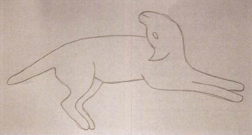
Opisthotonus is sometimes observed just before cardiopulmonary arrest but can also be seen in head and spinal trauma cases (Figure 3).
The possibility of seizure activity must be anticipated and treated with appropriate medication (usually diazepam initially) and oxygen therapy. A quiet, dark kennel with plenty of padding is essential if seizures do develop.
The group of patients at a higher risk of seizure activity include those with depressed skull fractures and penetrating head wounds, and these cases clearly require a greater level of observation.
Fluids and drugs
The use of intravenous fluids in head trauma cases does need to be very carefully monitored. The aims are to maintain circulating volume and replace deficits, whilst avoiding overload. Hyperglycaemia should be avoided and this will influence the choice of fluids.
It may not be possible to utilise central venous pressure monitoring because of concerns regarding jugular occlusion, so it may be wiser to rely upon clinical signs, laboratory data and physical examination. Essential clinical data must include recording of urinary output, which should at least match fluid input.
Fluid pumps or burette sets must be used in these cases to prevent accidental overload, especially in smaller patients.
Mannitol is a hypertonic crystalloid frequently used in cases of severe head trauma. Its hyperosmolar effect helps to decrease ICP rapidly. It is usually given as a bolus dose as opposed to a continuous rate.
The use of corticosteroids in head trauma is generally considered controversial and is not recommended. According to the Brain Trauma Foundation's Guidelines for the Management of Severe Traumatic Brain Injury (2007), in human patients the use of steroids is associated with increased mortality, does not improve outcome, or lower ICP particularly in cases of severe brain trauma.
Summary
The head-trauma patient may require close monitoring for a short period of time or many days of intensive care – each case is different.
Older individuals with existing disease are likely to take longer to recover from such incidents; but, in the author's experience, the majority of cats and dogs prove to be incredibly robust in their recuperation from a quite shocking head injury.
Additional information
Notes on contributors

Emma Opperman
Emma Opperman RVN
Emma has worked in veterinary practice for the last 24 years – in general practice and private referral practice, as well as in the role of ICU nurse at Bristol Veterinary School (Langford). She has lectured nationally and internationally and has written articles for veterinary nursing journals for many years. Her main interests are emergency nursing and teaching. Emma is on the VNJ editorial board and currently works as head nurse in the Cotswolds at a busy small animal hospital with a fantastic team of VNs.
References
- BRAIN TRAUMA FOUNDATION. (2007). Guidelines for Management and Prognosis of Traumatic Brain Injury. [Online] Available from: http//www.braintrauma.org [Accessed May 2014].
- BRAIN TRAUMA FOUNDATION. (2007). Guidelines for the Management of Severe Traumatic Brain Injury. 3rd Ed. New Rochelle. Mary Ann Leibert Inc.
- GREMMELT A., & BRAUN, U. (1995). Analgesia and Sedation in Patients with Head-Brain Trauma. Anaesthetist. 44 (Supp 3): 559–565.
- MARION, D. W., FIRLIK. A., & MCLAUGHLIN, M. R. (1995). Hyperventilation Therapy for Severe Traumatic Brain Injury New Horizon. 3(3): 439–447.
- O'DWYER, L. (2013) Nursing The Head Trauma Patient. The Veterinary Nurse. 4(5): 284–289.
- SATO, T., IKEBATA, Y., KOIE, H., SHIBUYA, H., SHIRAI, W., & NOGAMI, S. (2003). Magnetic Resonance Imaging and Pathological Findings in a Cat with Brain Contusions. Journal of Veterinary Medicine, Series A, Physiology, Pathology, Clinical Medicine. 50(4): 222–224.
Further Reading
- BEDELL, E. A., DEWITT D. S., & PROUGH, D. S. (1998) Fentanyl Infusion Preserves Cerebral Blood Flow During Decreased Arterial Blood Pressure After Traumatic Brain Injury In Cats. Journal of Neurotrauma. 15 (11): 985–992.
- DEWEX C. W. (2000) Emergency Management of the Head Trauma Patient; Principles and Practice. Veterinary Clinics of North America Small Animal Practice. 30(1): 207–225.
- DHUPA, N. (2002) Critical Care: Respiratory Focus. Veterinary Clinics of North America: Small Animal Practice. 32(5): 11–13.
- MACINTYRE, D. K., DROBATZ, K. J., HASKINS, S. C., & SAXON, W. D. (2005). Spinal Cord Trauma. Manual of Small Animal Emergency and Critical Care Medicine. 2nd Ed. Oxford. Wiley Blackwell.
- WHEELER, S. J. Ed (1995). BSAVA Manual of Small Animal Neurology 2nd Ed. Gloucester British Small Animal Veterinary Association.


