Corneal endothelium
The cornea is the outermost optical window of the eye and must remain clear to achieve excellent vision. The cornea consists of three major layers: the epithelium, stroma and endothelium. The innermost layer of the cornea, the endothelium, is responsible for maintaining the clarity of the entire cornea by its barrier and pump function. Human corneal endothelial cells are arrested in the G1 phase of the cell cycle and do not show proliferation in vivoCitation[1,2]. Due to this lack of proliferative capability, the standard therapy for corneal endothelial dysfunction has long been full-thickness penetrating keratoplasty (PKP). Although this approach has a relatively high rate of success, many complications, such as hemorrhage, infection, graft rejection, glaucoma and astigmatism, still occur. While human corneal endothelial cells are growth‑arrested in vivo, these cells can be induced to proliferate under certain culture conditions Citation[1,2]. Thus, the fabrication of transplantable endothelial cell sheets offers a possible ultimate treatment for many pathological cases where the corneal endothelial layer is the only component requiring replacement, especially in cases of insufficient donor tissue supply Citation[3,4]. This transplantation technique using cultured cells has been intensively studied; however, at this point, a large number of issues remains to be solved before it can be put into practice.
Corneal transplantation
In the 1990s, several new techniques for endothelial layer replacement were developed. One such procedure, identified as deep lamellar endothelial keratoplasty, was first performed in the USA in 2000 Citation[5]. Deep lamellar endothelial keratoplasty eliminated surface corneal sutures and incisions, while the advantages of normal corneal topography and faster wound healing were obtained, leading to faster visual rehabilitation and a more stable globe for the patient.
Another radical modification of the PKP technique utilizes the stripping of Descemet’s membrane and has been popularized as Descemet’s stripping endothelial keratoplasty. Descemet’s stripping endothelial keratoplasty has the advantages of being easier for the surgeon to perform and providing a smoother interface on the recipient side for the visual axis. Preparation of the donor tissue in endothelial keratoplasty has also been facilitated by the utilization of a microkeratome or, recently, femtosecond laser. The addition of this component to the surgical procedure has been popularized as Descemet’s stripping automated endothelial keratoplasty (DSAEK) Citation[6,7].
Disadvantages of DSAEK
The preliminary clinical results of the relatively new DSAEK procedure are encouraging, with DSAEK-patients showing fewer complications, faster healing and a significantly better visual acuity outcome Citation[8,9]. However, as we all know, DSAEK donor tissues must undergo several additional steps from tissue preparation to the completion of DSAEK procedure compared with PKP donor tissues, although variations occur in each step depending on the surgeons’ preferences. These steps include:
• Setting the tissue on the artificial anterior chamber (AC)
• Microkeratome/femtosecond laser cutting
• Tissue detachment from the artificial AC
• Marking on the tissue with gentian violet Citation[10]
• Tissue delivery and/or transportation Citation[11]
• Trephination, handling and/or folding with forceps Citation[12]
• Unfolding in the AC
• Tissue exposure to the air Citation[13]
• Tissue manipulation with an instrument for its centration
Therefore, donor buttons should be handled and transported very carefully to avoid additional damaging effects on the endothelial layer.
Current DSAEK tissue-insertion techniques usually use smaller incision sizes (typically 3–5 mm) than those of the donor-tissue diameter (often ≥8 mm in diameter). Therefore, the donor tissue must be folded prior to or at the insertion step. This folding requires additional handling of the donor tissue, which must be performed without inflicting damage to the endothelial cells of the donor tissue, resulting in the unfolding of the donor tissue in the proper orientation in the AC. In general, the handling of the donor tissue is accomplished with nontoothed and nonappositional forceps that attempt to manipulate the donor tissue by grasping the posterior stromal tissue edge. It has been recognized that this maneuver undoubtedly causes endothelial damage at the point of peripheral contact Citation[12]. The process of inserting the donor corneal button may be among the most traumatic to endothelial cells and may possibly cause higher failure rates for the corneal transplant, especially since endothelial cell density is the most crucial element determining the success of a corneal transplant. Therefore, any increase in cell loss that is incurred during surgery reduces the success of the procedure. The advantages of DSAEK surgery should be apparent, but DSAEK can result in an increased risk of endothelial cell damage with the current forceps-insertion technique.
Requirements & solutions in inserting DSAEK donor tissues
To decrease endothelial damage and improve the success rate and safety of DSAEK, there is a strong need for a new device to deliver the DSAEK donor tissue safely into the AC. In this single-insertion step alone, several critical complications may occur and affect the corneal endothelial viability and the success of DSAEK surgery. The representative concerns are described in the following sections.
Damage by forceps insertion
As reported previously, the DSAEK donor tissue that was grasped by inserting forceps demonstrated parallel bands and orthogonal wrinkles of scattered blue devitalized nuclei in a parallel arrangement Citation[12]. The orthogonal wrinkles of scattered blue devitalized nuclei presumably represent donor-tissue crumpling secondary to the insertion process caused by the corneal/scleral wound compression and the slipperiness under the forceps arms. Sometimes regrasping the tissue is unavoidable, owing to the slipperiness of the DSAEK tissue under the forceps arms, which then, of course, adds to the endothelial damage.
Anterior chamber collapse
In contrast to the intraocular lens insertion process in cataract surgery, the aim is to aspirate the ophthalmic viscoelastic device (OVD) completely before delivering the DSAEK donor tissue into the AC, in order to decrease the residual interface OVD between the host and donor graft in DSAEK surgery. In addition, the intraocular lens injector can seal the wound and maintain the AC depth, as surgeons usually make a tight wound incision. However, in DSAEK surgery, AC collapse during the conventional insertion process with the forceps is common. When this occurs, it is extremely difficult to remove the forceps from the wound without dragging and damaging the DSAEK tissue. To reduce this potential damage, surgeons can use an AC maintainer or smaller-sized incision. However, the former irrigation flow can cause tissue movements in the small AC space, which can then cause contact between the endothelial layer and the intraocular tissues, such as the iris and lens. Furthermore, the smaller-sized incision can cause additional endothelial damage, as reported by Terry et al.Citation[14].
High intraocular pressure
During and/or after the tissue insertion, the intraocular pressure can be higher owing to the AC maintainer, balanced salt solution injection or air injection. These types of AC-pressurizing processes should usually be performed after suturing the main incision. However, they are sometimes performed prior to the wound closure owing to the unstable AC or risk of donor tissue damage due to a collapsed AC. The higher intraocular pressure can then force the tissue out through the wound.
Tissue orientation
Corneas requiring DSAEK surgery usually have edema and the cornea (donor tissue) is transparent in nature. Therefore, it is difficult to recognize the orientation of the DSAEK tissue through edematous corneas under a surgical microscope without tissue marking. When unfolding the donor tissue in the AC, surgeons can have upside-down (endothelial side up) tissue orientation without knowing it. To avoid this, techniques such as making asymmetrical marks on the stromal side of the donor tissue Citation[10] and 60/40 or 40/60 tissue folding Citation[15], are utilized.
Wound compression pressure
To avoid AC collapse and induced astigmatism, smaller incisions (3–5-mm incision size) are employed in DSAEK. Through this small wound, we have to insert larger tissue grafts (usually of >8mm diameter and 150–250 µm thickness). As long as we use nonprotected methods, such as the forceps-insertion technique, the DSAEK tissue will not be able to avoid wound-compression damage.
Comparisons of DSAEK injectors & glides
The hot area of evolution in DSAEK is the use of instruments other than forceps to implant the donor graft. Largely, the DSAEK tissue-inserting devices can be categorized into injectors and glides, differentiated by the method with which the tissue is introduced into the AC.
With the glide techniques developed by Busin (Busin glide from Moria), Tan (sheet-glide technique) and Kobayashi (Kobayashi double-glide method, combining the Busin and sheet-glide techniques) Citation[16–18], surgeons pull the tissue from the far side of the AC with the forceps. There are some advantages to using glide techniques in DSAEK; for example, the donor tissue always enters in the correct orientation and is easier to unfold than with a pair of forceps. The process also appears to be less traumatic than using forceps for the insertion. However, higher intraocular pressure can push the tissue away from the glide and cause AC collapse, especially when combined with the AC maintainer. The glide-insertion techniques require surgeons to use both hands throughout the insertion process. This may be problematic if, for example, AC collapse occurs when the tissue is just crossing the wound area. In this case, it is practically impossible for the surgeon to control the tissue and the AC simultaneously. In addition, the sheet-glide technique cannot avoid the wound-compression pressure completely, even with the aid of a large quantity of cohesive OVD. This thicker OVD can move around the stromal side and cause an interface space between the host and donor cornea, potentially resulting in postoperative tissue detachment or dislocation. Donald Tan from Singapore recently developed a disposable DSAEK inserter (Network Medical Products, North Yorkshire, UK). Tan claims that the technique of gliding and pulling the donor tissue through the sclera wound is more efficient with their new device and should result in even less endothelial damage. The key concept in their new device is that they obviate contact between the previously mentioned sheet glide and the endothelial surface of the donor, as well as avoiding endothelium-to-endothelium contact in order to significantly reduce cell loss. This concept has something in common with that of the Busin glide. In brief summary, it appears to be less traumatic than using forceps for the insertion. While only the complication of damage by forceps insertion can be solved with the sheet-glide technique, damage by forceps insertion, tissue orientation and wound-compression pressure can be dealt with by Busin’s, Kobayashi’s and Tan’s new glides.
Five DSAEK injectors are summarized in . All injectors have an outer layer in common to protect the DSAEK tissue from wound-compression pressure.
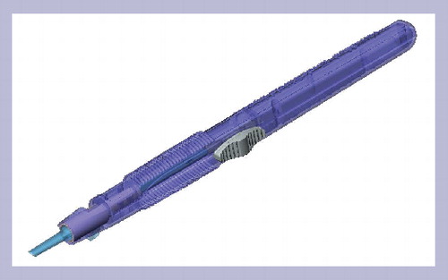
Keramed Shiuey Endoshield
The Endoshield uses a cartridge approach to insert the graft into the AC such as that used to insert an intraocular lens . However, rolling up a DSAEK graft like a newspaper risks damage to the endothelium. Keramed is currently making a cartridge that will curl the graft inward so that it will fit through a smaller wound than the 5-mm incision that is currently used in DSAEK. The resulting column strength allows you to push against it. An analogy would be trying to push a thin piece of paper by just pushing on one end. It would be difficult because it is so floppy and thin. However, if you roll up that piece of paper, it has column strength that you’re able to push against to force it to move. This technique is familiar to surgeons who perform cataract surgery with injectors Citation[19].
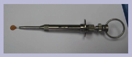
Rhein Medical/Lens Tec Harvey–Steinert injector
This device uses a reusable, stainless steel, single-handed hand-piece and a disposable carrier that acts as a sled for the graft to ride on . This sled is attached to a syringe-like piston that allows the cornea to be loaded very close to the tip, retracted into the shooter and then injected through the incision to allow delivery of the donor graft. This injector is also very similar to the intraocular lens injectors that most surgeons are familiar with Citation[19].
Ension Al-Ghoul vacuum-assisted injector
This device employs the novel idea of using the existing vacuum technology of a phaco machine and using it to hold the graft. Basically, this is an injector that has an acrylic cartridge and a tip that is mobile; surgeons can move it in and out of the cartridge. The injector also has an irrigation system, supplied at a point distal from the vacuum occlusion site, which maintains the stability of the AC as the tissue is inserted. To release the tissue, the surgeon stops the vacuum and presses reverse vacuum on the foot pedal, which sends fluid out through the vacuum ports, releasing the donor tissue into the AC, endothelial side down Citation[19].
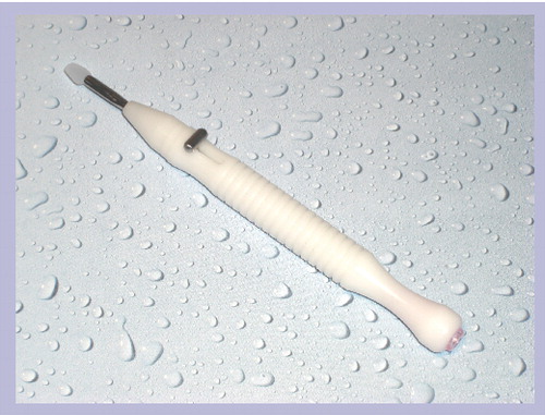
Fischer Surgical Neusidl Corneal Inserter
This device uses the similar carrier system to the Harvey–Steinert Injector. However, like the Al-Ghoul Vacuum-assisted Injector, this system has the AC-maintainer function .
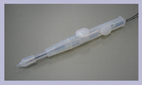
Kaneka Ide DSAEK injector
This injector is distinctive in that this has irrigation, air injection and pressure control functions in this single device . The tiny cannula in the center of the injector functions as both irrigation and air injection. The inner surface of the injector is coated with special biocompatible polymer to allow the tissue to slide more easily. These ideas are derived from the cardiac catheter. The air injection and pressure-releasing functions in particular are very helpful in DSAEK surgery because with other devices (e.g., glides, other injectors and forceps), the tissue is floating in the AC without any support. When surgeons pull the instruments out of and inject air into the AC, the tissue endothelium can touch the intraocular tissues, such as the iris or lens. This devastating event negates the merit of using an injecting device (i.e., glide or injector). Furthermore, the sudden intraocular pressure change during the pulling of the injecting device out from the wound can cause the tissue to be forced out. To avoid this, it is critical to control and release the pressure before removing the injecting device. Compared with the Rhein Medical/Lens Tec Harvey–Steinert injector, the Ension Al-Ghoul vacuum-assisted injector and the Fischer Surgical Neusidl corneal inserter devices described previously, the volume of the cannula that enters the AC is much smaller. This can be an advantage for handling the device in a small AC space. Unfortunately, however, this device requires surgeons to create the largest wound incision size (5.5 mm) among these five injectors.
Conclusion
In summary, DSAEK has been a revolutionary change in the treatment of endothelial disorders, avoiding the need for the more invasive PKP. However, it is not without its problems, one of which is the trauma that can befall the delicate endothelial graft when it is manipulated by forceps. As a possible solution, surgeons and companies are developing injectors and/or glides that may be able to insert the graft with less trauma to the endothelial cells. At this point, there are five innovative injectors and two glides that may hit the market in the near future. Depending on the surgeons’ preferences, their surgical techniques and economical situations, the requirements for the inserting device may vary. However, to make the best use of a limited supply of donor corneas and to achieve good long-term results, surgeons should perform the least traumatic surgeries on donor endothelium in any situation. In this respect, the injectors look more promising than the glides. Although it is controversial if surgeons have to use these devices (especially DSAEK experts), we have to examine the fact that the number of DSAEK surgeons is increasing due to its apparent simple techniques. Even in the hands of a DSAEK expert, the endothelial damage is reported to be more severe than that in PKP. We believe the future of donor insertion for posterior lamellar grafting will lie in using an injection system so that the surgeon can rely on a more reproducible and safer means of delivering the necessary tissue. No matter how careful one is while using forceps, the point of forceps is to squeeze and that is not what we want to do to a donor graft. These innovations on DSAEK inserting glide and injector may obviate all five complications previously described and we are looking forward to having ex vivo and in vivo study results in the future, although none of these injectors have been US FDA 510(K)-cleared yet.
Table 1. Five Descemet’s stripping automated endothelial keratoplasty injectors.
Financial & competing interests disclosure
Takeshi Ide has the nonpersonal educational grant related to the Descemet’s stripping automated endothelial keratoplasty injector discussed in this manuscript. The author has no other relevant affiliations or financial involvement with any organization or entity with a financial interest in or financial conflict with the subject matter or materials discussed in the manuscript apart from those disclosed.
No writing assistance was utilized in the production of this manuscript.
References
- Joyce NC, Meklir B, Joyce SJ, Zieske JD. Cell cycle protein expression and proliferative status in human corneal cells. Invest. Ophthalmol. Vis. Sci.37, 645–655 (1996).
- Joyce NC. Proliferative capacity of the corneal endothelium. Prog. Retin. Eye Res.22(3), 359–389 (2003).
- Sumide T, Nishida K, Yamato M et al. Functional human corneal endothelial cell sheets harvested from temperature-responsive culture surfaces. FASEB J.20, 392–394 (2006).
- Ide T, Nishida K, Yamato M et al. Structural characterization of bioengineered human corneal endothelial cell sheets fabricated on temperature-responsive culture dishes. Biomaterials27, 607–614 (2006).
- Terry MA, Ousley PJ. Deep lamellar endothelial keratoplasty in the first United States patients: early clinical results. Cornea20, 239–243 (2001).
- Gorovoy MS. Descemet-stripping automated endothelial keratoplasty. Cornea25, 886–889 (2006).
- Cheng YY, Pels E, Nuijts RM. Femtosecond-laser-assisted Descemet’s stripping endothelial keratoplasty. J. Cataract Refract. Surg.33, 152–155 (2007).
- Chen ES, Terry MA, Shamie N, Hoar KL, Friend DJ. Descemet stripping automated endothelial keratoplasty: six-month results in a prospective study of 100 eyes. Cornea27, 514–520 (2008).
- Koenig SB, Covert DJ. Early results of small-incision Descemet’s stripping and automated endothelial keratoplasty. Ophthalmology114, 221–226 (2007).
- Ide T, Yoo SH, Kymionis GD, Perez VL, Goldman JM, O’Brien TP. Descemet stripping automated endothelial keratoplasty (DSAEK): effect of nontoxic gentian violet marking pen on DSAEK donor tissue viability by using vital dye assay. Cornea27, 562–564 (2008).
- Ide T, Yoo SH, Kymionis GD, Goldman JM, Perez VL, O’Brien TP. Descemet stripping automated endothelial keratoplasty: effect of anterior lamellar corneal tissue-on/-off storage condition on Descemet-stripping automated endothelial keratoplasty donor tissue. Cornea27, 754–757 (2008).
- Ide T, Yoo SH, Goldman JM, Perez V, O’Brien TP. Descemet stripping automated endothelial keratoplasty: effect of inserting forceps on DSAEK donor tissue viability by using an in vitro delivery model and vital dye assay. Cornea26, 1079–1081 (2007).
- Cekiç O, Ohji M, Hayashi A, Fang XY, Kusaka S, Tano Y. Effects of humidified and dry air on corneal endothelial cells during vitreal fluid-air exchange. Am. J. Ophthalmol.134, 75–80 (2002).
- Terry MA, Saad HA, Shamie N et al. Endothelial keratoplasty: the influence of insertion techniques and incision size on donor endothelial survival. Cornea28(1), 24–31 (2009).
- Chen ES, Terry MA, Shamie N, Phillips PM, Friend DJ, McLeod SD. Descemet stripping automated endothelial keratoplasty: insertion using a novel 40/60 underfold technique for preservation of donor endothelium. Cornea27(8), 941–943 (2008).
- Mehta JS, Por YM, Beuerman RW, Tan DT. Glide insertion technique for donor cornea lenticule during Descemet’s stripping automated endothelial keratoplasty. J. Cataract Refract. Surg.33, 1846–1850 (2007).
- Busin M, Bhatt PR, Scorcia V. A modified technique for descemet membrane stripping automated endothelial keratoplasty to minimize endothelial cell loss. Arch. Ophthalmol.126, 1133–1137 (2008).
- Kobayashi A, Yokogawa H, Sugiyama K. Descemet stripping with automated endothelial keratoplasty for bullous keratopathies secondary to argon laser iridotomy – preliminary results and usefulness of double-glide donor insertion technique. Cornea27, S62–S69 (2008).
- Bethke W. Injecting innovation into DSEK. Rev. Ophthalmol.15(8), 1 (2008).