
Protein expression studies in Dictyostelium discoideum have been hampered by the lack of suitable loading controls for quantitative immunoblotting. Typical loading controls, such as actin, that are used for mammalian immunoblots are not appropriate for Dictyostelium studies due to the massive changes in protein expression levels that occur during development. Compounding the problem, commercial antibodies for most Dictyostelium proteins are lacking. In this month's issue, King and colleagues at the Beatson Institute for Cancer Research (Glasgow, UK) describe a possible solution to this dilemma—using labeled streptavidin conjugates to detect a highly expressed biotinylated protein as a loading control for quantitative immunoblotting of Dictyostelium proteins. While Dictyostelium is predicted to have up to 20 endogenously biotinylated proteins, the authors discovered that probing immunoblots of Dictyostelium whole-cell lysates with fluorescently labeled streptavidin revealed only a single prominent band of 80 kDa, which they subsequently confirmed to be 3-methylcrotonyl-CoA carboxylase alpha (MCCC1), the Dictyostelium homolog of a well-characterized human mitochondrial protein that is also heavily biotinylated. The authors realized that labeled-streptavidin detection of MCCC1 would be ideal as an immunoblot loading control. MCCC1 is extremely robust, being able to withstand harsh protein extraction procedures, and since it is directly detected with a fluorophore- or horseradish peroxidase-conjugated streptavidin and does not require a secondary antibody, its detection is extremely linear, as was shown for a 100-fold range of protein concentrations. Another advantage is the fact that various labeled streptavidin conjugates are commercially available and inexpensive. Finally, the MCCC1 expression levels remain constant throughout Dictyostelium development, making it well-suited to developmental studies. All of these advantages establish MCCC1 as a robust control protein for quantitative Dictyostelium immunoblots when directly detected with labeled streptavidin conjugates. As an additional benefit, the authors also demonstrated that fluorescently conjugated streptavidin strongly labeled the lumen of mitochondria in fixed Dictyostelium cells, making it a convenient mitochondrial stain that does not require blocking.
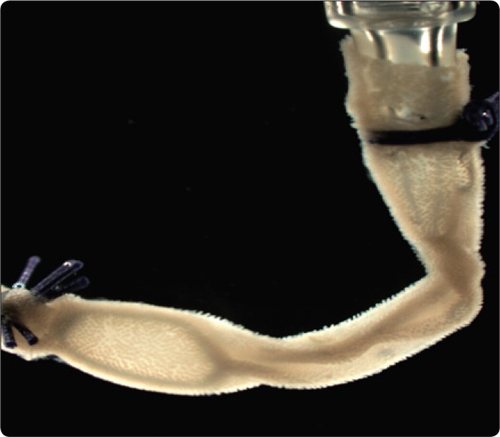
Numerous laboratory procedures rely upon cells or tissues that have been purified from the surrounding milieu. Naturally, the procedures for isolating these tissues vary according to tissue type, but they tend to be lengthy and complex owing to the fact that the material of interest must remain intact and functional while surrounding “contaminant” cells are removed. This is most often accomplished using either chemical or mechanical dissociation methods that must be carefully performed and optimized to avoid damaging the target tissue. Interest in isolating intact intestinal and pancreatic tissue is growing as more researchers focus on understanding host-microbiota interactions and the molecular biology of diabetes. Established procedures for isolating intestinal epithelia and pancreatic islets of Langerhans are complicated, requiring lengthy centrifugation steps. In this issue of BioTechniques, two research groups present significantly improved approaches for isolating these tissues. For intestinal epithelial isolation, Nik and Carlsson from the University of Gothenburg (Gothenburg, Sweden) describe a new procedure that reduces both mechanical cell damage and contamination with connective tissue as well as significantly decreasing the time required for purification. A stretch of intestine was first inverted and cleaned, then attached to a syringe and submerged in cold BD Cell Recovery Solution to dissolve the basement membrane. Using the syringe, the intestine was then inflated and deflated repeatedly over a 30 minute time course to push ephithelial crypts out of the mesenchymal crypt beds. Epithelium was then collected in a coherent sheet with intact crypts and villi.
Koh et al. from the University of Washington (Seattle, WA) faced similar cell contamination issues, but in their case, the contaminant was acinar cells that are difficult to separate from pancreatic islet cells. The established approach relies upon collagenase treatment of the pancreas, mechanical segregation of islets, and finally separation of islet and acinar cells by hand with a pipet. Koh et al. sought to simplify this procedure by using a peristaltic pump. The authors mathematically modeled the effects of flow rate, drag, buoyancy, and gravitation on the islets and thereby determined an equilibrium condition wherein the islet would be sucked into the pipet and remain suspended in the tip as solution was moved by the pump at a constant rate. The authors experimentally measured equilibrium positions for islets, which matched theoretical predictions, enabling them to collect undamaged islets quickly and efficiently with significant reductions in effort and time.
The advances in tissue isolation methodology reported in this issue of BioTechniques will not only help scientists working with intestinal epithelium and pancreatic islet cells, but may serve as starting points for researchers experimentally optimizing isolation conditions for other tissue types as well.

