
Introduction
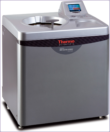
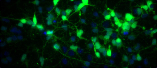
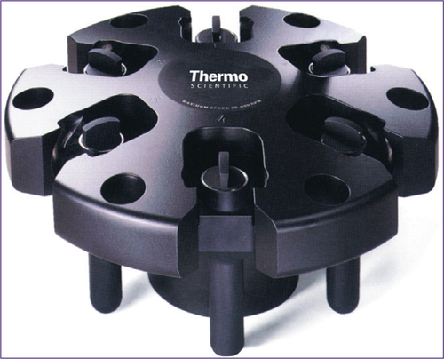
Protein function can be determined by using gain- or loss-of-function experiments, including the induction of exogenous gene expression in multiple cell lines and in vivo models. The utilization of viral transduction to induce exogenous gene expression is becoming an increasingly common technology. Viral transduction is more efficient than transfection since you can selectively target cell populations by using multiple virus types (e.g., retrovirus, adenovirus, lentivirus), gene expression is increased in difficult to transfect cell types, and gene expression in infected cells is permanent (Cepko and Pear, (Citation1996). Multiple methods can be used to conduct viral transduction, including incubation of target cell populations with dilute or concentrated viral supernatant. Furthermore, concentrated virus can be used for the rapid induction of genetic expression in vivo, which can eliminate the need for costly and time-consuming transgenic mouse models (Gaiano et al., (Citation1999). The virus can be concentrated by ultracentrifugation of viral supernatant harvested from transfected packaging cell lines. The procedure described below outlines the process for the production and concentration of retrovirus using the Thermo Scientific™ Sorvall™ WX ultracentrifuge and the Thermo Scientific™ SureSpin™ 630 swinging bucket titanium rotor. By incubating cortical progenitors with concentrated retrovirus produced by this method, we were able to selectively target gene expression (identified by green fluorescent protein, GFP) to dividing neural progenitors, a primary cell line that has difficulty with exogenous gene expression.
Procedure
Production of dilute virus
Transfect GP2-293 derived packaging cell line (Clontech) at 90% confluency with 5 µg each of pCLE-IRES2-eGFP (Anjen Chenn, Northwestern University, Chicago, IL) and pVSV-G (Clontech) using Lipofectamine™ and PLUS™ Reagent (Invitrogen)
Approximately 18 h post-transfection, replace the culture media with fresh media according to manufacturer's instructions
Place transfected GP2-293 cells at 32°C and 5% CO2 for virus production
Harvest virus by removing viral supernatant from confluent monolayer of transfected cells 5 to 6 times at 12 h intervals and placing into conical tube
Centrifuge conical tube in Thermo Scientific low-speed centrifuge at 200 × g for 5 min at room temperature
Syringe tube filter viral supernatant through a 45 µm filter into new conical tube
Freeze each filtered harvest at −80°C
Concentration of dilute virus
Thaw viral harvest(s) at room temperature
Remove viral harvest(s) into Thermo Scientific PA Thin-Walled Tubes (36 mL, cat #: 03141)
Place balanced tubes in Thermo Scientific SureSpin 630 (36 mL) swinging bucket titanium rotor
Centrifuge tubes in Thermo Scientific WX ultracentrifuge at 112,700 × g (25,000 rpm) at 4°C for 1.5 h
Remove supernatant from tubes, leaving viral pellet at the bottom
Resuspend viral pellet in 40 µL sterile PBS overnight at 4°C and aliquot into small volume working stocks in microcentrifuge tubes
Store aliquots at −80°C
Discussion
The procedure above describes a method to produce concentrated retrovirus using the WX ultracentrifuge and SureSpin 630 swinging bucket titanium rotor. Using concentrated retrovirus, we were able to selectively infect dividing neural progenitor cells as detected by GFP expression in a small volume without disturbing the culture conditions of the cells (, Noles and Chenn, (Citation2007). The above procedure can also be used to produce concentrated retrovirus to induce rapid exogenous gene expression in multiple in vivo models (e.g., mouse or chicken) without the need to create expensive and time consuming transgenic mouse models.
References
- Cepko, Constance and PearWarren. 1996. Transduction of Genes Using Retrovirus Vectors. Current Protocols in Molecular Biology, p. 9.9.1–9.9.16.
- Gaiano, N, JDKohtz, DHTurnbull, and G.Fishell. 1999. A method for rapid gain-of-function studies in the mouse embryonic nervous system.Nat Neurosci.9:812–9.
- Noles, SR and DAnjen Chenn. 2007. Cadherin inhibition of beta-catenin signaling regulates the proliferation and differentiation of neural precursor cells.Mol. Cell. Neurosci.35:549–58.

