
Abstract
Herceptin, the monoclonal antibody, was successfully immobilized on gold nanoparticles (GNPs) to improve their precise interactions with breast cancer cells (SK-BR3). The mean size of the GNPs (29 nm), as determined by dynamic light scattering, enlarged to 82 nm after herceptin immobilization. The in vitro cell culture experiment indicated that human skin cells (FB) proliferated well in the presence of herceptin-conjugated GNP (GNP–Her), while most of the breast cancer cells (SK-BR3) had died. To elucidate the mechanism of cell death, the interaction of breast cancer cells with GNP–Her was tracked by confocal laser scanning microscopy. Consequently, GNP–Her was found to be bound precisely to the membrane of the breast cancer cell, which became almost saturated after 6 hours incubation. This shows that the progression signal of SK-BR3 cells is retarded completely by the precise binding of antibody to the human epidermal growth factor receptor 2 receptor of the breast cancer cell membrane, causing cell death.
Introduction
For more than a decade, nanometer-sized gold nanoparticles (GNPs) have attracted considerable attention not only because of their size- and shape-dependent optical and electronic properties but also due to their latent uses in thermal imaging, catalysis, scattering analysis, photoelectronic devices, biomedical diagnostics, and other related fields.Citation1–Citation3 GNPs offer important new possibilities in cancer diagnosis and therapyCitation4 and can be used in the imagingCitation5,Citation6 of tumors in their primary and secondary locations, as delivery agents for antitumor drugs, and for nonaggressive amputation treatments. The conveyance of the NPs to the tumor is a multistage technique.Citation7 Systemically controlled NPs with tumor-binding ligands can accumulate in the cancer cell due to the additional disordered vasculature relative to noncontaminated tissue.Citation6,Citation7
Cancer is challenging to treat, so active diagnosis approaches in the early phases of cancer are life threatening. In this respect, imaging has become an essential instrument in cancer medical trials and therapeutic replication.Citation8 Fluorescent bioimaging is also of extreme importance for visualizing the manifestation and movement of specific particles, cells, and biological processes that affect the performance of tumors and/or their reaction to therapeutic medications.Citation9 Therefore, an extensive series of fluorescent modules has been explored in in vitro bioimaging studies, including the biomarking of cancer tissues,Citation10 angiogenic vasculature, and sentry lymph nodes.Citation11 In this respect, numerous nanomaterials such as quantum dots, noble metal NPs, upconverted NPs, and new fusion nanocomposites of reduced graphene oxide and GNPs have great potential for highly sensitive optical imaging of cancer in both in vitro and in vivo experiments. From this viewpoint, GNPs are innovative biocompatible nanoprobes, offering surfaces and cores that possess physicochemical properties (eg, optical chirality,Citation12,Citation13 fluorescence,Citation14 near-infrared photoluminescence,Citation15 and ferromagnetismCitation16) that offer novel openings for clinical diagnostics. As such, these tools will certainly play a vital part in the initial analysis and responsive recognition of tumors.
The multifunctional behavior of NPs offers exclusive benefits for the tumor-specific transfer of imaging and medicinal agents.Citation17 Monoclonal antibodies with substantially assorted structures and properties are economical to produce and have great feasibility as a class of tracking moieties.Citation18 One of the most broadly studied monoclonal antibodies used as a targeting moiety for the distribution of agents is herceptin. Herceptin is a water-soluble antibody that binds to the human epidermal growth factor receptor 2 (HER2) found on the membrane of SK-BR3 cells.
In this study, SK-BR3 cells were attacked with herceptin-conjugated GNPs (GNP–Her) for tumor treatment and diagnosis. Mercaptosuccinic acid (MSA)–immobilized GNPs were prepared by citrate reduction, followed by the coupling reaction of GNP with acid-terminated MSA. Herceptin was then immobilized on the surface of GNP (GNP–Her) to enrich the anticancer effects of chemotherapeutic agents.Citation19–Citation22 The surface properties of GNP and GNP–Her were characterized by Fourier transform infrared (FT-IR) spectroscopy, ultraviolet–visible (UV–vis) spectrometry, dynamic light scattering (DLS), and transmission electron microscopy (TEM). To estimate the cell compatibility and cytotoxicity of the GNPs and GNP–Her conjugates, human breast cancer cells (SK-BR3) were cultured in the presence of GNPs. The intracellular uptake of GNP–Her into the cells was also observed by confocal laser scanning microscopy (CLSM) and inductively coupled plasma mass spectrometry (ICP–MS).Citation23
Materials and methods
Auric chloride (HAuCl4), sodium citrate (Na3C6H5O7), MSA, and herceptin were purchased from Hoffman-La Roche Ltd (Basel, Switzerland). Cell culture reagents, fetal bovine serum, Dulbecco’s Modified Eagle’s Medium (DMEM), penicillin–streptomycin mix, trypsin–ethylene diamine tetraacetic acid (EDTA) solution, Dulbecco’s phosphate-buffered saline (PBS), and cell viability staining reagents were provided by Gibco BRL (Thermo Fisher Scientific, Waltham, MA, USA), and the cells (SK-BR3 and FB) were purchased from the Korean Cell Line Bank.
Synthesis of monodispersed GNPs
GNPs were prepared according to the method outlined by Anshup et al.Citation24 Briefly, in a 250 mL round-bottomed flask, 100 mL of 0.05% HAuCl4 was added and heated. Under prompt stirring, 3.5 mL of sodium citrate (1%) was supplemented and further rapidly stirred for 15 minutes at 120°C. After 30 minutes of stirring, the solution was then steadily cooled to room temperature. The citrate concentration was adjusted to attain different particle sizes. After 15 minutes, the liquid was removed and cooled to room temperature and filtered. The prepared GNPs were resuspended and purified by centrifugation and double precipitation from distilled water.
Preparation of hydrophilic GNPs
MSA (0.16 M) was dissolved in 70 mL of water in a 250 mL three-necked flask containing GNPs. The solution was stirred for 3 hours in nitrogen atmosphere. After the reaction was complete, the water was separated by vacuum, and the residue was transferred into a centrifuge tube and mixed with 4 mL of water. Subsequently, 20 mL of water was added to the mixed solution, and the precipitated product was separated and subjected to centrifugation at 3,000 rpm (revolutions per minute) for 15 minutes and washed with water. The prepared MSA-conjugated GNPs were dissolved and purified by centrifugation and double precipitation from water.
Immobilization of herceptin on GNPs
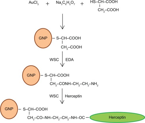
The immobilization of herceptin on GNPs (GNP–Her) was carried out by the reaction of water-soluble GNPs with herceptin (). Water-soluble GNPs (1.2 mmol) and herceptin (7 mmol) were added to a two-necked flask and dissolved in 10 mL of PBS buffer. 1-Ethyl-3-(3-dimethylaminopropyl) carbodiimide (EDC; 2 mmol) and N-hydroxysuccinimide (NHS; 2 mmol) were supplemented to the reaction solution, followed by stirring for nearly 5 hours at room temperature. The reaction product was filtered to remove the precipitate and added to a 100,000 molecular weight cutoff dialysis membrane in deionized water for 24 hours to discard the unreacted EDC, herceptin, and NHS. Lastly, the solution was filtered through a 0.45 μm membrane and dried under vacuum for 24 hours.
Figure 1 Schematic diagram showing the immobilization of herceptin on mercaptosuccinic acid-coated gold nanoparticles.
Preparation of cyanine dye–GNP–Her conjugates
Cyanine dye 5 (Cy5)-conjugated GNP–Her was obtained by reacting 1.5×10⁻³ mol/L GNP–Her with 4.8×10⁻⁵ mol/L fluorescent Cy5 in a carbonate buffer solution. The concentration of Cy5 dye was examined by UV–vis spectroscopy to calculate the number of NPs internalized into the cytoplasm of SK-BR3.Citation25
Results
Characterization of surface-modified GNPs
The immobilization of herceptin and Cy5 dye on surface-modified GNPs was recorded by UV–vis absorption spectrum from aqueous solution at room temperature using a Hitachi U-3000 spectrophotometer. The core diameter and the surface morphology of GNPs were observed by TEM (Philips CM-200, 120 kV). The samples for TEM observations were diluted with distilled water and deposited on Formvar-coated 400-mesh copper grids. After drying the GNP–fluid thin film on the copper grid, a thin carbon film, approximately 10–30 nm in thickness, was deposited on the NP film. The size and hydrodynamic diameter distribution of the GNPs were determined by DLS using a light-scattering spectrometer BI90 (Brookhaven, Long Island, NY, USA).
Cell culture
SK-BR3 (breast cancer cells) was used as the target cell, and FB (human skin cells) was used as the control cell line. In a humidified atmosphere containing 5% CO2, the cells were cultured routinely in a polystyrene dish at 37°C using 10 mL of McCoy medium or DMEM, supplemented with 10% fetal bovine serum and 1% penicillin–streptomycin G sodium. The medium was changed every 3 days. For subculture, the cells were washed twice with PBS and incubated with a trypsin–EDTA solution (0.25% trypsin, 1 mM EDTA) for 10 minutes at 37°C to detach the cells. Complete media were then added to the polystyrene dish at room temperature to prevent the effects of trypsin. The cells were washed thrice by centrifugation method and resuspended in a new culture dish. To observe the morphology of cells, the cells were seeded in a 10 mL Petri dish at a concentration of 1×10⁵ cells/mL and incubated for 3 days with GNP and GNP–Her. The surface morphology of adhered cells was observed by Nikon Eclipse TS100 optical microscopy.
Cell viability
To study the cytotoxic effects of GNP–Her, after 3 days of culture, the SK-BR3 and FB cells were suspended in PBS at a density of 1×10⁵ cells/mL. Subsequently, the cell suspension was mixed with 100 μL of the assay solution (10 μL propidium iodide–calcein AM) and incubated for 15 minutes at 37°C. The cells were then observed by fluorescence microscopy (Axioplan 2; Carl Zeiss Meditec AG, Jena, Germany) for the simultaneous observation of live and dead cells.
Intracellular uptake of GNPs
For the observation of effective cellular uptake of NPs via CLSM and fluorescence microscopy, the cells were seeded at a concentration of 1×10⁵ cells/mL in a 10 mL culture dish and incubated for 24 hours. After 1 day, the medium was swapped with GNP and GNP–Her, containing a particle concentration of 50 μg/mL, and incubated for a certain period (2–7 hours) for the specific internalization of the NPs into the cells. The cells were then washed three times with PBS, and images were taken using a confocal laser microscope. A Carl Zeiss LSM 410 (Oberkochen, Germany) confocal laser scanning microscope was used to obtain the confocal images. The location and integrity of the internalized GNP–Her conjugates were estimated by CLSM using 4,6-diamidino-2-phenylindole dihydrochloride (DAPI, blue) as a marker, which stains the nuclei of the cells. The cell nuclei were stained by the addition of DAPI solution (10 μL) with proper mixing and incubated for 10 minutes. To track the GNP–Her NPs, GNP–Her and DAPI (488 nm) were added to the cells. The stained cells were washed at least three times with 1 mL of fresh DMEM and images were then taken by CLSM. To measure the intracellular uptake of the NPs, cells were grown in 24-well culture plates with roughly 105 cells in 1 mL of the medium. After 20 hours’ incubation at 37°C, the cells were reseeded with culture medium containing GNP–Her at a concentration of 2×10⁻⁴ cells/mL. The intracellular gold concentration was quantified using ICP–MS. Cells were washed with PBS, separated, resuspended, counted, and centrifuged, and the cell pellets were dissolved in 37% HCl aqueous solution at 60°C–85°C for 40 minutes. The experiment was repeated three times, and the results were averaged with standard deviation.
Statistical analysis
The cell multiplying experiment was conducted in triplicate and the results are presented as means ± standard deviations. The Student’s t-test was used to estimate the statistically significant difference between the results. Difference was considered statistically significant at P<0.05.
Discussion
Surface characterization
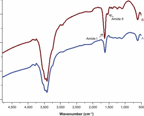
The surface modification of GNP with herceptin was confirmed by UV–vis spectra, as shown in . GNPs showed an absorption onset at 386 nm and after herceptin immobilization, they exhibited a red shift to 389 nm.Citation26 This red shift was produced by strong quantum confinement due to the increase in particle size after metal-to-ligand charge transfer.Citation27,Citation28 The surface modification of GNP with herceptin was also confirmed by FT-IR, as displayed in . In the case of the GNP spectrum, the introduction of MSA on the surface of GNP was confirmed by the characteristic peak at 1,700 cm−1 and 3,500 cm−1 (), which can be attributed to the free carboxyl (–COOH) and hydroxyl (–OH) groups.Citation26,Citation29 Again, after reaction of GNP with herceptin, two new peaks at positions around 1,648 cm−1 and 1,540 cm−1 were observed in the spectrum of GNP–Her (), which are assigned to the amide I (–CO–NH–) and amide II (–CO–NH–) bonds, respectively.Citation29 After the reaction of the free amine groups of GNP with herceptin, absorption at 1,648 cm−1 and 1,540 cm−1 appeared, due to the amide groups of GNP–Her, proving the effective immobilization of herceptin over the GNPs.
Figure 2 UV spectra of GNP and GNP–Her.
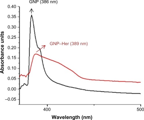
Figure 3 FT-IR spectra of GNP (A) and GNP–Her (B) measured using the KBr method.
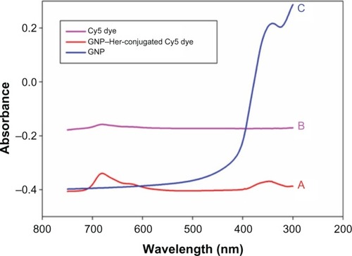
represents the TEM images of GNP and GNP–Her. The GNP has a spherical morphology, with a mean diameter of ~7.34 nm. In the case of GNP–Her, the particles had a mean diameter of ~8.14 nm, were spherical in shape, and showed significantly monodispersed particles (). The larger particle size and nonaggregated particle morphology was attributed to the conjugation of herceptin on the surface of the GNP. shows the typical sizes and size distributions of the synthesized GNPs and GNP–Her conjugates measured by DLS. The mean size of the GNPs, as determined by DLS, was ~29 nm. On the other hand, the mean size of GNP–Her was almost ~82 nm. The particle size, as determined by DLS, was significantly larger than that determined by TEM. This is because the DLS technique measures the mean hydrodynamic diameter of the GNP core bounded by the organic and solvation layers, and this hydrodynamic diameter is affected by the viscosity and concentration of the medium. TEM, however, gives the diameter of the core alone.Citation27 The synthesis of Cy5 dye-immobilized GNP–Her was also confirmed by UV–vis absorption spectroscopy, as shown in . The Cy5 dye-immobilized GNP–Her showed an absorption onset at 263 nm () and after Cy5 dye immobilization, it exhibited a red shift to 680 nm (). This red shift was caused due to the increase in particle diameter.Citation28
Figure 4 TEM images of GNP (A) and GNP–Her (B).
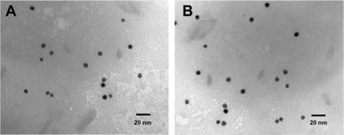
Figure 5 Particle-size distributions of GNP (A) and GNP–Her (B) measured by DLS.
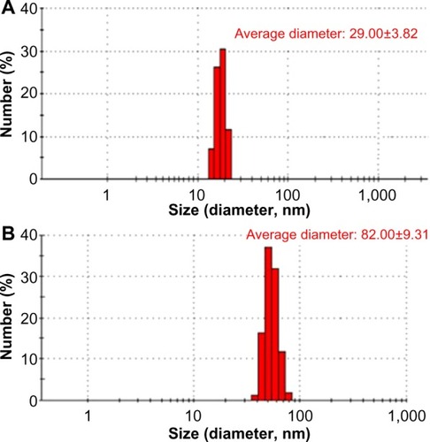
Figure 6 UV–vis absorption spectra of the GNP–Her-conjugated Cy5 dye (A), Cy5 dye (B), and GNP (C) in aqueous solution.
Evaluation of cytotoxicity
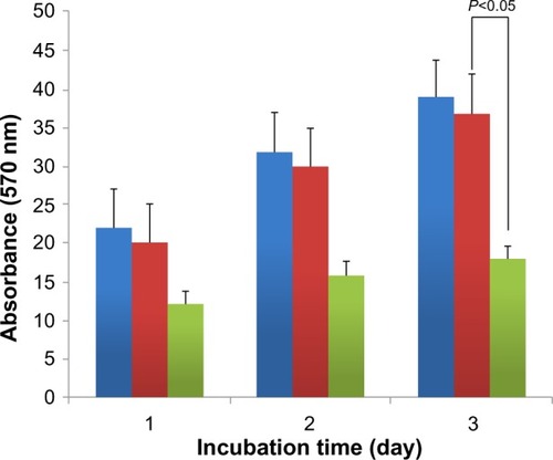
shows the distinction of the “live/dead” dye-stained SK-BR3 and FB cells cultured in the presence of GNP and GNP–Her after 3 days of incubation. Using this qualitative method, the living and the dead cells were stained green and red, respectively, and were visible by fluorescence microscopy. shows that all the FB cells remained viable after 3 days’ incubation, in the presence of NPs. On the other hand, after culturing for 3 days, in the presence of GNP–Her, most of the SK-BR3 cells had died, as shown in . shows the viability of SK-BR3 cells cultured for 1 day and 3 days in the presence of GNP–Her, as determined by the MTT (3-(4,5-dimethylthiazol-2-yl)-2,5-diphenyltetrazolium bromide)assay. After 1 day and 3 days of incubation, the level of SK-BR3 cell proliferation in the presence of GNP was similar to that of the cells cultured in the absence of NPs (polystyrene dish). On the other hand, cell propagation in the presence of GNP–Her was significantly lower than that in the presence of GNP. Therefore, the GNP conjugated with herceptin could increase the death of SK-BR3 cells considerably compared to the GNP without herceptin.
Figure 7 Fluorescence microscopy.
Abbreviations: GNP, gold nanoparticle; GNP–Her, herceptin-conjugated GNP; FB, fibroblast cell line.
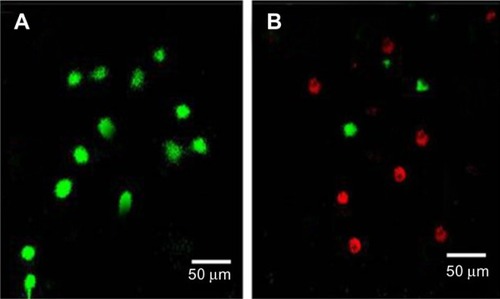
Figure 8 MTT assay.
Abbreviations: GNP, gold nanoparticle; GNP–Her, herceptin-conjugated GNP; MTT, 3-(4,5-dimethylthiazol-2-yl)-2,5-diphenyltetrazolium bromide.
A probable explanation of this large decrease in cell viability in the case of GNP–Her is that intracellularly distributed herceptin shows significant apoptotic activity by networking with several transcription factors related to cell propagation. Formerly, Bae et alCitation30 stated that degradable heparin nanogels and heparin/chitosan polyelectrolyte nanocomplexes could effectively induce apoptosis via receptor-mediated endocytosis through specific herceptin–HER2–integrin interaction. The endocytosed GNP–Her within the cells discharge free herceptin molecules in the cytoplasm by slicing the GNP–herceptin linkage under the reductive intracellular environment, which has a 300 times greater glutathione concentration than extracellular medium.Citation28 Glutathione is the main abundant reducing agent in the cytoplasm, helping in the dissipation of herceptin from the GNP by breaking the MSA–herceptin linkage. Furthermore, NPs are taken up by the cells through endocytosis, which disrupts the cell membrane, or weak cell adhesive interactions with GNP may encourage apoptosis. GNPs conjugated with herceptin may act as cellular markers and target the receptors expressed on the cell exterior, with cellular internalization.Citation31 The receptors are highly controlled by the cell surface proteins, which facilitate the specific communication between cells and their extracellular environments, and they are usually confined on the plasma membrane.Citation32
Estimation of intracellular uptake
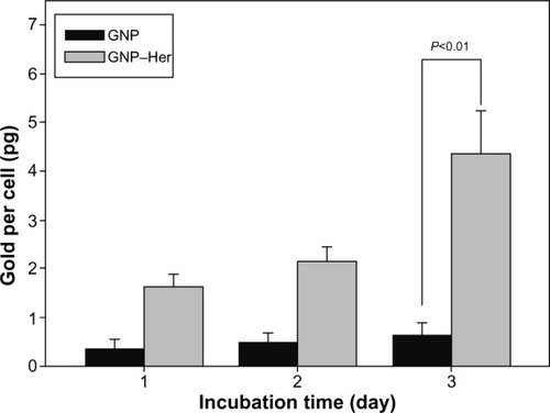
The quantity of GNP–Her taken up by SK-BR3 cells was estimated by ICP–MS and the outcomes are presented in . After herceptin conjugation, the NPs’ interaction with SK-BR cells was amplified significantly, in contrast to that of the original NPs, and increased to 1.4 pg per cell within the first day of culture, much higher than that of the original NPs.Citation33 The peak internalization of GNP–Her occurred at 4.6 pg per cell after 3 days of culture. The cause for the uptake enhancement might be ligand–receptor interactions on the cell membrane.Citation34 Moreover, the greater uptake of herceptin-modified GNPs indicates that the alteration not only enabled the NPs to target specific cells but also improved the cell internalization yield. Similar results obtained from (carboxymethyl)chitosan-modified superparamagnetic iron oxide nanoparticles were shown by Shi et alCitation35 in their reports; the quantity of NPs internalized in human muscle-derived cells is much more than that of superparamagnetic iron oxide nanoparticles, as observed by ICP–MS. The interaction between herceptin and SK-BR3 receptors expressed on the membrane might have contributed to the enhancement of GNP–Her internalization based on receptor-mediated endocytosis.Citation36
Figure 9 Amount of GNP (black bar) and GNP–Her (gray bar) taken up by SK-BR3 cells at different incubation times, as determined by ICP–MS.
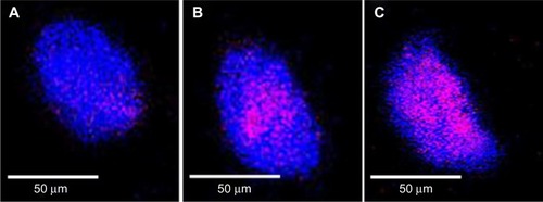
The internalization of GNP–Her into SK-BR3 cells was also confirmed by CLSM. shows that herceptin is an effective antibody, binding specifically to the HER2-bearing breast tumor cells. The internalization of GNP–Her into SK-BR3 cells was confirmed by CLSM to characterize the delivery of GNP–Her to the cytoplasm of the SK-BR3 cells. The fluorescence image was derived from the nucleus of the SK-BR3 cells (DAPI, blue) and the internalized GNP–Her (red). The cells were cultured in the presence of GNP–Her at various incubation times. Weak GNP–Her conjugates were observed in the fluorescence image (red color) after 1 hour () and slightly higher fluorescence was observed after 3 hours (). Intense fluorescence was noted after 6 hours (). On the other hand, the blue fluorescence derived from the nuclei stained with DAPI was strong after 1 hour of incubation, but it decreased with increasing incubation time and almost disappeared after 6 hours of incubation. In particular, the interaction of SK-BR3 with GNP–Her began after 1 hour of incubation and was accelerated and saturated after 6 hours. The CLSM images suggest that the NP-mediated delivery of monoclonal antibodies was achieved efficiently, resulting in cell death.Citation36 The mechanism of internalization involves endocytosis, followed by the release of herceptin-conjugated NPs to the cytoplasm.Citation29,Citation37 This suggests that the growth signal of SK-BR3 cells is withdrawn completely by the specific binding of herceptin to the receptor on SK-BR3 membrane, resulting in cell death.Citation35,Citation36
Figure 10 Fluorescence images of SK-BR3 cells.
Abbreviations: HER2, human epidermal growth factor receptor 2; DAPI, 4,6-diamidino-2-phenylindole dihydrochloride.
Conclusion
MSA-coated GNP was conjugated successfully with herceptin, which was confirmed by UV–vis and FT-IR spectroscopy techniques. The size of GNP determined by DLS was ~79 nm, whereas that of the GNP–Her was ~86 nm. GNP–Her had no cytotoxic effect on the control cells (FB) compared to the target cells (SK-BR3) and was internalized selectively into the target cells (SK-BR3); the free herceptin was delivered into the cytoplasm, which induced acute apoptosis. The GNP–Her NPs underwent endocytosis in breast cancer cells (SK-BR3) largely through a receptor-facilitated mechanism, whereby herceptin conjugated on the NPs targets the HER2 expressed on the membrane of the cancer cells.Citation29 Therefore, GNP–Her has potential use in the optical imaging and treatment of breast cancer.
Disclosure
The authors report no conflicts of interest in this work.
Acknowledgments
This work was supported by Institute of Biomedical Technologies, Auckland University of Technology, New Zealand. Dr Huh is grateful for the grant supported from the Research Foundation of Korea (NRF-2013R1A1A2A100062399) for technical support in cell culturing experiments.
References
- DavidMEChoiCHSeligsonDATolcherADordickJSAlabiCAEvidence of RNAi in humans from systemically administered siRNA via targeted nanoparticlesNature20104641067107020305636
- HanGYouCCKimBJForbesRTuringanRSMartinCTLight-regulated release of DNA and its delivery to nuclei by means of photolabile gold nanoparticlesAngew Chem Int Ed2006451931653169
- El-SayedIHHuangXEl-SayedMATolcherADordickJSAlabiCASelective laser photo-thermal therapy of epithelial carcinoma using anti-ER antibody conjugated gold nanoparticlesCancer Lett2006239112913516198049
- AllenTMLigand-targeted therapeutics in anticancer therapyAngew Chem Int Ed2002210705763
- ChaterjeeDKRufalhahAJZhangYTolcherAUpconversion fluorescence imaging of cells and small animals using lanthanide doped nanocrystalsBiomaterials200829793794318061257
- WangCSGold nanoclusters and graphene nanocomposites for drug delivery and imaging of cancer cellsAngew Chem Int Ed2011501164411648
- PanJFengSSTargeting and imaging cancer cells by folate-decorated, quantum dots (QDs) – loaded nanoparticles of biodegradable polymersBiomaterials2009301176118319062089
- JiangSGnanasammandhanMKZhangYOptical imaging-guided cancer therapy with fluorescentJ R Soc Interface20107131819759055
- Román-VelázquezCENoguezCGarzónILCircular dichroism simulated spectra of chiral gold nanoclusters: a dipole approximationJ Phys Chem B2003107441203512038
- PeyserLAVinsonAEBartkoAPDicksonRMPhotoactivated fluorescence from individual silver nanoclustersScience2001291550110310611141556
- YangYChenSSurface manipulation of the electronic energy of Sub nanometer-sized gold clusters: an electrochemical and spectroscopic investigationNano Lett2003317579
- ZhengJDicksonRMIndividual water-soluble dendrimer-encapsulated silver nanodot fluorescenceJ Am Chem Soc200212447139821398312440882
- HvkkinenHElectronic, and impurity-doping effects in nanoscale chemistry: supported gold nanoclustersAngew Chem Int Ed2003421112971300
- CrespoPPermanent magnetism, magnetic anisotropy, and hysteresis of Thiol-capped gold nanoparticlesPhys Rev Lett2004931872041872044
- BalabanRSNemotoSFinkelTMitochondria oxidants and agingCell2005120148349515734681
- LimCKChemiluminescence-generating nanoreactor formulation for near-infrared imaging of hydrogen peroxide and glucose level in vivoAdv Funct Mater2010201626442648
- MannervikBThe enzymes of glutathione metabolism: an overviewBiochem Soc Trans19871547177183315772
- ScottDToneyMMuzikarMHarnessing the mechanism of glutathione reductase for synthesis of active site bound metallic nanoparticles and electrical connection to electrodesJ Am Chem Soc2008130386587418166048
- McCarthyJRBhaumikJKarverMRSibel ErdemSWeisslederRTargeted nanoagents for the detection of cancersMol Oncol20104651152820851695
- LeeJFStovallGNPEllingtonADAptamer therapeutics advanceCurr Opin Chem Biol200610328228916621675
- LuWZhangGZhangRFloresLGHuangQGelovaniJGTumor site-specific silencing of NF-kappaB p65 by targeted hollow gold nanosphere-mediated photothermal transfectionCancer Res20107083177318820388791
- KarinMCaoYGretenFRLiZWNF-κB in cancer: from innocent by stander to major culpritNat Rev Cancer20022430131012001991
- LeeCMJeongHJKimEMKimDWLimSTKimHTSuperparamagnetic iron oxide nanoparticles as a dual imaging probe for targeting hepatocytes in vivoMagn Reson Imaging200962614401446
- AnshupAVenkatraamanJSChandramouliSPradeepTGrowth of gold nanoparticles in human cellsLangmuir20052125115621156716316080
- OgawaMReginoCASChoykePTIn vivo target-specific activatable near-infrared optical labeling of humanized monoclonal antibodiesMol Cancer Ther2009823223919139133
- XingZCParkMJHanSJChoiMJLeeBHKangIKIntracellular Uptake of magnetite nanoparticles conjugated with RGDS-peptideMacromol Res2011199897903
- LeeYKHongSMKimJSImJHParkSWEncapsulation of CdSe/Zns quantum dots in poly(ethyleneglycol)-poly(D,L-lactide) micelle for biomedical imaging and detectionMacromol Res2007154330336
- Kamuruzzaman SelimKMXingZCChoiMJChangYGuoHKangIKReduced cytotoxicity of insulin-immobilized CdS quantum dots using PEG as a spacerNanoscale Res Lett20116152853621943314
- LiJJiangFYangBSongXRLiYYangHTopological insulator bismuth selenide as a theranostic platform for simultaneous cancer imaging and therapySci Rep20133117
- BaeKHMokHParkTSynthesis characterization, and intracellular delivery of reducible heparin nanogels for apoptotic cell deathBiomaterials200829233376338318474396
- KooTBorahJSXingZCKangIKImmobilization of pamidronic acids on the nanotube surface of titanium discs and their interaction with bone cellsNanoscale Res Lett20138112413323497321
- GanJJiangSYangYImmobilization of homing peptide on magnetite nanoparticles and its specificity in vitroJ Bio Med Mater Res Part A2008271468476
- HongRHanGFernandezJKimMBForbesNSRotelloVMGlutathione-mediated delivery and release using monolayer protected nanoparticle carriersJ Am Chem Soc200612841078108916433515
- GomesPFGodoyMPFVelosoABMadhireiraJRExciton binding energy in type II quantum dotsPhys Status Solidi200742385388
- ShiZNeohKGKangETWangSCWangWCarboxymethyl chitosan-modified super magnetic iron oxide nanoparticles for magnetic resonance imaging of stem cellsACS Appl Mater Interfaces2009132833520353220
- HanSJPiersonRKangIKSpecific intracellular uptake of herceptin-conjugated CdSe/ZnS quantum dots into breast cancer cellsBio-Med Res Int201414954307954309
- GeelenTNicoloyKPaulisLMYeoSYStrijkersGJInternalization of paramagnetic phosphatidylserine-containing liposomes by macrophagesJ Biotechnol20121037111
- YamauraMCamiloRLSampaioLCPreparation and characterization of (3aminopropyl) triethosysilane-coated magnetite nanoparticlesJ Magn Magn Mater20041210217
- BaleSSKwonSJShahDABanerjeeADordickJSKaneRSNanoparticle-mediated cytoplasmic delivery of proteins to target cellular machineryJ Am Chem Soc20104314931500