
Abstract
Rumpel-Leede phenomenon is a rarely reported condition with an unknown prevalence. It is characterized by the acute development of non-blanchable purpuric macules resulting from dermal capillary rupture caused by compressive forces. We report a case of Rumpel-Leede phenomenon in both feet following the application of pneumatic compression in a 49-year-old woman who underwent en bloc spondylectomy for a giant cell tumor of the spine. The condition appeared after the application of pneumatic compression on both legs for venous thromboembolism prophylaxis, and the lesions spontaneously resolved after discontinuation of compression. Currently, most cases are reported in patients with a history of diabetes mellitus, hypertension, or thrombocytopenia. We report a case of Rumpel-Leede phenomenon in a patient without underlying medical conditions. In our patient, capillary fragility combined with increased intracapillary pressure was hypothesized as the underlying mechanism.
Introduction
Rumpel-Leede phenomenon is the acute development of non-blanchable purpuric macules resulting from dermal capillary rupture related to a proximal compressive force.Citation1 Its prevalence remains unknown, with rarely reported cases in the literature. The condition was first described in 1909 in a patient with scarlet fever who had petechiae on the arm distal to the tourniquet area for blood pressure monitoring.Citation1 The term was originally coined from the Rumpel-Leede capillary fragility test, also known as the tourniquet test, which is used for capillary fragility assessment.Citation2
The Rumpel-Leede phenomenon is commonly reported to occur after iatrogenic events, especially continuous blood pressure monitoring. Several predisposing factors, including thrombocytopenia, increased capillary fragility or permeability, diabetes mellitus, diabetes insipidus, hypertension, chronic renal failure, and use of antiplatelets or anticoagulants, have been demonstrated to be associated with this condition.Citation1–19 Here, we present the Rumpel-Leede phenomenon following prolonged use of pneumatic compression in a patient without underlying diseases who underwent en bloc spondylectomy for a giant cell tumor of the spine and developed sepsis.
Case Presentation
A 49-year-old woman diagnosed with a giant cell tumor of the spine underwent en bloc spondylectomy at the T2 level in a university-based hospital. She had no underlying medical conditions. After the operation, a pneumatic compression device, Kendall SCD 700 Series® (Covidien, Mansfield, MA, USA), was continuously applied to both legs for venous thromboembolism prophylaxis. Three weeks later, the patient developed high-grade fever (body temperature 38.5℃), tachycardia (pulse rate 140 bpm), and hypotension (blood pressure 90/60 mmHg). Laboratory results showed leukocytosis with predominant neutrophils [white blood cell (WBC) count 18,250/mm3 (neutrophils 90.3%), platelets 297,000/mm3]. Urinary analysis revealed the presence of numerous WBC. Hemoculture and urine culture revealed Escherichia coli (E. coli). The patient was diagnosed with acute pyelonephritis with E. coli septicemia, and intravenous ceftriaxone was prescribed.
Several hours after the onset of fever and hypotension, the patient developed an erythematous rash on both dorsal aspects of the feet distal to the pneumatic compression device. The rash had a sudden onset and was asymptomatic. The patient had no bleeding in other areas of the body. Dermatologic consultation was conducted one day after the onset of the rash. Physical examination revealed multiple non-blanchable erythematous macules that coalesced into patches on both dorsa of the feet (). There were no splinter hemorrhages or fingertip lesions.
Figure 1 Multiple non-blanchable erythematous macules and patches on both dorsa of feet.
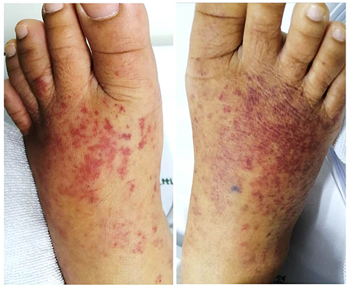
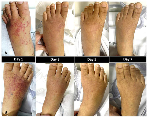
In our patient, small-vessel vasculitis, pigmented purpuric dermatosis (PPD), and sepsis-induced thrombocytopenia were included in the differential diagnosis. Small vessel vasculitis and PPD are often located on the lower extremities and show gradual onset. Small vessel vasculitis clinically presents with palpable purpura, whereas PPD demonstrates non-palpable purpura as in our patient. However, lesions of PPD frequently manifest with petechiae mixed with yellow-brown patches.Citation20 Sepsis-induced thrombocytopenia commonly manifests as generalized petechiae and purpura. Owing to the lesions located distal to the pressure area, lack of inflammatory signs, abrupt onset, and normal platelet counts, the Rumpel-Leede phenomenon was provisionally diagnosed. The pneumatic compressive device was then discontinued. The lesions gradually improved within one week ().
Figure 2 The improvement of the rash on left foot (A) and right foot (B) from day 1 to day 7.
Discussion
The Rumpel-Leede phenomenon is an acute capillary rupture that commonly occurs after non-invasive blood pressure monitoring or tourniquet application. It is characterized by a sudden onset, asymptomatic erythematous to purpuric rash distal to the compressive device, and is associated with predisposing factors of capillary fragility. Its prevalence is still unknown; however, it is considered a rarely reported condition owing to underrecognition.
The Rumpel-Leede phenomenon has been previously described under various conditions. The majority of cases were women, all of whom demonstrated a non-blanchable, petechial, or purpuric rash, distributed distally to the device band or cuff. Most cases resolved spontaneously within two weeks without any specific treatment. The sphygmomanometer was the most commonly used device.Citation15–19 Others included the tourniquet,Citation8 radial arterial wristband,Citation12 peritoneal insufflation during laparoscopic abdominal surgery,Citation13 knee stocking,Citation21 cloth material,Citation22 and car seat.Citation23 Regarding comorbidities, diabetes mellitus, hypertension, and thrombocytopenia are frequently reported.Citation24 All cases showed spontaneously improved after discontinuing the causative device, and there were no long-term sequelae.
The diagnosis of Rumpel-Leede phenomenon is based on patient history, clinical findings, and a self-limiting course after removal of the causative device. Recognition of this condition could help avoid unnecessary investigations and invasive diagnostic procedures. However, there is no specific treatment for this phenomenon. Cessation of the causative device, along with reassurance of the patient, are recommended. We discontinued the pneumatic compressive device in our patient, and the lesion resolved within one week.
Regarding the function of the pneumatic compression device applied in our patient, the pressure was gradually increased by pneumatic inflation to reach a pressure of 45 mmHg in 11s, and then was deflated automatically to allow for blood refilling. The next cycle was initiated when adequate blood refilling was detected by the device sensor. In our patient, the device was used continuously to increase venous blood flow. Currently, there is no previous report of Rumpel-Leede phenomenon following the application of a pneumatic compressive device, possibly owing to the low-pressure force generated by the device.
In our case, the patient had a history of sepsis without thrombocytopenia or pneumatic compression preceding the onset of petechiae. We hypothesized that the pathogenesis of the Rumpel-Leede phenomenon was dermal capillary rupture due to an increase in intracapillary pressure in association with the inflation of a pneumatic cuff after a pneumatic compression device, combined with endothelial dysfunction from sepsis, which could be a risk factor for increased capillary fragility and microangiopathy.Citation25,Citation26 Another explanation is that occurrence of hypotension had led to the inflation pressure of pneumatic compression being higher than venous pressure, resulting in increased intraluminal pressure and subsequent capillary rupture, although the device generated low pressure. To the best of our knowledge, this is the first report of the Rumpel-Leede phenomenon following the application of pneumatic compression.
Conclusion
The Rumpel-Leede phenomenon is an acute capillary rupture commonly observed in the area distal to pressure. This phenomenon is associated with capillary fragility and thrombocytopenia. Our report highlights the importance of recognizing this condition to make a prompt diagnosis, and also reveals its occurrence in a patient with no overt capillary fragility and normal platelet counts, despite the pneumatic compression device being considered safe and low-pressure.
Ethics Approval and Consent to Participate
This article was performed in accordance with the principles of Declaration of Helsinki. Ethical review and approval was not required to publish the case details in accordance with the local legislation and institutional requirements. Written informed consent was obtained from the patient for publication of this case report and any accompanying images as per our standard institutional rules.
Disclosure
The authors declare that this manuscript was prepared in the absence of any commercial or financial relationships that could be construed as a potential conflict of interest.
Additional information
Funding
References
- Wang K, Lee J. Images in clinical medicine. Rumpel-Leede sign. N Engl J Med. 2014;370(1):e1. doi:10.1056/NEJMicm1305270
- Chester MW, Barwise JA, Holzman MD, Pandharipande P. Acute dermal capillary rupture associated with noninvasive blood pressure monitoring. J Clin Anesth. 2007;19(6):473–475. doi:10.1016/j.jclinane.2006.12.010
- Jeon YS, Kim YS, Lee JA, Seo KH, In JH. Rumpel-Leede phenomenon associated with noninvasive blood pressure monitoring - a case report. Korean J Anesthesiol. 2010;59(3):203–205. doi:10.4097/kjae.2010.59.3.203
- Balamurugesan K, Viswanathan S. Rumpel-leede phenomenon in a hypertensive lady on amlodipine. J Clin Diagn Res. 2014;8(4):YD01–02. doi:10.7860/JCDR/2014/6574.4301
- Leerunyakul K, Suchonwanit P. Asian hair: a review of structures, properties, and distinctive disorders. Clin Cosmet Investig Dermatol. 2020;13:309–318. doi:10.2147/CCID.S247390
- Hartley A, Lim PB, Hayat SA. Rumpel-Leede phenomenon in a hypertensive patient due to mechanical trauma: a case report. J Med Case Rep. 2016;10(1):150. doi:10.1186/s13256-016-0950-3
- Sriphojanart T, Khunkhet S, Suchonwanit P. A retrospective comparative study of the efficacy and safety of two regimens of diphenylcyclopropenone in the treatment of recalcitrant alopecia areata. Dermatol Rep. 2017;9(2):7399. doi:10.4081/dr.2017.7399
- Lambdin JT, Mackenzie C. Rumpel-Leede phenomenon in a patient with adult-onset Still’s disease. BMJ Case Rep. 2017;2017. doi:10.1136/bcr-2016-217290
- Khoury Abdulla R, Safian RD. Rumpel-leede phenomenon after radial artery catheterization. Circ Cardiovasc Interv. 2018;11(4):e006507. doi:10.1161/CIRCINTERVENTIONS.118.006507
- Iamsumang W, Leerunyakul K, Suchonwanit P. Finasteride and its potential for the treatment of female pattern hair loss: evidence to date. Drug Des Devel Ther. 2020;14:951–959. doi:10.2147/DDDT.S240615
- van Rhijn BD, Vlachojannis GJ, Balak DMW. Petechial purpuric rash after non-invasive blood pressure measurement: Rumpel-Leede sign. BMJ Case Rep. 2019;12(10):e231541. doi:10.1136/bcr-2019-231541
- Malik J. Rumpel-Leede phenomenon after right radial catheterization procedure. Anatol J Cardiol. 2021;25(9):E33. doi:10.5152/AnatolJCardiol.2021.117
- Feeley TBM, Connolly LR. Facial petechiae following laparoscopic surgery: a case report of Rumpel-leede phenomenon. A a Pract. 2021;15(10):e01533. doi:10.1213/XAA.0000000000001533
- Suchonwanit P, Udompanich S, Thadanipon K, Chanprapaph K. Trichoscopic signs in systemic lupus erythematosus: a comparative study with 109 patients and 305 healthy controls. J Eur Acad Dermatol Venereol. 2019;33(4):774–780. doi:10.1111/jdv.15421
- Hsiao PJ, Liaw FY, Chan JS, Chen JS. Rumpel-Leede phenomenon in a diabetes mellitus patient. QJM. 2015;108(8):671–672. doi:10.1093/qjmed/hcv024
- Lee S, Jang YB, Kang KP, Lee TH, Kim W, Park SK. Rumpel-Leede phenomenon in a hemodialysis patient. Kidney Int. 2010;78(2):224. doi:10.1038/ki.2010.91
- Pritchard AE, Lockhart EL. Rumpel-Leede phenomenon in a patient being treated with prasugrel. Transfusion. 2017;57(7):1642. doi:10.1111/trf.13962
- Ramme AJ, Gales J, Stevens N, Verma V, Egol K. Rumpel-Leede phenomenon in a patient with laboratory markers positive for sjogren disease. A a Case Rep. 2016;6(11):352–354. doi:10.1213/XAA.0000000000000309
- Leerunyakul K, Suchonwanit P. Evaluation of hair density and hair diameter in the adult Thai population using quantitative trichoscopic analysis. Biomed Res Int. 2020;2020:2476890. doi:10.1155/2020/2476890
- Spigariolo CB, Giacalone S, Nazzaro G. Pigmented purpuric dermatoses: a complete narrative review. J Clin Med. 2021;10(11):2283. doi:10.3390/jcm10112283
- Bossory Goike L, Dean S. Images in vascular medicine. Rumpel-Leede phenomenon in a patient with chronic lymphocytic leukemia treated with acalabrutinib. Vasc Med. 2020;25(3):281–282. doi:10.1177/1358863X19901166
- Nguyen TA, Garcia D, Wang AS, Friedlander SF, Krakowski AC. Rumpel-Leede phenomenon associated with tourniquet-like forces of baby carriers in otherwise healthy infants: baby carrier purpura. JAMA Dermatol. 2016;152(6):728–730. doi:10.1001/jamadermatol.2015.6270
- Ho VPY, Chong JH, Bishnoi P, Koh MJA, Wong SMY. Rumpel-Leede Phenomenon - an alarming rash with benign etiology in 4 healthy infants. Pediatr Dermatol. 2020;37(1):150–152. doi:10.1111/pde.14010
- Varela D, Tran D, Ngamdu KS, Trullender B, Mukherjee D, Abbas A. Rumpel-Leede phenomenon presenting as a hypertensive urgency. Proc. 2016;29(2):200–201.
- Suchonwanit P, McMichael AJ. Alopecia in association with malignancy: a review. Am J Clin Dermatol. 2018;19(6):853–865. doi:10.1007/s40257-018-0378-1
- Opal SM, van der Poll T. Endothelial barrier dysfunction in septic shock. J Intern Med. 2015;277(3):277–293. doi:10.1111/joim.12331