Abstract
Acute hemorrhagic edema of infancy (AHEI) is a benign type of leukocytoclastic vasculitis. It is a benign phenomenon although it manifests with fever, large palpable purpuric skin lesions, and edema. The presentation of AHEI can often resemble that of Henoch-Schönlein purpura. Since AHEI is a self-limited disease, conservative management is the most commonly followed approach. Our patient had complete resolution of AHIE without medical treatment.
Keywords:
Introduction
Acute hemorrhagic edema of infancy (AHEI) is a benign type of leukocytoclastic vasculitis. It has also been described as Finkelstein’s disease, Seidlmayer’s disease, and postinfectious cockade purpura.Citation1 AHEI is considered a benign phenomenon and it usually resolves spontaneously in a few weeks.Citation2 We are presenting a 14 month old male with AHEI who presented with rash on the face and extremities, as well as edema in the left lower extremity.
Case report
A 14 month old boy presented to our emergency department with intermittent low grade fever of 2 days duration associated with a rash, swelling of the left leg, and an inability to bear weight. The mother stated that the patient had a runny nose and a mild cough 1 week prior to admission.
There was no diarrhea, change in behavior, toxic ingestion, medication use, or any recent vaccinations. Past medical history and family history were unremarkable and immunizations were up to date. The patient was admitted to the ward for fever and rash. On admission, his temperature was 38°C, he had a respiratory rate of 25 breaths per minute, his heart rate was 110 beats per minute, and his blood pressure was 100/70 mmHg.
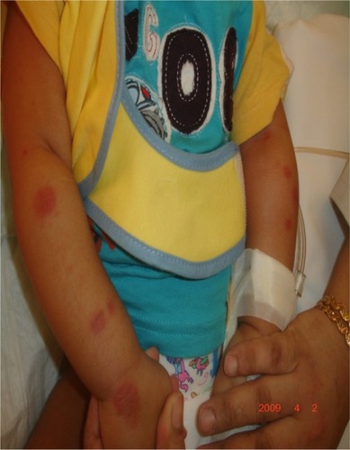
On examination, the patient was not looking toxic, his neck was supple, and the neurological exam was unremarkable. The skin showed ecchymotic, purpuric, targetoid plaques with oval and round shapes on the face and extremities (–). The right auricle of the ear was mildly edematous and purple in color. There was mild swelling on the hands and lower extremities without skin induration and the range of motion of all joints was normal. The rest of the physical exam was unremarkable. The complete blood count and urine analysis were both unremarkable.
Figure 1 Purpuric and ecchymotic lesions on the left lower extremity.
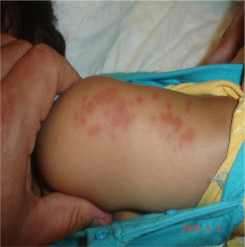
Figure 2 Oval, purpuric, and targetoid lesion on the right lower extremity.
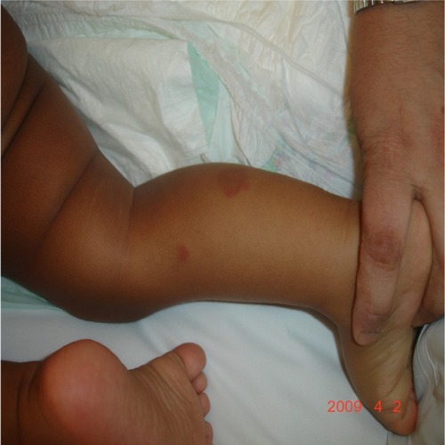
Figure 3 Circular and oval plagues on the upper extremities with swelling more prominent on the right side.
A decision was made to biopsy one of the lesions from the left forearm and it showed inflammatory infiltrate surrounding dermal blood vessels. The infiltrate was a mixture of neutrophils, lymphocytes, and scattered eosinophils with fragmented nuclei of neutrophils, and there was focal necrosis of the vessel wall. On direct immunofluorescence there was no immunoglobulin A (IgA) deposition. The patient was discharged after the diagnosis and the rash resolved spontaneously in 3 weeks.
Discussion
AHEI is more common in males,Citation2 without racial predominance.Citation3 The age group is typically between 4 and 24 months.Citation1 In the majority of the cases, AHEI is a self-limited disease (1–3 weeks), but there have been reports of recurrent attacks (up to four attacks).Citation4
Patients with AHEI are usually nontoxic on presentationCitation2 and they classically present with fever, large palpable purpuric skin lesions, and edema. The skin changes are characterized as well demarcated, annular, medallion-like, rosette shaped purpuric plaques, and tend to appear suddenly in the face and extremities.Citation1 However, the skin lesion does not always present in a typical way and it has been reported that it can appear as hemorrhagic blisters.Citation3 The edema is typically on the face, extremities, and auricles.Citation2
Many organisms including adenovirus, vermicelli zoster virus, cytomegalovirus, herpes simplex virus, tuberculosis, streptococci, and staphylococci are associated with AHEI.Citation4 Almost 75% of cases of AHEI are preceded by infections like otitis media, urinary tract infection, upper respiratory infection, and pneumonia. An immune complex hypersensitivity complex has been suggested as the etiology of AHEI since vaccines and medications are trigger factors.Citation2
Extracutaneous and viscera involvement are rare with AHEI. However, Fiore et al documented two cases with intestinal engagementCitation5 and Wanatabe and Sato reported a case with renal involvement and hypocomplementemia.Citation6
Blood laboratory results are usually nonspecific. In a complete blood count there might be thrombocytosis, leukocytosis with eosinophilia, or lymphocytosis. In addition, C-reactive protein and the erythrocyte sedimentation rate may be high.Citation7 In some cases, patients with AHEI can present with hypocomplementemia affecting complement C4, complement C1q, and complement CH50,Citation8 and transient abnormal liver function.Citation9
A skin biopsy for histopathology and immunofluorescence testing is useful if diagnosis is unclear.Citation10 The typical description of the histology in the patient with AHEI is perivascular neutrophilic infiltration with several nuclear fragments in the vascular wall, resulting in fibrinoid necrosis.Citation7 The rare serious complications of AHEI are renal involvement, gastrointestinal bleeding, epididymo-orchitis, and cartilage damage.Citation11
Since AHEI is a self-limited disease, conservative management is the most commonly followed approach.Citation2 Steroid use is warranted when there is a severe or persistent gastrointestinal symptom or complication of the disease. The use of immunosuppressive medications like cyclophosphamide or azathioprine to prevent renal damage has been reported as futile.Citation10 Antibiotics can be used if there is a concomitant bacterial infection.Citation7
AHEI can present as erythema multiforme, but the latter does not show leukocytoclastic vasculitis. AHEI can also mimic urticarial vasculitis, but the former usually has an acute presentation with rare remissions.Citation8 The presentation of AHEI can often resemble that of Henoch-Schönlein purpura (HSP) and some authors consider AHEI as a cutaneous variant of HSP.Citation1 It is crucial to differentiate between AHEI and HSP since the latter requires systemic corticosteroid therapy. AHEI and HSP are similar in that both have skin lesions and leukocytoclastic vasculitis. However, the differences are as follows. AHEI presents in a younger population (3 to 24 months old) in comparison to HSP (3 to 6 years old).Citation11 The skin lesions in AHEI manifest as ecchymotic changes with large purpura and edema involving the face, extremities and scrotum, while the purpura in HSP is usually smaller and involves the buttocks and the legs.Citation12 Involvement of renal, gastrointestinal, and viscera are rare in AHEI when compared to HSP. IgA levels are usually normal in AHEI, but elevated in HSP.Citation11 There is usually no IgA deposition in AHEI compared to HSP. Finally, complement C1q deposition is more diagnostic of AHEI.Citation2
Conclusion
AHEI is a self-limited disease that usually resolves spontaneously. It is crucial to make the diagnosis correctly to avoid unnecessary work up and therapy, while simultaneously monitoring for rare but severe complications.
Disclosure
The authors report no conflicts of interest in this work.
References
- JindalSRKuraMMAcute hemorrhagic edema of infancy-a rare entityIndian Dermatol Online J20134210610823741666
- SavinoFLupicaMMTarascoVAcute hemorrhagic edema of infancy: a troubling cutaneous presentation with a self-limiting coursePediatr Dermatol [Epub ahead of print]
- KarremannMJordanAJBellNWitschMDürkenMAcute hemorrhagic edema of infancy: report of 4 cases and review of the current literatureClin Pediatr (Phila)200948332332618772356
- FotisLNikorelouSLariouMSDelisDStamoyannouLAcute hemorrhagic edema of infancy: a frightening but benign diseaseClin Pediatr (Phila)201251439139321357201
- FioreERizziMRagazziMAcute hemorrhagic edema of young children (cockade purpura and edema): a case series and systematic reviewJ Am Acad Dermatol20085968469518656284
- WatanabeTSatoYRenal involvement and hypocomplementemia in a patient with acute hemorrhagic edema of infancyPediatr Nephrol2007221979198117704952
- FreitasPBygumAVisual impairment caused by periorbital edema in an infant with acute hemorrhagic edema of infancyPediatr Dermatol
- BansalSGhateSJerajaniHRSudden onset purpura in a healthy infant: acute hemorrhagic edema of infancyIndian J Dermatol201156334935121772612
- ObeidMHaleyJCrewsJParhizgarRJohnsonLCampTAcute hemorrhagic edema of infancy with abdominal pain and elevated transaminasesPediatr Dermatol200825664064119067875
- YuJEManciniAJMillerMLIntussusception in an Infant with acute hemorrhagic edema of infancyPediatr Dermatol200724616417300653
- MoradinejadMHEntezariPMahjoubFZiaeeVAcute hemorrhagic edema of infancy; a report of five Iranian infants and review of the literatureIran J Pediatr201121110711223056774
- JavidiZMalekiMMashayekhiVTayebi-MaybodiNNahidiYAcute hemorrhagic edema of infancyArch Iran Med200811110310618154430