
 ?Mathematical formulae have been encoded as MathML and are displayed in this HTML version using MathJax in order to improve their display. Uncheck the box to turn MathJax off. This feature requires Javascript. Click on a formula to zoom.
Abstract
Purpose
To determine and compare the shear bond strength (SBS) of bracket-bonding system cured with light-emitting diode (LED) and halogen-based light-curing unit at various polymerization times.
Materials and methods
Ninety six human maxillary premolar teeth extracted for orthodontic purpose were divided into four groups, according to the light-curing unit and exposure times used. In the halogen group, the specimens were light cured for 20 and 40 seconds. In the LED group, the specimens were light cured for 5 and 10 seconds. Stainless steel brackets were bonded with Enlight bonding system, stored in distilled water at 37°C for 24 hours and then submitted to SBS testing in a universal testing machine at a crosshead speed of 0.5 mm/minute. Adhesive remnant index (ARI) was used to evaluate the amount of adhesive remaining on the teeth determined by stereomicroscope at 10× magnification.
Results
The highest mean SBS was obtained with the halogen 40 seconds (18.27 MPa) followed by halogen 20 seconds (15.36 MPa), LED 10 seconds (14.60 MPa) and least with LED 5 seconds (12.49 MPa) group. According to analysis of variance (ANOVA) and Tukey’s multiple-comparison test, SBS of halogen 20 seconds group was not significantly different from halogen 40 seconds group, LED 5 seconds group and LED 10 seconds group, whereas halogen 40 seconds group was significantly different from LED 5 seconds and LED 10 seconds group. The method of light curing did not influence the ARI, with score 2 being predominant.
Conclusion
Polymerization with both halogen and LED resulted in SBS values that were clinically acceptable for orthodontic treatment in all groups. Hence, for bonding orthodontic brackets, photoactivation with halogen for 20 seconds and LED for 5 seconds is suggested.
Introduction
Orthodontic appliances may be attached by cementing bands or by bonding brackets directly to the enamel surface using a retentive base. The development of firm attachment of brackets directly to the teeth is one of the prominent milestones in the orthodontic field as it enables the efficient movement of the teeth. Firm attachment was initially achieved by cementing bands on all the teeth.
Direct bonding of brackets using acid etching has become contemporary in orthodontics fields.Citation1 This was a revolution in the practice of clinical orthodontics, and since then, there has been a rapid rate of product development in terms of adhesives, brackets and their technical properties.
Orthodontic bonding to the enamel surface leads to significant improvement in treatment by increasing patient comfort, better esthetics and decreasing periodontal problems.Citation2–Citation4
The advent of light cure adhesives in 1979 by Tavas and WattsCitation5 became popular because of their various advantages compared with self-cured adhesive materials.
These advantages are application of a single paste, control of working time, easy removal of excess bonding material, reduced risk of contamination and immediate insertion of the arch wire,Citation6,Citation7 as they provide increased working time for precise bracket placement and ease of manipulation. Currently, four different technologies are available for curing of dental composites by light that are halogen lamps, lasers, plasma arc lamps and light-emitting diode (LED).
Halogen lamps were the first to be introduced as the source of light. Though it was a boon for the clinical achievements in the dental practice in the initial stages it had many demerits. Quick overheating of the filament of the halogen bulbs made its use restricted for lengthy procedures, especially in orthodontics.
Mills et alCitation8 in 1995 presented the solid-state LED. Since then, LED has attracted increasing attention as a new source for light-activated polymerization. Many light-curing devices have become popular and it is important to determine the light-curing unit that is most efficient as well as give the desired bond strength. Various studies have been carried out to evaluate the bond strength of bracket-bonding systems using shear testing.Citation9–Citation17
The objective of this study was to determine and compare the shear bond strength (SBS) of the bracket-bonding system cured with LED and halogen-based light-curing unit at various polymerization times.
Materials and methods
In this study, 96 human bicuspid teeth extracted for orthodontic purpose were obtained. The sampling method used was convenient sampling. Extracted teeth were collected from Oral and Maxillofacial Surgery Unit, Tribhuvan University Teaching Hospital, Kathmandu, and stored in distilled water at room temperature to eliminate dehydration and bacterial growth.Citation18,Citation19 All patients had provided written informed consent at the time of treatment that their extracted teeth may be used for future research purposes.
Inclusion criteria were extracted teeth with intact buccal enamel and teeth extracted not more than 3 months until testing, while teeth subjected to caries and any pretreatment chemical agent, and that had cracks caused by the pressure of extraction forceps, which is visualized by naked eye, and developmental defects such as hypoplastic enamel were excluded. This study was approved by the Institutional Review Board, Institute of Medicine, Kathmandu.
The teeth were then mounted on an acrylic block in such a way that the roots were embedded completely into the acrylic up to cementoenamel junction and leaving the crown exposed. The labial surface of the tooth was kept perpendicular to the bottom surface of mold.Citation20 Cuts were made on both sides of the acrylic block with acrylic trimmer for the purpose of securing it in universal testing machine at the time of debonding.
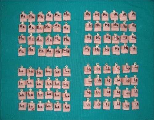
The mounted specimens were randomly divided into four groups (two halogen and two LED groups) of 24 in each according to the exposure time and the type of light unit and named accordingly as halogen 20 seconds (Ha), halogen 40 seconds (Hb), LED 5 seconds (La) and LED 10 seconds (Lb) groups.Citation21 All specimens were immersed in distilled water except at the time of bonding and bebonding procedures.
Before bonding, the buccal surfaces of all the teeth were cleaned with fluoride-free fine pumice powder in water using a brush at slow-speed micromotor hand piece for 10 seconds for removal of any dirt\calculus\deposits or stains. The teeth were then rinsed thoroughly with water for 10 secondsCitation23,Citation24 and dried with oil- and moisture-free compressed air. The buccal surface of each tooth was etched for 30 seconds with 37% phosphoric acid in gel form.Citation25 Each tooth was then rinsed with a distilled water spray for 5 seconds and dried with oil-free air till the etched tooth appeared chalky white.
A thin coat of light-cured adhesive primer Orthosol (Enlight, Ormco Corp., USA) was applied to acid-etched enamel. Enlight composite resin was applied on the 0.022″ slot roth stainless steel double mesh premolar bracket base (Minidiagonale, Leone Co., Sesto, Florentine, Florence, Italy) having a surface area of 8.8 mm2, which was then placed on the teeth with a holding pincer near the center of the buccal surfaces with sufficient manual pressure that lead the excess material to flow at the margins of the bracket, which was then removed with an exploratory probe before polymerization.
Conventional halogen light-curing system and LED light-curing system for curing orthodontic bracket adhesive were used (). The light intensity was monitored with radiometers (CM300-2000, APOZA, Taiwan; ).
Table 1 Characteristics of the halogen and LED units
In the Ha group, light was placed only in an occlusal direction for 20 seconds, while in the Hb group, light was placed both in mesial and distal directions for 20 seconds each.
In the La group, light was placed only in an occlusal direction for 5 seconds, while in the Lb group, light was placed both in mesial and distal directions for 5 seconds each.
Light curing was performed by maintaining the curing tip as close to the bracket as possible with the formation of an angle of 45° with the buccal surface of the tooth.
The specimens () were then stored in distilled water at 37°C for 24 hoursCitation26 and submitted to the SBS testing in a universal testing machine (AG-IC/100 KN, Shimadzu, Japan).
Figure 1 All samples of four experimental groups.
Before debonding, the specimen mounted in its acrylic block with cuts on both side was secured to the lower grip of the machine such that the buccal surface of the tooth with bonded bracket was parallel to the plunger that was attached to the moving crosshead of the universal testing machine ().
Figure 2 Close view of crosshead of universal testing machine with sample in situ.
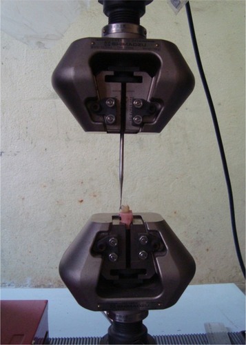
A chisel-edge-shaped plunger was attached in the movable crosshead of the universal testing machine and placed in such a way that the leading edge aimed the enamel–adhesive interface that moves at a crosshead speed of 0.5 mm/min. Load was applied occlusogingivally to the bracket, which produced a shear force at the bracket–tooth interface. A computer connected with the testing machine showed the results of each test that were recorded.
The force required to dislodge the brackets was measured in Newton, and the SBS (MPa) was determined by dividing the force by the bracket base area (8.8 mm2).
After bond strength testing, all specimens were collected and visually examined with a stereomicroscope (Olympus SZX12, Olympus Corp., Japan) at 10× magnification to assess the adhesive remnant index (ARI)Citation21 available at Nepal Agricultural Research Council, Khumaltar, Kathmandu. The ARI was used to evaluate the amount of resin remaining on the tooth after debonding. The criteria for ARI scoring given by Artun and BerglandCitation28 were used in this study ().
Table 2 Grading of ARI
The data were processed and analyzed by using the SPSS software version 16.0 (SPSS Inc., Chicago, IL, USA). Data were then subjected to analysis of variance (ANOVA) to identify differences in various groups. Tukey’s post hoc multiple-comparison test was used to identify where differences occurred.
Kappa test was performed for method error calculation. Chi-square test was performed to determine the differences in ARI scores of all four groups. A p-value of <0.05 was considered to be significant with a 95% confidence limit.
Results
SBS values including mean and SD obtained for the four groups are shown in .
Table 3 Shear bond strength mean values (MPa) in the experimental groups
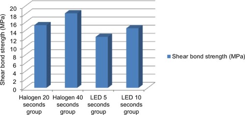
The highest mean SBS was obtained with the Hb group (18.27 MPa) followed by Ha (15.36 MPa), Lb (14.60 MPa) and least with La (12.49 MPa) groups.
The graphical representation of mean SBS values is shown in .
Figure 3 Shear bond strength mean values (MPa) in the experimental groups.
ANOVA was used to compare the mean values of SBS obtained in each group. The test showed that the difference in the mean values of SBS was statistically significant (p=0.001).
Tukey’s post hoc multiple-comparison test was used for intergroup comparisons. The test showed that the SBS of Ha group was not significantly different from Hb, La and Lb groups. La group was not significantly different from Lb group, whereas Hb group was significantly different from La and Lb groups.
Adhesive index scores based on the amount of resin left on the tooth after debonding of the four groups are shown in .
Table 4 ARI scores in the experimental groups
The standard error of method, as calculated using kappa test, was 0.933, which shows the good intraobserver reliability.
The chi-square test detected no statistically significant difference in the ARI scores of all the four groups (p=0.065), which means that the method of light curing did not influence the ARI, with the score 2 being predominant.
Discussion
Among different technologies for light curing, halogen lamps and LED are the most commonly used ones. Light power is the important factor that contributes to the level of polymerization. According to Rueggeberg,Citation29 higher the light power, greater the number of photons reaching the resin and in turn higher number of free radicals available for polymerization. Halogen unit of mean power of 620 mW/cm2 and the LED unit of mean power of 800 mW/cm2 were used in this study.
As during tooth formation there is rapid development of bovine enamel and dentin, bovine enamel has larger crystal grains and more lattice defects compared to human enamel.Citation30 It was reported that bond strength values of permanent bovine enamel arê35% and 44% below that of the human enamel.Citation31 Though bovine teeth can be used as a substitute for human teeth, the latter is the best substrate for bonding.Citation32
Bond strength studies mostly used distilled water for storing the testing specimens.Citation18,Citation19,Citation33 To standardize the shear bond test protocols, Fox et alCitation34 recommended distilled water as a prebonding storage media in the shear bond studies.
Studies regarding the effect of the storage media on dentin bond strength have found that distilled water storage did not adversely affect the bond strength of the teeth stored for up to 6 months.Citation35,Citation36 In vitro bond strength testing comprise shear, tensile and torsion tests. As failure of the bracket-tooth adhesion is assumed not to be a result of pure tensile and torsional stress, these tests are less often performed, while shear testing is most popular as there is similarity with the clinical debonding situation.Citation37
A methodologic review of SBS studies revealed that crosshead speeds of 0.5 and 1 mm/min are often used in SBS tests.Citation38
Previous studies have shown that LED-curing units are as effective as halogen-curing units. According to Dunn and Taloumis,Citation24 there was no significant difference in bond strength with LED or halogen-based light-curing units.
In this study, the highest mean SBS was obtained with the Hb (18.27 MPa) followed by Ha (15.36 MPa), Lb (14.60 MPa) and least with La (12.49 MPa) groups.
All LED groups presented lower mean SBS value compared with the halogen groups. Statistically, mean SBS of Ha group was not significantly different from Hb, La and Lb groups. La group was not significantly different from Lb group, whereas Hb group was significantly different from La and Lb groups.
SBS of Hb group of this study was higher than that in the study done by Dunn and Taloumis,Citation24 Abtahi and Khamverdy,Citation39 Banerjee S and Banerjee RCitation25 and Balasubramanian et alCitation20 while the study of Turkkahraman and KucukesmenCitation22 and Cerekja and CakirerCitation18 showed similar results.
SBS of Ha group of this study was higher than that in the study by Hui-Ping et al.Citation19
SBS of Hb and Lb groups of this study were higher than those in the study done by Usumez et alCitation15 whereas study by Dall’Igna et alCitation21 showed the similar results with Lb while higher with La group when compared with this study.
In this study, La group, with a shorter exposure time, showed the lowest mean SBS value, which was adequate bond strength for majority of the clinical situations, as values of 5.9–7.8 MPa proposed by Reynolds and von FraunhoferCitation3. According to Lopez,Citation40 the SBS recommended for successful clinical bonding was estimated to be 7 MPa.
Results of this study are in accordance with the study of Mavropoulos et alCitation41 and Gronberg et al,Citation16 that is, a minimum time of 5 seconds resulted in SBS values that are sufficient to resist the forces exerted in a clinical situation, while study by Yu et alCitation17 showed divergent results as light curing with an LED for 4 and 6 seconds showed lower SBS values; when cured for 8 seconds, then only the values were considered to be satisfactory.
The results of our study showed that as light-curing time increased for both light units, there was a gradual increase in mean SBS. Peutzfeldt and AsmussenCitation42 study also is in accordance with this study. Various authors have also reported the similar findings.Citation15–Citation17,Citation41,Citation43
There was no significant difference in ARI scores among the groups, which is supported by the literature,Citation41,Citation44,Citation45 with the predominant score being 2. It means that failures occurred mostly at the bracket–adhesive interface after debonding, with the material remaining adhered to the tooth surface that allows removal of composite resin and preservation of the enamel from possible trauma.Citation17,Citation46,Citation47
The results of our study showed that the light unit does not affect the location of orthodontic bond failures because the majority of these occurred at the bracket–composite resin interface (scores 2 and 3) with both light units. According to Gronberg et alCitation16 and Yu et al,Citation17 this failure location could indicate incomplete resin polymerization at the base of bracket as a result of the short period of light exposure. This diminishes the probability of damage to the enamel during bracket debonding, which is advantageous in the orthodontic treatment.
One limitation of this study was that being an ex vivo study, it cannot completely reproduce the complex interaction processes that occur in the oral cavity.Citation48,Citation49 In a contest of SBS measurement, several factors can influence the results. This in vitro study fails to simulate factors such as intraoral aging of resin composites, PH and temperature fluctuation based on individual’s dietary intake and oral hygiene, complex cyclic loading, microbial attack and enzymatic degradation. Pickett et alCitation50 and Murray and HobsonCitation51 found that as the biodegradation that occurs in the oral cavity, bond strength values in vivo tend to be lower than those found ex vivo.
Another limitation was the use of a constant crosshead speed of 0.5 mm/minute during debonding in a universal testing machine. According to Eliades and Brantley,Citation52 debonding in vivo occurs at a higher speed though this load speed is generally used.
Despite the above mentioned limitations, the results of our study suggest that both halogen- and LED-curing unit can be used in bracket bonding with reduced light-curing time without affecting the SBS. This reduction in curing time leads to various advantages like shorter chair time and lower risk of contamination of saliva.
The LED light units are smaller, cordless and lighter with estimated lifetimes of over 10,000 hours, and that do not require a noisy cooling fan.Citation15 Therefore, it seems that they are a better choice as compared to halogen sources.
Conclusion
Bond strength values between the La and Lb groups with the Ha group were statistically equivalent.
Bonding of orthodontic brackets by photoactivation with halogen for 20 seconds and LED for 5 seconds resulted in SBS values that are clinically acceptable for orthodontic treatment and hence suggested as it requires a reduced clinical chair time.
The method of light curing did not influence the ARI, with predominant score 2 among all groups.
Acknowledgments
This study was conducted as a part of a thesis under the guidance of Professor Dr Shrestha.
Disclosure
The authors report no conflicts of interest in this work.
References
- De MunckJVan LanduytKPeumansMA critical review of the durability of adhesion to tooth tissue: methods and resultsJ Dent Res200584211813215668328
- BuonocoreMGA simple method of increasing the adhesion of acrylic filling materials to enamel surfacesJ Dent Res195534684985313271655
- ReynoldsIRvon FraunhoferJADirect bonding of orthodontic attachments to teeth: the relation of adhesive bond strength to gauze mesh sizeBr J Orthod1976329195779822
- OzturkBMalkocSKoyuturkAECatalbasBOzerFInfluence of different tooth types on the bond strength of two orthodontic adhesive systemsEur J Orthod200830440741218678760
- TavasMAWattsDCA visible light-activated direct bonding material: an in vitro comparative studyBr J Orthod198411133376367816
- SfondriniMFCacciafestaVKlersyCHalogen versus high-intensity light-curing of uncoated and pre-coated brackets: a shear bond strength studyJ Orthod2002291455011907309
- SfondriniMFCacciafestaVScribanteAKlersyCPlasma arc versus halogen light curing of orthodontic brackets: a 12-month clinical study of bond failuresAm J Orthod Dentofacial Orthop2004125334234715014412
- MillsRWJandtKDAshworthSHDental composite depth of cure with halogen and blue light emitting diode technologyBr Dent J1999186838839110365460
- HobsonRSMcCabeJFRugg-GunnAJThe relationship between acid-etch patterns and bond survival in vivoAm J Orthod Dentofacial Orthop2002121550250912045768
- De MunckJVan MeerbeekBYoshidaYFour-year water degradation of total-etch adhesives bonded to dentinJ Dent Res200382213614012562888
- WhittakerDKStructural variations in the surface zone of human tooth enamel observed by scanning electron microscopyArch Oral Biol19822753833926956250
- Sharma-SayalSKRossouwPEKulkarniGVTitleyKCThe influence of orthodontic bracket base design on shear bond strengthAm J Orthod Dentofacial Orthop20031241748212867901
- GraberTMVanarsdallRLKatherineWLOrthodontics: Current Principles and Techniques4th edSt Louis, MOElsevier Mosby Publisher2005
- SfondriniMFCacciafestaVScribanteABoehmeAJost-BrinkmannPGEffect of light-tip distance on the shear bond strengths of resin-modified glass ionomer cured with high-intensity halogen, light-emitting diode, and plasma arc lightsAm J Orthod Dentofacial Orthop2006129454154616627181
- UsumezSBuyukyilmazTKaramanAIEffect of light-emitting diode on bond strength of orthodontic bracketsAngle Orthod200474225926315132454
- GronbergKRossouwPEMillerBHBuschangPDistance and time effect on shear bond strength of brackets cured with a second-generation light-emitting diode unitAngle Orthod200676468268816808578
- YuHSLeeKJJinGCBaikHSComparison of the shear bond strength of brackets using the led curing light and plasma arc curing light: polymerization timeWorld J Orthod20078212913517580506
- CerekjaECakirerBEffect of short curing times with a high-intensity light-emitting diode or high-power halogen on shear bond strength of metal brackets before and after thermocyclingAngle Orthod201181351051621261490
- ChenHui-PingHsuHsiu-MingLiuJia-KuangLeeTzer-MinBond strengths of fluoride-releasing orthodontic resins using plasma ARC and halogen lightsJ Taiwan Assoc Orthod20112321420
- BalasubramanianMRRaviKKrishna RajRDilipSArul PrakashKComparison of shear bond strength of orthodontic bracket using three different curing lights-in vitro studyRGUHS J Dent Sci20113238
- Dall’IgnaCMMarchioroEMSpohrAMMotaEGEffect of curing time on the bond strength of a bracket-bonding system cured with a light-emitting diode or plasma arc lightEur J Orthod2011331555920558589
- TurkkahramanHKucukesmenHCOrthodontic bracket shear bond strengths produced by two high-power light-emitting diode modes and halogen lightAngle Orthod200575585485716283816
- SwansonTDunnWJChildersDETaloumisLJShear bond strength of orthodontic brackets bonded with light-emitting diode curing units at various polymerization timesAm J Orthod Dentofacial Orthop2004125333734115014411
- DunnWJTaloumisLJPolymerization of orthodontic resin cement with light-emitting diode curing unitsAm J Orthod Dentofacial Orthop2002122323624112226603
- BanerjeeSBanerjeeRA comparative evaluation of the shear bond strength of five different orthodontic bonding agents polymerized using halogen and light-emitting diode curing lights: an in vitro investigationIndian J Dent Res2010225731732
- MarquezanMLauTRodriguesCShear bond strengths of orthodontic brackets with a new LED cluster curing lightJ Orthod2010371374220439925
- OyaBHacerDABagdagulHKSaraSEffect of different light curing systems on the shear bond strength of resin-modified glass ionomer cement and polyacid-modified composite resinJ Dent Oral Hyg2009121721
- ArtunJBerglandSClinical trials with crystal growth conditioning as an alternative to acid-etch enamel pretreatmentAm J Orthod19848543333406231863
- RueggebergFContemporary issues in photocuringCompend Contin Educ Dent Suppl199925S4S15 quiz S73
- MoriwakiYKaniTKozataniTTsutsumiSShimodeNYamagaRThe crystallinity change of bovine enamel during maturationJ Dent Mat196897885
- BarkmeierWWEricksonRLShear bond strength of composite to enamel and dentin using Scotchbond Multi-PurposeAm J Dent1994731751797993609
- RueggebergFASubstrate for adhesion testing to tooth structure – review of the literatureDent Mater1991712102015994
- FinnemaKJOzcanMPostWJRenYDijkstraPUIn-vitro orthodontic bond strength testing: a systematic review and meta-analysisAm J Orthod Dentofacial Orthop20101375615.e3622.e320451780
- FoxNAMcCabeJFBuckleyJGA critique of bond strength testing in orthodonticsBr J Orthod199421133438199163
- GoodisHEMarshallGWWhileJMGeeLHornbergerBMarshallSJStorage effects on dentin permeabilityDent Mater19939279848595846
- MirandaWGPlacidoEMouraSKCardosoPEInfluence of postextraction substrate aging on the microtensile bond strength of a dental adhesive systemJ Adhes Dent20057319319616240959
- MitchellDLBandless orthodontic bracketJ Am Dent Assoc19677411031105225485
- NetoJulio Orrico de Aragão Pedra e CalMiguelJosé Augusto MendesUma análise dos testes in vitro de força de adesão em Ortodontia [An analysis of the in vitro adhesion in Orthodontics]Rev Dent Press Ortodon Ortop Facial2004944451 Portuguese
- AbtahiSMKhamverdyZA comparison of the shear bond strength of orthodontic brackets bonded with light-emitting diode and halogen light-curing unitsJ Dent200633107111
- LopezJIRetentive shear strengths of various bonding attachment basesAm J Orthod19807766696786992590
- MavropoulosAStaudtCBKiliaridisSKrejciILight curing time reduction: in vitro evaluation of new intensive light-emitting diode curing unitsEur J Orthod200527440841215961571
- PeutzfeldtAAsmussenEResin composite properties and energy density of light cureJ Dent Res200584765966215972597
- StaudtCBMavropoulosABouillaguetSKiliaridisSKrejciILight-curing time reduction with a new high-power halogen lampAm J Orthod Dentofacial Orthop2005128674975416360916
- DavariARYassaeiSDaneshkazemiARYosefiMHEffect of different types of enamel conditioners on the bond strength of orthodontic bracketsJ Contemp Dent Pract200781364317211503
- Cal-NetoJPMiguelJAZanellaEEffect of a self-etching primer on shear bond strength of adhesive precoated brackets in vivoAngle Orthod200676112713116448282
- ThindBSStirrupsDRLloydCHA comparison of tungsten-quartz-halogen, plasma arc and light-emitting diode light sources for the polymerization of an orthodontic adhesiveEur J Orthod2006281788216199410
- PrietschJRSpohrAMLima da SilvaINPinheiro BeckJCSilva OshimaHMDevelopment of a device to measure bracket debonding force in vivoEur J Orthod200729656457017804426
- OiloGBiodegradation of dental composites/glass-ionomer cementsAdv Dent Res1992650541292463
- EliadesTEliadesGWattsDCStructural conformation of in vitro and in vivo aged orthodontic elastomeric modulesEur J Orthod199921664965810665194
- PickettKLSadowskyPLJacobsonALacefieldWOrthodontic in vivo bond strength: comparison with in vitro resultsAngle Orthod200171214114811302591
- MurraySDHobsonRSComparison of in vivo and in vitro shear bond strengthAm J Orthod Dentofacial Orthop200312312912532055
- EliadesTBrantleyWAThe inappropriateness of conventional orthodontic bond strength assessment protocolsEur J Orthod2000221132310721241