Abstract
Background and aim
Gastric cancer (GC) is a fatal malignancy with high rate of recurrence and metastasis. Here, we investigated the correlations between the expression of autophagic protein LC3B and 2 epithelial–mesenchymal transition-related proteins (E-cadherin and Vimentin) and the clinicopathological factors and prognosis of patients with GC.
Materials and methods
The expression of LC3B, E-cadherin, and Vimentin in GC samples (110 cases) and paracarcinoma tissues (40 cases) was analyzed using the Oncomine databases and further detected by immunohistochemistry. The correlations among these markers expression and clinicopathological features in GC were analyzed. The patients were followed for survival analysis.
Results
Compared to the nontumor tissues, the expression of LC3B and Vimentin proteins were significantly elevated in GC tissues, but the E-cadherin expression was decreased (all p<0.05). Interestingly, LCB expression was positively correlated with Vimentin (r=0.320, p=0.001) and negatively associated with E-cadherin expression (r= −0.484, p<0.001) in GC. The expression of these markers was closely related to tumor differentiation, T classification, TNM stage, and lymph node metastasis (all p<0.05). Furthermore, survival analyses and screening Kaplan–Meier plotter database revealed that GC patients with high LC3B and Vimentin expression levels had a poorer clinical outcome than those with low expression. Conversely, high E-cadherin expression was linked with favorable overall survival (all p<0.05, log-rank test). Multivariate survival analysis demonstrated that LC3B, E-cadherin, and Vimentin expression were independent prognostic factors of GC patients.
Conclusion
LC3B, E-cadherin, and Vimentin may play an important role in the tumorigenesis of GC, and these marker expressions may serve as additional prognostic indicators for overall survival of patients. The interactions of autophagy and epithelial–mesenchymal transition in GC merits further investigation.
Introduction
Gastric cancer (GC) represents one of the most aggressive malignant diseases with a high mortality rate globally. It still remains the leading causes of cancer-related death in Asia, in spite of its declining incidence worldwide.Citation1 Surgery is the cornerstone of therapy in early stage GC patients. However, most patients are in advanced or distant metastatic disease at diagnosis, and thus surgery is limited, resulting in a 5-year overall survival (OS) rate as low as 29%.Citation2 Clinicopathological parameters (TNM staging, grade of differentiation, and histological type) are used to predict patients with prognosis and help guide therapeutic decisions, which, however, have limitation because of heterogeneity of patients.Citation3 Therefore, it is of great clinical significance to identify new additional prognostic markers in order to improve individual patient care.
Autophagy is a self-degradative process for the degradation and recycling of cytoplasmic components such as proteins and damaged organelles. The process is tightly regulated and highly conserved to ensure homeostasis.Citation4 Autophagy has complex and context-dependent roles in cancer, which depends on tumor stage, genetic context, and tumor microenvironment.Citation5 The modulation of autophagy has been considered as a therapeutic approach for targeting cancer. In addition, studies reported that autophagy-related markers were associated with prognosis of cancer.Citation6,Citation7 LC3B is widely used as autophagy marker. Immunohistochemical LC3B staining is considered to be indicative of basal autophagy inpatient tissue.Citation8,Citation9 LC3B protein has been proposed as potential prognostic biomarker for various tumors, including melanoma,Citation10 esophageal adenocarcinomas,Citation8 and breast cancer.Citation6 However, there is sparse data on the prognostic role of LC3B in patients with GC.
The process whereby epithelial cells take on characteristics of mesenchymal cells is termed epithelial to mesenchymal transition (EMT).Citation11 The EMT process involves downregulation of the expression of E-cadherin, which is a well-established epithelial marker, and upregulation of Vimentin.Citation12 Studies have reported that EMT has a critical role for contributing to invasive and metastatic potential of cancer. Recent researches in ovarian cancer, nonsmall-cell lung cancer, colorectal cancer, and GC Citation13–Citation15 have shown that EMT-related markers were implicated as critical predictors for prognosis. Previously, EMT- and autophagy-related markers were separately reported regarding their prognostic effects on GC.Citation15,Citation16 Recent observations indicate that the interaction between autophagy and EMT exists in cancer.Citation17 However, the complex relationship about the 2 processes are not definitive yet in GC. In addition, their mutual associations in a clinical setting remain elusive.
Therefore, we conducted the present study to determine the correlations between the expression of autophagic protein LC3B and 2 EMT-related proteins E-cadherin and Vimentin, and the clinicopathological factors and prognosis of GC.
Materials and methods
Patients
GC samples (110 cases) and paracarcinoma tissues (40 cases), obtained from 3201 Affiliated Hospital of Medical College of Xi’an Jiaotong University from 2010 to 2014, were used in the current study. All the patients with GC underwent a radical resection from the same surgical team and were histologically confirmed to have gastric adenocarcinoma. The patients did not receive preoperative chemotherapy and/or radiotherapy. Two independent gastroenterology pathologists assessed the tumor-related clinicopathologic factors by pathological examination. The clinicopathological parameters, including age, gender, tumor differentiation, stage, and lymph node metastasis, were retrospectively collected. These variables are listed in . Follow-up information was obtained by registered telephone, mail, or outpatient service. OS was calculated as the time from the date of surgery to death from any cause or the last follow-up date. Survival data were collected until October 2017 or until the date of patient death.
Table 1 Relationship between LC3B, E-cadherin, and Vimentin expression and clinicopathological characteristics in GC
Immunohistochemistry and evaluation of immunostaining
Tissue samples were formalin fixed and paraffin embedded. streptavidin-perosidase (SP) immunohistochemistry staining was performed in strict accordance with the instructions of the kit (SP Staining Kits; BIOSS Biotechnology Co., Ltd., Beijing, People’s Republic of China). Antigen retrieval was done by a combination of heat and pressure in sodium citrate buffer (pH 6.0). Sections were incubated with rabbit polyclonal primary antibody anti-LC3B (dilution, 1:500; cat. no. bs-4843R; BIOSS), E-cadherin (dilution, 1:500; cat. no. bs-1519R; BIOSS), and Vimentin (dilution, 1:200; cat. no. bs-0756R; BIOSS) overnight at 4°C. After incubation with coordinate secondary antibody, staining was displayed with DBA solution. The nuclei were lightly counterstained with hematoxylin and sections were visualized through a light microscope (Olympus BH-12, Olympus Optical Co., Ltd., Tokyo, Japan) at magnification 400×. The immunoreactivity was evaluated according to the cell staining intensity and percentage of positive areas, as described previously.Citation18 The percentage of staining was semiquantitatively scored as follows: 0% number of positive stained cell scored 0, <10% scored 1, 10%–50% scored 2, and >50% scored 3. The intensity of staining was as follows: no staining (colorless) scored 0, weak staining (pale yellow) scored 1, moderate staining (yellow) scored 2, and strong staining (brown) scored 3. The staining index was generated by multiplying the intensity score and the score of the percentage of positive cells. The final scores of <4 were noted as low expression, and the remaining were designated as high expression. Two pathologists who were blinded to patient clinicopathological data independently assessed the immunostaining results.
Bioinformatics analysis
To determine the expression pattern of the LC3B, E-cadherin, and Vimentin genes in GC, a search of the publicly available Oncomine database (http://www.oncomine.com, Compendia Bioscience, Inc, Ann Arbor, MI, USA) was initially conducted. Briefly, the 3 genes were queried in the database, and the results were displayed by using Box chart. The data type was restricted to mRNA. GC versus normal tissues were compared in the differential analysis. We set up the p-value as 0.05 and gene rank to top 10%. The prognostic value of the LC3B, E-cadherin, and Vimentin mRNA expression in GC was also analyzed using the Kaplan–Meier Plotter (http://kmplot.com/analysis/). Currently, the database encompasses 54,675 genes, and their effect on survival is assessed using 10,461 cancer samples.Citation19 The database was used as previously described.Citation20 Briefly, the genes were entered into the database of GC to obtain Kaplan–Meier survival plots. The Multivariate Cox regression model was used and HR, 95% CI, and log-rank p-value were calculated and displayed.
Statistical analysis
Categorical variables were compared using the chi-squared test or Fisher’s exact test. Spearmen’s rank correlation analysis was applied to analyze the relationship between LC3B- and EMT-related markers. OS curves were estimated using the Kaplan–Meier method and evaluated using the log-rank test. Multivariate analysis on prognostic variables was performed using the Cox proportional hazard regression model with stepwise forward selection. Statistical analyses were performed using SPSS 23.0 software (IBM Corporation, Armonk, NY, USA). Statistical significance was defined as p<0.05.
Ethics statements
The Institutional Review Board of 3201 Affiliated Hospital of Medical College of Xi’an Jiaotong University (Hanzhong, People’s Republic of China) approved this research. Written informed consent was obtained from all participants.
Results
Expression of LC3B, E-cadherin, and Vimentin in GC and adjacent normal tissues
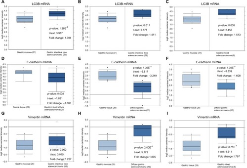
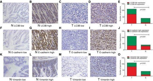
First, we evaluated transcription levels of LC3B, E-cadherin, and Vimentin in GC and normal gastric mucosa using the Oncomine database. In D’Errico et al’sCitation21 and Chen et al’sCitation22 data set, the results revealed that LC3B and Vimentin mRNA expression was higher in intestinal, diffuse, and mixed gastric adenocarcinoma compared with gastric mucosa (, all p<0.05). In Cho’s data set,Citation23 E-cadherin mRNA expression in the gastric adenocarcinoma tissue was markedly lower than in normal tissue (, all p<0.05). Then, as shown in , we detected LC3B, E-cadherin, and Vimentin protein expression in GC and adjacent tissues using immunohistochemistry (IHC). Consistent with mRNA expression, as shown in , the high expression rates of LC3B and Vimentin in GC tissues were 64.5% (71/110) and 70.9% (78/110), respectively, which were higher than those in adjacent tissues (LC3B, 20% [8/40], p<0.001; Vimentin, 30% [12/40], p<0.001). The most adjacent normal tissues (77.5%, 31/40) showed higher levels of E-cadherin protein compared with the corresponding GC tissues (30.9%, 34/110, p<0.001).
Table 2 Expressions of LC3B, E-cadherin, and Vimentin in GC tissue and N tissue
Figure 1 Box and whiskers plots of Oncomine data on LC3B (A–C), E-cadherin (D–F), and Vimentin (G–I) mRNA levels in GC and normal tissues.
Abbreviation: GC, gastric cancer.

Figure 2 Representative examples of LC3B, E-cadherin, and Vimentin expression (200×).
Notes: Low LC3B expression in paired nontumor (A) and GC tissues (C), high LC3B expression in adjacent normal (B) and GC tissues (D), comparisons of LC3B expression in adjacent normal and GC tissues (E), low E-cadherin expression in adjacent normal (F) and GC tissues (H), high E-cadherin expression in adjacent normal (G) and GC tissues (I), comparisons of E-cadherin expression in adjacent normal and GC tissues (J), low Vimentin expression in adjacent normal (K) and GC tissues (M), high Vimentin expression in adjacent normal (L) and GC tissues (N), comparisons of Vimentin expression in adjacent normal and GC tissues (O).
Abbreviations: GC, gastric cancer; N, adjacent normal tissue; T, GC tissue.

Correlations between LC3B, E-cadherin, and Vimentin expression and clinicopathological factors in GC
As listed in , the LC3B expression was associated with the T classification (p=0.006), lymph node metastasis (p=0.026), TNM stage (p=0.001), and the degree of differentiation (p=0.007). Significant associations between E-cadherin expression and T classification (p=0.033), nodal involvement (p=0.032), TNM stage (p=0.007), and histological differentiation (p=0.024) were identified. Vimentin expression was closely related to T classification (p=0.039), nodal involvement (p=0.013), and TNM stage (p=0.000). LC3B, E-cadherin, and Vimentin expression were unrelated to age, gender, and tumor size (all p>0.05).
LC3B expression correlated with E-cadherin and Vimentin in GC
We next evaluated the correlations between LC3B and E-cadherin and Vimentin expression in GC tissues. As shown in , the Spearman rank correlation analysis indicated that the expression of LC3B was negatively correlated with E-cadherin expression (r= −0.484, p<0.001) and was positively associated with Vimentin expression (r=0.320, p=0.001).
Table 3 Correlations between LC3B with E-cadherin and Vimentin in GC
Survival analysis
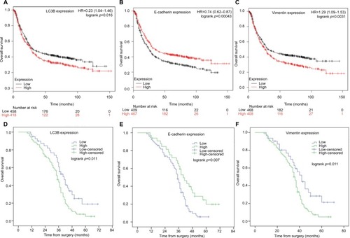
We next examined the prognostic value of LC3B, E-cadherin, and Vimentin mRNA expression in GC using the Kaplan–Meier plotter database (http://kmplot.com/analysis/). The following Affymetrix IDs are valid: 208786_s_at (LC3B), 201131_s_at (E-cadherin), and 201426_s_at (Vimentin). Survival analysis revealed that high expression of LC3B and Vimentin predicted worse OS in GC patients (N=876, HR =1.23 [95% CI: 1.04–1.46], p=0.016, ; HR =1.29 [95% CI: 1.09–1.53], p = 0.003; ). However, high E-cadherin mRNA high expression was found to be correlated to better OS (N=876, HR = 0.74 [95% CI: 0.62–0.87], p< 0.001, ). In our cohort, overall, 91 patients died and 19 survived after a median follow-up of 36 months (range, 8–71 months). Consistent with the aforementioned Kaplan–Meier analysis, patients with high LC3B and Vimentin expression exhibited shorter survival than those with low expression (all p< 0.05, ). High E-cadherin expression was associated with favorable prognosis in GC patients (p< 0.05, ). Multivariate Cox analysis identified tumor differentiation, TNM stage, T classification, and lymph node metastasis as significant prognostic factors for OS. In particular, LC3B, E-cadherin, and Vimentin expression were also independently prognostic factors for OS in patients with GC. These analyses are presented in .
Table 4 Multivariate analysis of OS for GC patients
Figure 3 The prognostic value of LC3B, E-cadherin, and Vimentin expression in GC patients.
Notes: OS curves of are plotted for patients with tumors expressing low or high levels LC3B (A), E-cadherin (B), and Vimentin (C) mRNA in Kaplan–Meier plotter database (N=876). OS curves are plotted for patients with tumors expressing low or high levels LC3B (D), E-cadherin (E), and Vimentin (F) in our retrospective cohort (N=110). P-value was calculated by log-rank test and p<0.05 was regarded as statistically significant.
Abbreviations: GC, gastric cancer; OS, overall survival.

Discussion
In current study, we evaluated the expression of LC3B- and EMT-related markers (E-cadherin and Vimentin) in GC and the corresponding adjacent normal tissues. In addition, we investigated their relationships with clinicopathological factors and clinical outcomes. Our data showed that LC3B, E-cadherin, and Vimentin were universally expressed in GC and corresponding normal tissues. Specifically, LC3B and Vimentin expression were significantly higher in GC tissues than that in the counterpart normal tissues in both mRNA and protein levels. The expression of E-cadherin in GC tissues was significantly reduced in comparison with normal tissues. LC3B was positively associated with Vimentin and negatively correlated with E-cadherin in GC tissues by Spearman rank correlation analysis. The expression of these markers was closely related to tumor differentiation, T classification, TNM stage, and nodal involvement. Furthermore, Kaplan–Meier survival analyses revealed that GC patients with high LC3B and Vimentin expression levels had a worse clinical outcome than those with low expression levels. Conversely, high E-cadherin expression was linked with favorable OS. Multivariate survival analysis demonstrated that LC3B, E-cadherin, and Vimentin expression levels were independent prognostic factors of GC patients.
GC continues to pose a major challenge in clinical practice with a poor prognosis and limited treatment options. Therefore, it is urgent to identify new additional prognostic markers to guide surveillance and improve individual treatment strategies. Mounting studies have indicated that autophagy has an important role in GC development. One study has investigated the prognostic value of autophagy-related proteins in GC and suggested LC3B was not associated with OS.Citation24 Yoshioka et alCitation25 has examined LC3 expression in gastrointestinal cancers and found that high protein expression of LC3 in 58% of GC (22/38 cases). A recent study by Masuda et alCitation16 evaluated clinicopathological and prognostic significance of LC3 in GC. Consistent with the current study, Masuda et alCitation16 report that LC3 was associated with T-stage (depth of tumor invasion) and lymph node metastasis, and suggested that LC3-positive indicated worse outcome in GC than that of patients negative by univariate analysis. Tumor metastasis renders most GC inoperable, resulting in a dismal prognosis. Studies found that autophagy facilitates tumor metastasis through affecting EMT,Citation26 tumor angiogenesis,Citation27 and inflammatory responses,Citation28 which may partially explain the LC3B high expression associated with more aggressive behavior of patients with GC. However, some studiesCitation29,Citation30 revealed that high LC3B expression predicted the poor outcome of triple-negative breast cancer and oral squamous cell carcinoma patients. These findings suggest that autophagy serves multifaceted roles in different types of cancer.
EMT is a process by which epithelial cells dedifferentiate and acquire mesenchymal phenotype. EMT exhibits changes at the molecular level as observed by the decreased expression of E-cadherin and increased expression of Vimentin. Although the role of EMT in cancer is complicated and tissue-specific,Citation31 numerous studies that have shown that EMT is a key GC progression driver and facilitates GC invasion and metastasis.Citation32 In line with the previous report,Citation33 the present study showed that E-cadherin and Vimentin were prognostic factors of GC. Higher Vimentin and lower E-cadherin protein levels in GC patients predicted worse OS. Interestingly, the findings of current study suggested that LC3B was linked to EMT-related markers expression in GC. Remarkably, LC3B was positively associated with Vimentin and negatively correlated with E-cadherin in GC tissues. Recent evidence from basic research indicate that autophagy and EMT in cancer are linked in an intricate relationship.Citation34 The 2 processes share common molecular mediators and signaling pathways, such as TGFβ, STAT3, and PI3K/AKT/mTOR signaling cascade. Autophagy is a survival-promoting pathway. On the one hand, aberrant EMT activation requires enhanced autophagy to survive during the cancer metastatic spreading under micro-environmental stress. On the other side, autophagy acting as tumor suppression mechanism inhibits the early phases of cancer progression by destabilizing crucial mediators of EMT.Citation34 Qiang and HeCitation35 reported that autophagy deficiency promotes EMT by SQSTM1-mediated TWIST1 stabilization. Zhao et alCitation36 found that the inhibition of autophagy promotes EMT through increasing ROS/HO-1 signaling pathway in ovarian cancer cells. At present, the precise interactions of autophagy and EMT in GC still remain obscure. Thus, the pivotal molecular mechanism behind the regulation of autophagy and EMT should be further explored.
Several potential limitations should be acknowledged for the current study. First, the numbers of samples are limited, which influences the power of statistical analysis. Second, this was a retrospective study using single-institutional medical information. Certain inherent biases exist. Future larger prospective studies with large samples size may be needed to validate our current data. Finally, the results of our study relied solely on histological examination, and many more in vivo interactions would need to be further researched.
Conclusion
In summary, LC3B, E-cadherin, and Vimentin may serve as potential prognostic biomarkers for GC; however, much more evidence is needed to prove this. There are correlations between autophagy and EMT in GC based on our study, but their complex interactions require further studies to explore the molecular mechanisms involved. The modulation of autophagy and EMT could be promising targets for the treatment of GC.
Acknowledgments
The present study was supported by grants from the scientific research project of 3201 Affiliated Hospital of Medical College of Xi’an Jiaotong University (No. 3201ky201629).
Disclosure
The authors report no conflicts of interest in this work.
References
- Van CutsemESagaertXTopalBHaustermansKPrenenHGastric cancerLancet2016388100602654266427156933
- MillerKDSiegelRLLinCCCancer treatment and survivorship statistics, 2016CA Cancer J Clin201666427128927253694
- WongSSKimKMTingJCGenomic landscape and genetic heterogeneity in gastric adenocarcinoma revealed by whole-genome sequencingNature Commun20145547725407104
- LevyJMMTowersCGThorburnATargeting autophagy in cancerNature Rev Cancer201717952854228751651
- KimmelmanACThe dynamic nature of autophagy in cancerGenes Dev201125191999201021979913
- LadoireSPenault-LlorcaFSenovillaLCombined evaluation of LC3B puncta and HMGB1 expression predicts residual risk of relapse after adjuvant chemotherapy in breast cancerAutophagy201511101878189026506894
- GiatromanolakiANCharitoudisGSBechrakisNEAutophagy patterns and prognosis in uveal melanomasMod Pathol20112481036104521499230
- AdamsODislichBBerezowskaSPrognostic relevance of autophagy markers LC3B and p62 in esophageal adenocarcinomasOncotarget2016726392413925527250034
- LadoireSChabaKMartinsIImmunohistochemical detection of cytoplasmic LC3 puncta in human cancer specimensAutophagy2012881175118422647537
- TangDYLEllisRALovatPEPrognostic impact of autophagy biomarkers for cutaneous melanomaFront Oncol2016623627882308
- ZhengXCarstensJLKimJEpithelial-to-mesenchymal transition is dispensable for metastasis but induces chemoresistance in pancreatic cancerNature2015527757952553026560028
- OkabeHMimaKSaitoSEpithelial-mesenchymal transition in gastroenterological cancerJ Cancer Metastasis Treat201513183189
- TakaiMTeraiYKawaguchiHThe EMT (epithelial-mesenchymal-transition)-related protein expression indicates the metastatic status and prognosis in patients with ovarian cancerJ Ovarian Res201477625296567
- YangZWangHXiaLOverexpression of PAK1 correlates with aberrant expression of EMT markers and poor prognosis in non-small cell lung cancerJ Cancer2017881484149128638464
- XuGFZhangWJSunQXuXZouXGuanWCombined epithelial-mesenchymal transition with cancer stem cell-like marker as predictors of recurrence after radical resection for gastric cancerWorld J Surg Oncol20141236825441488
- MasudaGOYashiroMKitayamaKClinicopathological correlations of autophagy-related proteins LC3, Beclin 1 and p62 in gastric cancerAnticancer Res201636112913626722036
- GugnoniMSancisiVManzottiGGandolfiGCiarrocchiAAutophagy and epithelial-mesenchymal transition: an intricate interplay in cancerCell Death Dis2016712e252027929542
- YangZWangHXiaLOverexpression of PAK1 correlates with aberrant expression of EMT markers and poor prognosis in non-small cell lung cancerJ Cancer2017881484149128638464
- SzászAMLánczkyANagyÁCross-validation of survival associated biomarkers in gastric cancer using transcriptomic data of 1,065 patientsOncotarget2016731493224933327384994
- LiuZYWuTLiQNotch signaling components: diverging prognostic indicators in lung adenocarcinomaMedicine20169520e371527196489
- D’ErricoMde RinaldisEBlasiMFGenome-wide expression profile of sporadic gastric cancers with microsatellite instabilityEur J Cancer2009453461469
- ChenXLeungSYYuenSTVariation in gene expression patterns in human gastric cancersMol Biol Cell20031483208321512925757
- ChoJYLimJYCheongJHGene expression signature-based prognostic risk score in gastric cancerClin Cancer Res20111771850185721447720
- CaoQHLiuFYangZLPrognostic value of autophagy related proteins ULK1, Beclin 1, ATG3, ATG5, ATG7, ATG9, ATG10, ATG12, LC3B and p62/SQSTM1 in gastric cancerAm J Trans Res20168938313847
- YoshiokaAMiyataHDokiYLC3, an autophagosome marker, is highly expressed in gastrointestinal cancersInt J Oncol200833346146818695874
- SuZYangZXuYChenYYuQApoptosis, autophagy, necroptosis, and cancer metastasisMol Cancer2015144825743109
- DingYPYangXDWuYXingCGAutophagy promotes the survival and development of tumors by participating in the formation of vasculogenic mimicryOncol Rep20143152321232724626574
- YangXYuDDYanFThe role of autophagy induced by tumor microenvironment in different cells and stages of cancerCell Biosci201551425844158
- ZhaoHYangMZhaoJWangJZhangYZhangQHigh expression of LC3B is associated with progression and poor outcome in triple-negative breast cancerMed Oncol201330147523371253
- TangJYHsiEHuangYCHsuNCChuPYChaiCYHigh LC3 expression correlates with poor survival in patients with oral squamous cell carcinomaHum Pathol201344112558256224055091
- BrabletzTKalluriRNietoMAWeinbergRAEMT in cancerNat Rev Cancer201818212813429326430
- HuangLWuRLXuAMEpithelial-mesenchymal transition in gastric cancerAm J Transl Res20157112141215826807164
- KimMALeeHSLeeHEKimJHYangHKKimWHPrognostic importance of epithelial-mesenchymal transition-related protein expression in gastric carcinomaHistopathology200954444245119309396
- GugnoniMSancisiVManzottiGGandolfiGCiarrocchiAAutophagy and epithelial–mesenchymal transition: an intricate interplay in cancerCell Death Dis2016712e252027929542
- QiangLHeYYAutophagy deficiency stabilizes TWIST1 to promote epithelial-mesenchymal transitionAutophagy201410101864186525126736
- ZhaoZZhaoJXueJZhaoXLiuPAutophagy inhibition promotes epithelial-mesenchymal transition through ROS/HO-1 pathway in ovarian cancer cellsAm J Cancer Res20166102162217727822409
