
Abstract
Introduction
Circular RNAs, as a class of long-time-ignored non-coding RNA, have been revealed as multifunctional RNAs in recent years, especially in the cancer research. However, the mechanism of most circular RNAs and their clinical application values in human cancers remain unknown, including in colorectal cancer (CRC).
Methods
In this study, we focused on the expression pattern and clinical values of hsa_circ_0000711 in CRC. The expression level of hsa_circ_0000711 in 101 paired CRC tissues and 3 CRC cell lines (HCT116, COLO205, and H-T29), as well as human normal colon epithelial cell line NCM460, were measured by quantitative real-time polymerase chain reaction.
Results
Our results revealed that the expression level of hsa_circ_0000711 was significantly downregulated in CRC tissues (P=9.35E-16) and CRC cell lines (P<0.01). In addition, the area under the receiver characteristic curves was 0.81. The sensitivity and specificity were 0.91 and 0.58, respectively. Meanwhile, our study showed that low expression of hsa_circ_0000711 could serve as an independent prediction biomarker associated with poor overall survival of CRC patients (hazard ratio =2.409; 95% CI: 1.276–4.547; P=0.004).
Conclusion
All of these results indicated that hsa_circ_0000711 may play a crucial role in CRC carcinogenesis and could be a potential effective biomarker for the diagnosis and prognosis of CRC.
Introduction
Colorectal cancer (CRC) is one of the most common gastrointestinal cancers and the fourth leading cause of cancer-related death in the world.Citation1 The highest incidence of CRC is reported in countries of Europe, North America, and Oceania.Citation2 According to epidemiologic data from the American Cancer Society, about 135,430 newly diagnosed and 502,600 deaths due to CRC were projected to occur only in the USA in 2017.Citation3 Moreover, the rapidly increasing incidence of CRC in previously low-risk areas, such as Eastern Europe and East Asia, has been noted, which is attributed to the changes in dietary patterns and risk factors toward a so-called western lifestyle.Citation4 Currently, the cornerstones of therapy for CRC are surgery, neoadjuvant radiotherapy, and adjuvant chemotherapy. However, the outcome of CRC remains unsatisfactory and the 5-year survival rate is <65%.Citation5 Local recurrence and distant metastasis are the main causes leading to poor outcome of CRC.Citation6 The 5-year survival rate of CRC dramatically declines from ~90% in early-stage non-metastatic tumors to about 5% in cases with distant metastasis.Citation7 Therefore, precise prediction at the molecular level of relapse and identify effective early biomarkers for CRC are urgently needed for individual diagnosis and therapy.
Circular RNAs (circRNAs) are a novel class of non-coding RNAs (ncRNAs) and single-stranded covalently closed circular transcripts without 5′ caps and 3′ tails.Citation8,Citation9 In the beginning, circRNAs were regarded as splicing errors or by-products of RNA processing with low abundance.Citation10 However, owing to the development of the next-generation RNA sequencing and bioinformatic analysis, researchers have discovered thousands of circRNAs and numerous studies have shed light on the biogenesis and function of circRNAs.Citation11,Citation12 Emerging evidence has showed that circRNAs are involved in the progression of tumorigenesis, invasion, and metastasis in numerous human carcinomas by regulating microRNA (miRNA) function as miRNA sponges.Citation13 Additionally, compared with the linear RNAs, circRNAs expression is characterized as tissue-specific and stable which can not be degraded by RNase.Citation14,Citation15 Furthermore, numerous evidence supported that some circRNAs can stably exist in human body fluid.Citation16 All these indications suggested that cir-cRNAs could be served as a promising biomarker for cancer diagnosis and prognosis.Citation17–Citation19
Searching CRC-associated circRNAs by synthetically bioinformatic analysis in 2 circRNA databases: CircBase and circ2Traitsby,Citation20,Citation21 we focused on circRNA-hsa_circ_0000711 in this study. The position of hsa_circ_0000711 is located at chr16:68155889–68160513 with 4,624 nts of the spliced sequence length in gene symbol NFATC3. In this study, we first investigated the hsa_circ_0000711 expression characteristics in CRC tissues and corresponding normal tissues, as well as cell lines, and evaluated the association between the hsa_circ_0000711 expression level and clinicopathological characteristics of patients with CRC. Moreover, we also explored its diagnostic and prognostic potential for CRC.
Materials and methods
Patient characteristics and tissue specimen collection
A total of 101 CRC tissues and paired adjacent nontumorous tissues were collected from CRC surgical specimens at the Department of gastrointestinal surgery in Ningbo Lihuili Hospital between June 2010 and April 2015. None of the patients underwent chemotherapy or radiotherapy before surgery. All the specimens were immediately preserved in RNA-fixer reagent after surgical excision and stored in liquid nitrogen. The final diagnosis was confirmed by 2 experienced pathologists. There were 2 well-differentiated, 83 moderately differentiated, and 16 poorly differentiated cases. Tumors were staged according to the TNM stage system of the American Joint Committee on Cancer seventh edition (2010). There were 21 Stage I, 32 Stage II, 40 Stage III, and 8 Stage IV cases. Clinicopathologic characteristics were available for each of the 101 patients. Patients were followed up for 60 months. Median follow-up was 39 months, with an interquartile range of 29–50 months. During followup, 5 patients were lost and 42 died. All patients agreed to participate in the study and signed the informed consent for the tissue collection. The study was approved by the Human Research Ethical Committee of Ningbo Lihuili Hospital.
Cell culture
Human normal colon epithelial cell line NCM460, as well as human CRC cell lines HCT116, COLO205, and HT29 were obtained from Shanghai Institute of Biochemistry and Cell Biology, Chinese Academy of Sciences (Shanghai, China). Cells were cultured in Roswell Park Memorial Institute-1640 Medium (HyClone, Logan, UT, USA) supplemented with 10% fetal bovine serum (ExCell Biology, Shanghai, China) with 50 U/mL penicillin and 50 g/mL streptomycin (HyClone). The incubators were maintained at 37°C in a 5% CO2 atmosphere. Exponentially growing cells were used for experiments.
Total RNA extraction and quantitative real-time polymerase chain reaction (PCR) assay
Total RNA was first extracted from tissues and cells using TRIzol reagent (Thermo Fisher Scientific, Waltham, MA, USA), and then reversed transcription into cDNA by GoS-cript Reverse Transcription (RT) System (Promega Corporation, Fitchburg, WI, USA) following the manufacturer’s protocol. The divergent primers for amplified sequences of hsa_circ_0000711 were: forward, 5′-CACTAGACTGGCCTTTACC-3′ and reverse, 5′-CACAATCATCTGGCTCAA-3′. Glyceraldehyde-3-phosphate dehydrogenase (GAPDH) was simultaneously amplified as internal control.Citation22 Each reaction mixture (20 µL) contained 5 µL cDNA, 1 µL forward primer, 1 µL reverse primer, 10 µL SYBR Green I Master (Hoffman-La Roche Ltd., Basel, Switzerland), and 3 µL DNA/RNA-free water (Hoffman-La Roche Ltd.). The PCR amplification was performed in 384-well plates using the RocheLightCycler 480II instrument (Hoffman-La Roche Ltd.). The PCR reaction was conducted using the following conditions: 95°C for 10 minutes, amplification for 45 cycles at 95°C for 20 seconds, 60°C for 30 seconds, and 72°C for 40 seconds. A melting curve step was performed at 95°C for 15 seconds, 1 minute at 60°C, and then increasing temperature at 0.11°C per second for up to 95°C to measure fluorescence signal. The cycle threshold (Ct) values were recorded for both hsa_circ_0000711 and GAPDH. The expression level of hsa_circ_0000711 was calculated using the ΔCt method, higher ΔCt means lower expression. All the samples were assayed in triplicate.
Immunohistochemical analysis of tissue carcinoembryonic antigen (CEA) and carbohydrate antigen 19–9 (CA19-9)
We incubated the paraffin tissue sections in primary anti-CEA or anti-CA19-9 (Dako Denmark A/S, Glostrup, Denmark) for 1 hour at room temperature, and then, the tissues were incubated in diaminobenzidine (Dako Denmark A/S) for color development after incubation with broad-spectrum second antibody K5007 (Dako Denmark A/S). The standard for determination of the results was according to the 2010 American Society of Clinical Oncology/the College of American Pathologists guideline.
Statistical analyses
All statistical analyses were performed by using SPSS Version 18.0 (SPSS Inc., Chicago, IL, USA). Differences in continuous data between pairs of groups were detected by Student’s t-test, one-way analysis of variance test was applied between multiple groups (>2 groups). The diagnostic value of hsa_circ_0000711 was assessed using the receiver operating characteristic (ROC) curve and the area under the curve (AUC). Survival analyses were conducted by Kaplan–Meier and Cox proportional hazard regression analyses. A 2-tailed P-value of <0.05 was considered statistically significant. All the figures were drawn using the GraphPad Prism 6 software (GraphPad Software, Inc., La Jolla, CA, USA).
Results
Expression of hsa_circ_0000711 in CRC tissues and cell lines
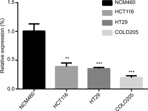
We explored hsa_circ_0000711 expression levels in 101 paired CRC tissues and corresponding normal tissues, the sequence of the qRT-PCR product was coincident with that in circBase (http://circbase.org/; ). The result showed that its expression in CRC tissues was significantly lower than that in adjacent normal tissues (P=9.35E-16, ). Among 101 paired CRC samples tested in our study, 76 cases (79.2%) exhibited significant downregulation of has_circ_0000711 in cancerous tissues (). Then, we found that the expression levels of hsa_circ_0000711 in 3 CRC cell lines, HCT116, COLO205, and HT29, were significantly downregulated than those in normal colon epithelial cell line NCM460 ().
Figure 1 DNA sequencing results of hsa_circ_0000711 in colorectal cancer tissues.
Note: The arrow points to the cyclation site.
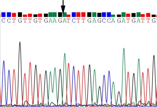
Figure 2 hsa_circ_0000711 expression levels in 101 paired colorectal tissues and corresponding normal tissues.
Abbreviation: Ct, cycle threshold.
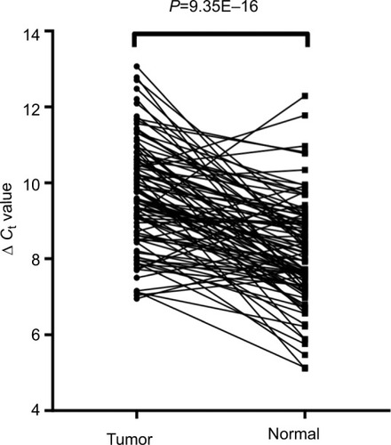
Figure 3 The expression level of hsa_circ_0000711 was significantly downregulated in 79.2% (76/96) colorectal cancer tissues compared with the adjacent normal tissues.
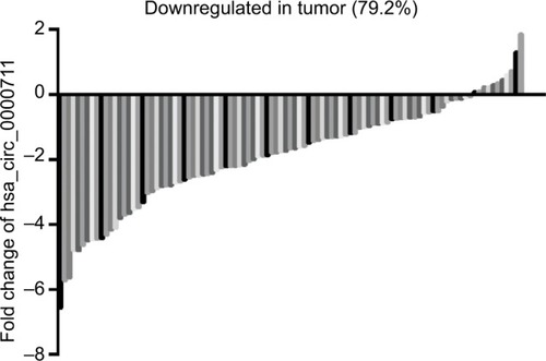
Figure 4 Relative expression of hsa_circ_0000711 in colorectal cancer cell lines (HCT116, COLO205 and HT29) and normal colon epithelial cell line (NCM460).
Notes: Data are expressed as mean ± SD of 3 independent experiments. Asterisks indicate P-values that are significant (*P<0.05, ** P<0.01, *** P<0.001).
Association between hsa_circ_0000711 expression levels and clinicopathological factors in CRC patients
Based on the aforementioned findings, we analyzed the association between hsa_circ_0000711 expression and clinicopathological characteristics of patients with CRC, such as age, gender, diameter, differentiation, invasion, lymph node metastasis, clinical stage, distant metastases, location, perineural invasion, and CEA and CA19-9 levels. However, no difference of hsa_circ_0000711 expression was found among these clinicopathological characteristics of CRC patients ().
Table 1 The association of hsa_circ_0000711 expression levels with clinicopathological characteristics of CRC patients
The diagnostic value of hsa_circ_0000711 in CRC
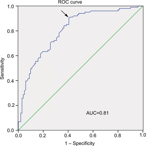
The ROC curve was used to evaluate the diagnostic potential of hsa_circ_0000711 in CRC patients. The ROC curve is a comprehensive index that can reflect on the specificity and sensitivity of continuous variables. The maximum Youden index was used as a cutoff point. In current study, showed the AUC was up to 0.81 at a cutoff ΔCt value of 3.37. The sensitivity and specificity were 0.91 and 0.58, respectively. Hence, hsa_circ_0000711 might be a diagnostic indicator of CRC.
Figure 5 ROC curve.
Abbreviations: AUC, area under the curve; ROC, receiver operating characteristic.
The prognostic value of hsa_circ_0000711 in CRC
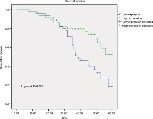
We constructed survival curves to investigate the prognostic value of hsa_circ_0000711 for CRC (). The median expression was used as a cutoff point. A total of 51 patients were assigned to the low-expression group, and the others to the high-expression group. The Kaplan–Meier survival analysis revealed that low-expression level of hsa_circ_0000711 was significantly associated with poor outcome of CRC (log-rank P=0.003). In univariate Cox proportional hazards analysis, patients with low expression had a significantly increased risk of death compared with patients with high expression of hsa_circ_0000711 (hazard ratio [HR]=2.418; 95% CI: 1.315–4.446; P=0.005). Subsequently, a multivariate Cox proportional hazard analysis was performed by adjusting for T classification and lymphatic metastasis. The results indicated low expression of hsa_circ_0000711 (HR=2.409; 95% CI: 1.276–4.547; P=0.004) and lymphatic metastasis (HR=2.045; 95% CI: 1.105–3.785; P=0.023) was an independent predictive biomarker of overall survival (OS) of CRC patients ().
Table 2 Multivariate Cox proportional hazards analysis of the 101 colorectal cancer patients
Figure 6 The Kaplan–Meier survival curve.
Note: The median ΔCt value was used as a cutoff point.
Abbreviation: Ct, cycle threshold.
Discussion
Owing to advancements in high-throughput sequencing technologies and bioinformatics, circRNAs, once perceived as the by-products of splicing errors, recently were found to be extensively expressed in human cells and play roles in many biological processes.Citation15,Citation23 Although, circRNAs are commonly grouped into a type of ncRNA, recent study showed that circRNAs can be translated into proteins,Citation24,Citation25 which enriched circRNAs’ critical roles in life activity. In addition, mounting evidence shows that circRNAs are widely involved in the initiation and progression of multiple diseases, especially cancers.Citation26 For example, Deng et al disclosed that hsa_circ_0009910 promotes carcinogenesis by promoting the expression of miR-449a target IL6R in osteosarcoma.Citation27 Yao et al demonstrated that circZKSCAN1 inhibits growth, migration, and invasion of hepatocellular carcinoma via several cancer-related signaling pathways.Citation28 Due to lack of the 5′cap and polyA tail, circRNAs are highly conserved and remarkably stable.Citation29,Citation30 In addition, circRNAs are generally expressed in a tissue- and cell-type-specific manner and could help distinguish different tumors or predict treatment responses.Citation31 Taken together, these characteristics exhibit that circRNAs could be potential to serve as a kind of valuable biomarker of cancer diagnosis and prognosis.
Up to now, increasing researches and achievements reported that circRNAs have involved in CRC progression. Xie et al revealed that hsa_circ_001569 was a positive regulator in cell proliferation and invasion of CRC by as a sponge of miR-145.Citation32 Wang et al found that hsa_circ_001988 was decreased expression and correlated with differentiation and perineural invasion of CRC.Citation33 In the current study, our findings first showed that hsa_circ_0000711 expression level was significantly downregulated in both CRC tissues and CRC cell lines. Although previous studies showed that circRNAs might be correlated with clinicopathological parameters, no difference in hsa_circ_0000711 expression levels were found for clinicopathological characteristics of CRC patients in our study. Future rigorous clinical research studies with larger sample sizes will be essential to validate our findings.
Diagnosis of CRC patients depends on clinical symptoms and auxiliary examinations, such as colonscopy, CT colonography, MRI, and PET/CT colonography.Citation34 However, due to non-specific symptoms in the early stage of CRC, and lack of effective diagnostic markers with high sensitivity and specificity, a low early diagnostic rate poses challenges for treatment. Because of the lack of free ends, circRNAs are resistant to exonucleases and more stable than linear RNA isoforms.Citation15 Besides, targeted validation of the accuracy of a circRNA is accomplished by PCR using outward-facing primers and Sanger sequencing for a semi-random primer, and the technology is well developed.Citation35 Due to their special structure, circRNAs can act as effective biomarkers for the early diagnosis of cancer.Citation36–Citation38 In this study, we also explored the value of hsa_circ_0000711 in CRC diagnosis by ROC curve, which is a comprehensive index used to reflect the sensitivity and specificity of continuous variables. The larger the area under the ROC curve (AUC) is, the higher diagnostic value is.Citation39 In our study cohort, we found that the AUC was 0.81 and the sensitivity was 0.91. Compared with the diagnostic value of conventional CRC-related biomarkers, such as CEA and CA199,Citation40 hsa_circ_0000711 had a higher AUC value. Meanwhile, we noticed that the specificity of hsa_circ_0000711 was not so high, suggesting that hsa_circ_0000711 cannot be used to diagnose CRC independently. However, given its near-perfect sensitivity, hsa_circ_0000711 might be an effective biomarker for the diagnosis of CRC if combined with other diagnostic technologies. Due to a relatively small sample size, the rigorous clinical research studies with larger sample sizes will be essential to corroborate our finding.
Although classification according to TNM and Union for International Cancer Control stage provides valuable prognostic information and guides therapy decisions, the response and outcome of individual patient’s therapy is not predicted enough.Citation41 Increasing evidence showed that the circRNAs could be prognostic markers for cancers, including CRC.Citation42 In our study, log-rank test showed that low expression level of hsa_circ_0000711 was significantly associated with poor OS of CRC patients, which was in accordance with our univariate Cox proportional hazards analysis results. In addition, a multivariate Cox proportional hazard analysis was performed by adjusting for T classification and lymphatic metastasis to confirm that low expression level of hsa_circ_0000711 was an independent unfavorable factor for CRC outcomes. All these findings supported the fact that hsa_circ_0000711 expression level could be a potential biomarker for prognosis of CRC and guide the individualized treatment programs.
Conclusion
To sum up, the present study indicates that circRNA0003906 was significantly downregulated in CRC and may play a crucial role in CRC carcinogenesis. Additionally, our finding supported that hsa_circ_0005986 might be a potential effective biomarker for the diagnosis and prognosis of CRC.
Acknowledgments
This work was supported by the Zhejiang Provincial Natural Science Foundation of China (grant number LY16H160005); the Project of Scientific Innovation Team of Ningbo (grant number 2015B11050); the Ningbo Natural Science Foundation (grant number 2014A610235 and 2017A610236).
Disclosure
The authors report no conflicts of interest in this work.
References
- BrennerHKloorMPoxCPColorectal cancerLancet201438399271490150224225001
- SiegelRLMillerKDFedewaSAColorectal cancer statistics, 2017CA Cancer J Clin201767317719328248415
- SiegelRLMillerKDJemalACancer statistics, 2017CA Cancer J Clin201767173028055103
- CenterMMJemalAWardEInternational trends in colorectal cancer incidence ratesCancer Epidemiol Biomarkers Prev20091861688169419505900
- MillerKDSiegelRLLinCCCancer treatment and survivorship statistics, 2016CA Cancer J Clin201666427128927253694
- ZippiMde TomaGMinerviniGDesmoplasia influenced recurrence of disease and mortality in stage III colorectal cancer within five years after surgery and adjuvant therapySaudi J Gastroenterol2017231394428139499
- PizziniSBisogninAMandruzzatoSImpact of microRNAs on regulatory networks and pathways in human colorectal carcinogenesis and development of metastasisBMC Genomics20131458923987127
- ChenLLYangLRegulation of circRNA biogenesisRNA Biol201512438138825746834
- EbbesenKKKjemsJHansenTBCircular RNAs: identification, biogenesis and functionBiochim Biophys Acta20161859116316826171810
- KosADijkemaRArnbergACvan der MeidePHSchellekensHThe hepatitis delta (delta) virus possesses a circular RNANature198632360885585602429192
- JeckWRSharplessNEDetecting and characterizing circular RNAsNat Biotechnol201432545346124811520
- ZhangXOWangHBZhangYLuXChenLLYangLComplementary sequence-mediated exon circularizationCell2014159113414725242744
- XiaWQiuMChenRCircular RNA has_circ_0067934 is upregulated in esophageal squamous cell carcinoma and promoted proliferationSci Rep201663557627752108
- BarrettSPSalzmanJCircular RNAs: analysis, expression and potential functionsDevelopment2016143111838184727246710
- MemczakSJensMElefsiniotiACircular RNAs are a large class of animal RNAs with regulatory potencyNature2013495744133333823446348
- ShaoYLiJLuRGlobal circular RNA expression profile of human gastric cancer and its clinical significanceCancer Med2017661173118028544609
- SunHTangWRongDHsa_circ_0000520, a potential new circular RNA biomarker, is involved in gastric carcinomaCancer Biomark201821229930629103021
- YangFLiuDYGuoJTCircular RNA circ-LDLRAD3 as a biomarker in diagnosis of pancreatic cancerWorld J Gastroenterol201723478345835429307994
- ZhuXWangXWeiShsa_circ_0013958: a circular RNA and potential novel biomarker for lung adenocarcinomaFEBS J2017284142170218228685964
- GlažarPPapavasileiouPRajewskyNcircBase: a database for circular RNAsRNA201420111666167025234927
- GhosalSdasSSenRBasakPChakrabartiJCirc2Traits: a comprehensive database for circular RNA potentially associated with disease and traitsFront Genet2013428324339831
- ShenZLiQDengHLuDSongHGuoJLong non-coding RNA profiling in laryngeal squamous cell carcinoma and its clinical significance: potential biomarkers for LSCCPLoS One201499e10823725243407
- LiYZhengQBaoCCircular RNA is enriched and stable in exosomes: a promising biomarker for cancer diagnosisCell Res201525898198426138677
- LegniniIdi TimoteoGRossiFCirc-ZNF609 is a circular RNA that can be translated and functions in myogenesisMol Cell2017661223728344082
- PamudurtiNRBartokOJensMTranslation of circRNAsMol Cell2017661921.e728344080
- QuSZhongYShangRThe emerging landscape of circular RNA in life processesRNA Biol201714899299927617908
- DengNLiLGaoJHsa_circ_0009910 promotes carcinogenesis by promoting the expression of miR-449a target IL6R in osteosarcomaBiochem Biophys Res Commun2018495118919629117539
- YaoZLuoJHuKZKSCAN1 gene and its related circular RNA (circZKSCAN1) both inhibit hepatocellular carcinoma cell growth, migration, and invasion but through different signaling pathwaysMol Oncol201711442243728211215
- HansenTBJensenTIClausenBHNatural RNA circles function as efficient microRNA spongesNature2013495744138438823446346
- JeckWRSorrentinoJAWangKCircular RNAs are abundant, conserved, and associated with ALU repeatsRNA201319214115723249747
- QuSLiuZYangXThe emerging functions and roles of circular RNAs in cancerCancer Lett201841430130929174799
- XieHRenXXinSEmerging roles of circRNA_001569 targeting miR-145 in the proliferation and invasion of colorectal cancerOncotarget2016718266802669127058418
- WangXZhangYHuangLDecreased expression of hsa_circ_001988 in colorectal cancer and its clinical significancesInt J Clin Exp Pathol2015812160201602526884878
- KijimaSSasakiTNagataKUtanoKLeforATSugimotoHPreoperative evaluation of colorectal cancer using CT colonography, MRI, and PET/CTWorld J Gastroenterol20142045169641697525493009
- SzaboLSalzmanJDetecting circular RNAs: bioinformatic and experimental challengesNat Rev Genet2016171167969227739534
- ChenSLiTZhaoQXiaoBGuoJUsing circular RNA hsa_circ_0000190 as a new biomarker in the diagnosis of gastric cancerClin Chim Acta201746616717128130019
- QinMLiuGHuoXHsa_circ_0001649: a circular RNA and potential novel biomarker for hepatocellular carcinomaCancer Biomark201616116116926600397
- WangJLiXLuLHeLHuHXuZCircular RNA hsa_circ_0000567 can be used as a promising diagnostic biomarker for human colorectal cancerJ Clin Lab Anal2018325e2237929333615
- JonesCMAthanasiouTSummary receiver operating characteristic curve analysis techniques in the evaluation of diagnostic testsAnn Thorac Surg2005791162015620907
- FengBZhengMHZhengYFNormal and modified urinary nucleosides represent novel biomarkers for colorectal cancer diagnosis and surgery monitoringJ Gastroenterol Hepatol200520121913191916336453
- KongXLiJCaiYA modified TNM staging system for non-metastatic colorectal cancer based on nomogram analysis of SEER databaseBMC Cancer20181815029310604
- HsiaoKYLinYCGuptaSKNoncoding effects of circular RNA CCDC66 promote colon cancer growth and metastasisCancer Res20177792339235028249903