
Abstract
Background
Metformin, a first-line antidiabetic drug, has recently been reported with anticancer activities in various cancers; however, the underlying mechanisms remain elusive. The aim of the present study was to investigate the role of cellular FADD-like IL-1β-converting enzyme (FLICE)-inhibitory protein large (c-FLIPL) in metformin-induced anticancer activity in non-small cell lung cancer (NSCLC) in vitro.
Materials and methods
Cell viability was measured by MTT assay. Quantitative real-time PCR was carried out to detect the level of mRNA of related genes. The expression of related proteins was detected by Western blot. siRNA was used to silence the expression of targeted proteins.
Results
Metformin significantly suppressed proliferation of both A549 and H460 cells in a dose-dependent manner. Mechanistic studies suggested that metformin killed NSCLC cells by inducing apoptotic cell death. Moreover, metformin greatly inhibited c-FLIPL expression and then promoted its degradation. Furthermore, metformin significantly activated Adenosine 5′-monophosphate (AMP)-activated protein kinase (AMPK) and its downstream glycogen synthase kinase 3beta (GSK-3β), block the expression of AMPK, and GSK-3β with siRNA partially reversed metformin-induced cytotoxicity and restored the expression of c-FLIPL in lung cancer cells. Metformin also suppressed the activity of AMPK downstream protein kinase A (PKA), PKA activators, both 8-Br-cAMP and forskolin, greatly increased c-FLIPL expression in NSCLC cells.
Conclusion
This study provided evidence that metformin killed NSCLC cells through AMPK/PKA/GSK-3β axis-mediated c-FLIPL degradation.
Introduction
As one of the most malignant cancers, lung cancer severely endangers the life and health of human beings. The incidence and mortality rates of this disease rank top of different types of tumors in People’s Republic of China.Citation1 Lung cancer mainly includes non-small cell lung cancer (NSCLC) and small cell lung cancer (SCLC). NSCLC accounts for about 85% lung cancer, the 5-year survival rate of which is <15%. Surgery, chemotherapy, and radiotherapy are the main options for late-stage NSCLC patients, but the effects are dissatisfying. Immune-oncology has recently changed the paradigms of NSCLC, which greatly improves 5-year survival rate and prognosis of NSCLC patients. Therefore, it is of great significance to explore novel anticancer agents for late-stage NSCLC treatment.
Metformin is a first-line antidiabetic drug and has been reported with antitumor activities in various cancers, including breast cancer,Citation2 hepatocellular cancer,Citation3 gastric cancer,Citation4 lymphoma,Citation5 melanoma,Citation6 esophageal squamous cell carcinoma,Citation7 and lung cancer.Citation8 Increasing studies have demonstrated that the combination of metformin with anticancer agents induced synergic anticancer effects in NSCLC, including radiation,Citation9 gefitinib,Citation10,Citation11 erlotinib,Citation12 sorafenib,Citation13 car-boplatin,Citation14 cisplatin,Citation15 and TRAIL.Citation16 Epidemiological studies have suggested that metformin reduces the incidence of lung cancer with diabetes and improves the survival of patients with type II diabetes of lung cancer.Citation17–Citation19 Adenosine 5′-mono-phosphate (AMP)-activated protein kinase (AMPK) is one of the main mechanisms by which metformin exerts anticancer activities.Citation20 It has been well established that AMPK and its downstream glycogen synthase kinase 3 beta (GSK-3β) and mTOR play a crucial role in metformin-induced anticancer effects.Citation2–Citation4 MicroRNA, c-Myc, and tumor microenvironment contribute to the antitumoral activities of metformin as well.Citation21–Citation23 However, the molecular mechanisms of metformin-induced anticancer effects warrants further investigation.
Cellular FADD-like IL-1β-converting enzyme (FLICE)-inhibitory protein (c-FLIP) is a key negative regulator of apoptosis pathways.Citation24 There are three isoforms of this protein, c-FLIPL, c-FLIPS, and c-FLIPR. c-FLIPL is a 55 kDa protein, including two N-terminal DED domains and a C-terminal caspase-like domain, which is structurally similar to pro-caspase 8.Citation25 c-FLIP is always overexpressed in various tumors and cell lines, including lung cancer.Citation26 Overexpression of c-FLIP confers increased resistance to apoptosis mediated by TRAIL and Fas. Previous studies have demonstrated that high levels of c-FLIP are correlated with poor prognosis in different cancers. Therefore, targeting c-FLIP might be a promising strategy for cancer treatment. Our previous studies have demonstrated that the combination of Chal-24 with anticancer agents results in synergistic cytotoxicity in lung cancer by downregulation of c-FLIPL, including cisplatinCitation27 and TRAIL.Citation28 Previous studies have reported that metformin decreases the expression of c-FLIPL in bladder cancer,Citation29 renal cell carcinomaCitation30 and lung cancer cells;Citation16 however, the molecular mechanisms underlying metformin-mediated c-FLIPL downregulation remain largely unknown.
In the present study, we investigated the role of c-FLIPL in metformin-mediated anticancer effects in NSCLC in vitro. We found that metformin induced apoptotic cell death in NSCLC cells, and it remarkably downregulated the expression of c-FLIPL. Mechanistic studies demonstrated that metformin mainly suppressed c-FLIPL expression through AMPK/PKA/GSK-3β pathway. These results provide novel insight into the role of c-FLIPL in metformin-induced anticancer effects in NSCLC in vitro.
Materials and methods
Reagents
Metformin (D150959) was purchased from Sigma-Aldrich (St Louis, MO, USA). Anti-phosphor-AMPKα (Thr172) (40H9) (2535) and AMPKα (5832), phosphor-cAMP-response element binding protein (CREB) (Ser133) (87G3) (9198) and CREB (48H2) (9197), phosphor-GSK-3β (Ser9) (9336) and GSK-3β (27C10) (9315), PARP (3493), cleaved Caspase-3 (9664) and GAPDH (2118) were from Cell Signaling Technology (Danvers, MA, USA). Antibody for β-tubulin (200608) was from Zen Bioscience (Chengdu, Sichuan, People’s Republic of China). Anti-Caspase 3 (ab32351) and c-FLIPL were from Abcam (Cambridge, UK). z-VAD (Ome)-FMK (600332) and chlorhexidine were from Cell Signaling Technology.
Cell culture
Lung cancer cell lines A549 and H460 were obtained from America Type Culture Collection (Manassas, VA, USA) and grown in RPMI-1640 medium supplemented with 10% FBS, 2 mM l-glutamine, 100 U penicillin, and 100 µg/mL streptomycin. All cells were cultured in standard incubator conditions at 37°C with 5% CO2. RPMI-1640 medium without glucose and FBS were purchased from Gibco (Thermo Fisher Scientific, Waltham, MA, USA).
Cell viability assay
Cells were seeded in triplicate data density of 5×10⁴/well and after staining with trypan-blue were counted every day for 3 days. In some experiments, cells were seeded in triplicate at a density of 2.5×10⁴/well. The next day and thereafter, cells were incubated with 20 mg/mL MTT for 3 hours. MTT was then dissolved in dimethyl sulfoxide and read at OD 570 nm. The results were expressed as relative cell number against the corresponding readings from the cells on day set to 100.
RNA extraction
Total RNA was extracted from cells using Transzol (Takara Bio Inc., Shiga, Japan), Chloroform (Sangon Biotech, Shanghai, People’s Republic of China), Isopropyl alcohol (Sangon Biotech), DEPC water (Sangon Biotech), and 75% ethanol (Sangon Biotech). The yield of RNA was determined using a NanoDrop 2000 spectrophotometer (Thermo Fisher Scientific), and the integrity was evaluated using agarose gel electrophoresis stained with ethidium bromide.
Real-time quantitative RT-PCR
Quantification was performed with a two-step reaction process: reverse transcription (RT) and PCR. The PrimeScript™ RT reagent Kit with gDNA Eraser (Takara Bio Inc.) was used for each RT reaction in a total volume of 10 µL. Reactions were performed in a GeneAmp® PCR System 9700 (Thermo Fisher Scientific) for 15 minutes at 37°C, followed by heat inactivation of RT for 5 seconds at 85°C. The 10 µL RT reaction mix was then diluted 10× in nuclease-free water and held at −20°C.
Real-time PCR was performed using ABI Prism 7900 Real-time PCR Instrument (Thermo Fisher Scientific) with the Trans-Start® Top Green qPCR SuperMix (+ Dye I/+ Dye II)-qPCR kit (Transgen Biotech Co. Ltd., Beijing, People’s Republic of China) in a 10 µL PCR reaction mixture. Reactions were incubated in a 96-well optical plate (Transgen Biotech Co. Ltd.) at 94°C for 30 minutes, followed by 40 cycles of 94°C for 50 seconds and 60°C for 30 seconds. Each sample was run in triplicate for analysis. At the end of the PCR cycles, melting curve analysis was performed to validate the specific generation of the expected PCR product. The primer sequences were designed in the laboratory and synthesized by Beijing TSINGKE, based on the mRNA sequences obtained from the NCBI database as follow. Primer sequences for real-time PCR are shown in .
Table 1 Primer sequences for real-time PCR
Western blotting
Cell lysates were prepared by suspending cells in M2 buffer (20 mM Tris–HCl pH 7.6, 0.5% NP40, 250 mM NaCl, 3 mM EDTA, 2 mM DTT, 0.5 mM phenylmethylsulfonylfluoride, 20 mM β-glycerophosphate, 1 mM sodium vanadate, and 1 µg/mL leupeptin). The samples were boiled for 5 minutes, equal amounts of proteins from each cell lysate were resolved by 8% or 12% SDS-PAGE and analyzed by Western blot. The proteins were visualized with enhanced chemiluminescence (Millipore) following the instructions of the manufacturer. Each experiment was repeated at least three times and representative results are shown.
Knockdown protein expression by RNAi
siRNA was purchased from RiboBio Co., Ltd (Guangzhou, Guangdong, People’s Republic of China). A549 and H460 cells were seeded in a 12-well plate and 48-well plate the day before transfection at 40%–50% confluence. siRNA was transfected with siRNA INTERFERin (polyplus transfection).Citation31 Twenty-four hours after transfection, metformin (80 mM) was added to the culture for 48 hours and MTT assay was carried out to measure cell viability, or metformin was added to the culture for 16 hours and expression of related proteins were detected by Western blot. Knockdown was confirmed by Western blot.
Statistical analysis
All data were expressed as mean ± SD. All data were analyzed using SPSS 17.0 for Windows (SPSS Inc., Chicago, IL, USA). P<0.05 was considered statistically significant.
Results
Metformin promoted NSCLC cells apoptosis
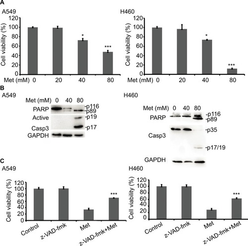
To investigate the anticancer effects of metformin, A549 and H460 cells were treated with increasing concentrations of metformin for 48 hours; cell viability was detected and quantified by MTT assay. As shown in , metformin suppressed NSCLC cell proliferation in a dose-dependent manner. Consistent with previous studies that metformin induced apoptosis in many types of cancer cells, metformin greatly induced caspase-3 activation and PARP cleavage in both A549 and H460 cell lines (hallmarks of apoptosis), indicating metformin induced apoptosis in NSCLC cells (), which was further validated by pan-caspase inhibitor z-VAD-fmk reversed metformin-induced cytotoxicity (). These results indicate that metformin mainly induces apoptotic cytotoxicity in lung cancer cells.
Figure 1 Metformin induces apoptotic cell death in lung cancer cells.
Notes: (A) A549 and H460 cells were treated with metformin (20, 40, and 80 mM) for 48 hours. Cell viability was measured and quantified by MTT assay. (B) A549 and H460 cells were treated with indicated concentrations of metformin for 24 hours. Then, indicated proteins were detected by Western blot, GAPDH or tubulin was detected as an input control. (C) The cells were pretreated with z-VAD-FMK (10 µM) for 1 hour, and then exposed to metformin for 48 hours. Cell viability was measured and quantified by MTT assay. N=3. *P<0.05, ***P<0.001.
Metformin suppressed the expression of c-FLIPL in NSCLC
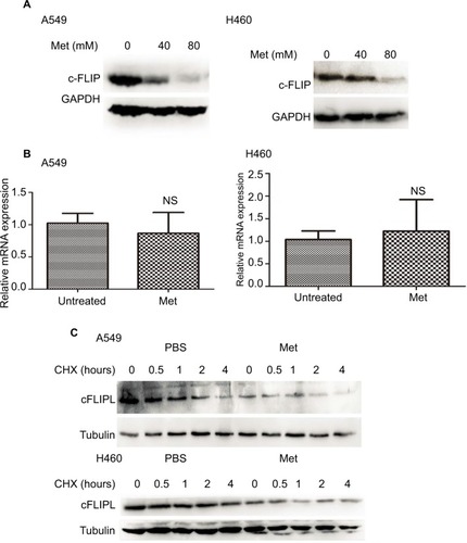
It has been well documented that c-FLIPL is one of the main inhibitors of cell death, and previous studies have reported that metformin promoted c-FLIPL degradation in bladder cancer and lung cancer, respectively, thus resulting in synergism of TRAIL-induced cytotoxicity, but whether metformin also influences the expression of c-FLIPL in NSCLC still remains elusive. As shown in , metformin also greatly suppressed c-FLIPL expression in both A549 and H460 cells. To further investigate the mechanism by which metformin suppressed the expression of c-FLIPL in lung cancer cells, we first investigated the c-FLIPL mRNA levels by qRT-PCR. Metformin did not influence the mRNA levels of this protein in both A549 and H460 cell lines (). Then, a cycloheximide chase assay was carried out to investigate the stability of c-FLIPL. Lung cancer cells were pretreated with metformin (80 mM) for 4 hours, then exposed to cycloheximide (200 µg/mL) for indicated times. As shown in , metformin significantly accelerated the degradation of c-FLIPL in both A549 and H460 cells compared with the control group. Taken together, these results strongly suggested that metformin kills lung cancer cells through degradation of c-FLIPL in lung cancer cells.
Figure 2 Metformin downregulated the expression of c-FLIPL in NSCLC.
Notes: (A) A549 and H460 cells were treated with metformin (80 mM) for 16 hours. Then, indicated proteins were detected by Western blot, and GAPDH or tubulin was detected as an input control. (B) Lung cancer cells were treated with metformin for 8 hours. Then, total RNA were extracted, and the mRNA level of c-FLIPL was examined by qRT-PCR, β-actin was detected as an input control. (C) A549 and H460 cells were pretreated with metformin (80 mM) or PBS for 4 hours, and then treated with CHX (200 µg/mL) for indicated times. Then, indicated proteins were detected by Western blot and tubulin was detected as an input control. N=3.
Abbreviations: c-FLIPL, cellular FLICE-inhibitory protein large; FLICE, FADD-like IL-1β-converting enzyme; NSCLC, non-small cell lung cancer.
Metformin downregulated c-FLIPL expression depending on AMPK activation
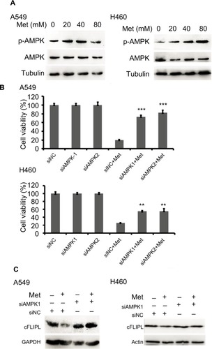
It has been well documented that AMPK activation is the main action of metformin in various cancer cells. However, the part of this pathway in apoptosis via c-FLIPL remains elusive in NSCLC. To elucidate whether metformin downregulates c-FLIPL expression through AMPK, we examined the AMPK activation by Western blotting. Metformin indeed induced AMPK activation in both A549 and H460 cell lines in a dose-dependent manner (). To further confirm the role of AMPK in metformin-induced c-FLIPL degradation in lung cancer cells, siRNA was used to silence the expression of AMPK. As shown in , siRNA partially reversed metformin-induced cell death in both A549 and H460 cell lines. Consistently, siRNA restored the expression of c-FLIPL in NSCLC cells (). These results support that metformin promotes c-FLIPL degradation in an AMPK-dependent manner.
Figure 3 Metformin inhibits c-FLIPL expression through AMPK.
Notes: (A) A549 and H460 cells were treated with metformin (20, 40, 80 mM) for 2 hours. Then, indicated proteins were detected by Western blot, and tubulin was detected as an input control. (B) The cells were transfected with siRNA (10 nM) for 24 hours, and then treated with metformin (80 mM) for 24 hours. Then, cell viability was detected by MTT assay. (C) A549 and H460 cells were transfected with siRNA (10 nM) for 24 hours, and then treated with metformin (80 mM) for 16 hours. Then, indicated proteins were detected by Western blot, and tubulin was detected as an input control. N=3. **P<0.01, ***P<0.001.
Abbreviations: AMPK, adenosine 5′-monophosphate-activated protein kinase; c-FLIPL, cellular FLICE-inhibitory protein large; FLICE, FADD-like IL-1β-converting enzyme.
Metformin downregulated the expression of c-FLIPL through the inhibition of PKA
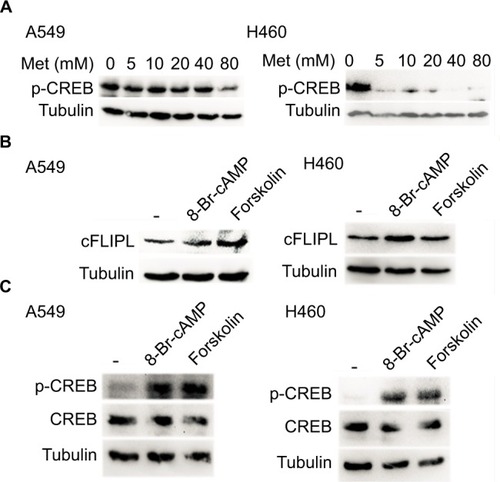
We further explored the mechanism by which metformin promoted c-FLIPL degradation in lung cancer cells. It has been reported that metformin increases the level of AMPK, then leads to inhibition of cAMP production and protein kinase A (PKA) activity.Citation20,Citation32,Citation33 We then examined the PKA activity by investigating the phosphorylation level of CREB, which is a downstream substrate of PKA. Metformin indeed suppressed the activity of PKA in lung cancer cells, and it greatly decreased the level of p-CREB (). To further confirm the role of PKA in metformin-mediated c-FLIPL suppression, PKA activators, 8-Br-cAMP and forskolin, remarkably increased the activity of PKA, meanwhile significantly increased the protein levels of c-FLIPL (). Meanwhile, both 8-Br-cAMP and forskolin greatly promoted the proliferation rate of lung cancer cells under microscope (data not shown). These results suggest that metformin downregulates c-FLIPL expression by inhibition of PKA activity.
Figure 4 Metformin suppressed the expression of c-FLIPL by inhibition of PKA activity.
Notes: (A) A549 and H460 cells were treated with metformin (5, 10, 20, 40, 80 mM) for 8 hours. Then, indicated proteins were detected by Western blot, and GAPDH was detected as an input control. (B, C) Lung cancer cells were treated with 8-Br-cAMP (1 mM) and forskolin (10 µM) for 8 hours. Then, indicated proteins were detected by Western blot, and tubulin was detected as an input control.
Abbreviations: c-FLIPL, cellular FLICE-inhibitory protein large; FLICE, FADD-like IL-1β-converting enzyme; PKA, protein kinase A.
Metformin downregulated c-FLIPL expression depending on GSK-3β activation
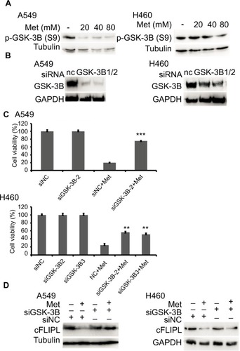
It has been reported that metformin induces AMPK activation, leading to PKA suppression; PKA phosphorylated GSK-3β at S9, then activates GSK-3β.Citation34,Citation35 Then, we continued to investigate whether metformin decreased the expression of c-FLIPL through GSK-3β in lung cancer cells. Metformin indeed decreased the levels of inhibitory p-GSK-3β (S9), indicating metformin induced activation of GSK-3β signaling pathway (). To further clarify whether metformin downregulated the expression of c-FLIPL through GSK-3β, siRNA was used to silence the expression of GSK-3β. As shown in , siRNA partially reversed metformin-induced anticancer activities in NSCLC cells (). Meanwhile, siRNA restored the expression of c-FLIPL in both A549 and H460 cell lines (). Collectively, these results strongly indicate that metformin promotes c-FLIPL degradation through AMPK/PKA/GSK-3β pathway in NSCLC.
Figure 5 Metformin promotes c-FLIPL degradation via GSK-3β.
Notes: Metformin inhibits c-FLIPL expression through AMPK. (A) A549 and H460 cells were treated with metformin (20, 40, 80 mM) for 8 hours. Then, indicated proteins were detected by Western blot, and tubulin was detected as an input control. (B, C) The cells were transfected with siRNA (10 nM) for 24 hours, and then treated with metformin (80 mM) for 24 hours. Then, cell viability was detected by MTT assay. (D) A549 and H460 cells were transfected with siRNA (10 nM) for 24 hours, and then treated with metformin (80 mM) for 16 hours. Then, indicated proteins were detected by Western blot, and tubulin was detected as an input control. N=3. **P<0.01, ***P<0.001.
Abbreviations: AMPK, adenosine 5′-monophosphate-activated protein kinase; c-FLIPL, cellular FLICE-inhibitory protein large; FLICE, FADD-like IL-1β-converting enzyme; GSK-3β, glycogen synthase kinase 3 beta.
Discussion
In the present study, we explored the role of c-FLIPL in metformin-induced anticancer activities in NSCLC in vitro. The results demonstrated that metformin induced apoptotic cytotoxicity in several human lung cancer cell lines through downregulation of c-FLIPL. These results established a novel mechanism for metformin-induced antitumor activity which involves a series of processes: metformin promoted c-FLIPL degradation to induce apoptotic cytotoxicity mainly through AMPK/PKA/GSK-3β axis.
As a novel anticancer agent, metformin has attracted great attention since it greatly decreases the incidence of different types of cancers in clinical trials. In vitro and in vivo studies have shown that metformin not only inhibits proliferation, but also suppresses metastasis in various cancers. Moreover, combination therapies of metformin and other anticancer drugs always result in synergic cytotoxicity, including radiation,Citation9 gefitinib,Citation10,Citation11 erlotinib,Citation12 sorafenib,Citation13 carboplatin,Citation14 cisplatin,Citation15 and TRAIL.Citation16 However, until now, the underlying mechanisms of metformin-induced anticancer activities remain unclear.
As one crucial negative regulator of cell death, c-FLIP is able to block caspase 8 activation induced by chemotherapy-induced apoptosis. c-FLIP is often highly expressed in different type of cancers, which confers poor prognosis of patients.Citation26,Citation36 Accumulating evidence has suggested that downregulation of c-FLIP restores sensitivity to anticancer agents, including TRAIL, Fas, and cisplatin.Citation31,Citation36 Previous studies have reported that metformin decreases the expression of c-FLIPL in bladder cancer,Citation29 renal cell carcinoma,Citation30and lung adenocarcinoma cells.Citation16 Consistently, in the present study, we demonstrated that metformin significantly suppressed the expression of c-FLIPL by decreasing the stability of c-FLIPL protein with little influence of mRNA level of c-FLIPL gene ().
Shumei Meng and Jia Cao have reported that low concentrations of metformin suppress glucose production by activating AMPK.Citation37,Citation38 AMPK is downregulated in hepatocellular carcinoma, low p-AMPK expression is associated with poor prognosis, and metformin inhibits hepatocellular carcinoma by inducing AMPK activation.Citation3 It has been reported that AMPK activation is the main pathway of metformin in cancer cells. Storozhuk et al have reported that metformin inhibits NSCLC cell growth depending on AMPK; suppression of AMPK with siRNA reversed metformin-induced anticancer effect.Citation9 The role of AMPK and its downstream pathway in metformin-mediated anticancer activities has also been reported in various cancers, including GSK-3βCitation2,Citation39 and mTOR.Citation4,Citation5 Tao Zhang et al firstly reported that metformin downregulated c-FLIPL expression in bladder cancer cells mainly through mTOR/S6K1 pathway.Citation29 Nazim et al demonstrated that metformin promoted c-FLIPL autophagic degradation in lung adenocarcinoma cells.Citation16 Recently, Jang et al reported that metformin promoted c-FLIPL degradation in renal cell carcinoma without affecting the mRNA level of c-FLIPL.Citation30 However, the role of c-FLIPL in metformin-induced anticancer activities remains largely unknown. In the present study, unlike previous report that metformin decreases the expression of c-FLIPL independent of AMPK in bladder cancer,Citation29 we demonstrated that metformin mainly downregulated c-FLIPL expression through AMPK and its downstream GSK-3β pathway since siRNA-targeted AMPK and GSK-3β not only reversed metformin-induced cell death, but also restored c-FLIPL expression ( and ). Peiguo Shi et al have reported that metformin inhibits the activity of PKA, which is a downstream substrate of AMPK, then leading to the suppression of TNBC stem cell.Citation2 Consistently, we also found that metformin suppressed the activity of PKA in NSCLC cells, PKA activators, both 8-Br-cAMP and forskolin, significantly increased the expression of c-FLIPL without affecting the level of c-FLIPL mRNA (). Taken together, the part of c-FLIPL in metformin-mediated anticancer activities may be specific in different types of cancers.
Conclusion
Our results established a novel mechanism for killing cancer cells by metformin that involves AMPK/PKA/GSK-3β signaling pathways-mediated c-FLIPL degradation and extended our knowledge of metformin-induced anticancer activities. Further investigation in vivo should be designed to confirm these observations.
Disclosure
This study was supported in part by National Natural Science Foundation of China (No. 81860412) to S. Shi, Yunnan Natural Science Foundation of China (2017FE468 [−151] to S. Shi, 2017FE468[−200] to Z. Luo, 2018FE001 [−031] to C. Wang), Yunnan Provincial Fund for High Level Reserve Talents in Health Science (H2017063) to Z. Luo and also partly supported by 100 young and middle-aged academic and technical backbone of Kunming Medical University (60117190451 to S. Shi, 60117190423 to Z. Luo). The authors report no other conflicts of interest in this work.
References
- LiuYHeCHuangXCancer statistics in China, 2015CA Cancer J Clin201666211513226808342
- ShiPLiuWTalaMetformin suppresses triple-negative breast cancer stem cells by targeting KLF5 for degradationCell Discov201731701028480051
- ZhengLYangWWuFPrognostic significance of AMPK activation and therapeutic effects of metformin in hepatocellular carcinomaClin Cancer Res201319195372538023942093
- HanGGongHWangYGuoSLiuKAMPK/mTOR-mediated inhibition of survivin partly contributes to metformin-induced apoptosis in human gastric cancer cellCancer Biol Ther2015161778725456211
- ShiWYXiaoDWangLTherapeutic metformin/AMPK activation blocked lymphoma cell growth via inhibition of mTOR pathway and induction of autophagyCell Death Dis20123e27522378068
- TomicTBottonTCerezoMMetformin inhibits melanoma development through autophagy and apoptosis mechanismsCell Death Dis20112e19921881601
- HsiehSCTsaiJPYangSFTangMJHsiehYHMetformin inhibits the invasion of human hepatocellular carcinoma cells and enhances the chemosensitivity to sorafenib through a downregulation of the ERK/JNK-mediated NF-κB-dependent pathway that reduces uPA and MMP-9 expressionAmino Acids201446122809282225245054
- PennatiMFoliniMZaffaroniNTargeting survivin in cancer therapy: fulfilled promises and open questionsCarcinogenesis20072861133113917341657
- StorozhukYHopmansSNSanliTMetformin inhibits growth and enhances radiation response of non-small cell lung cancer (NSCLC) through ATM and AMPKBr J Cancer2013108102021203223632475
- LiKLLiLZhangPA multicenter double-blind phase II study of metformin with gefitinib as first-line therapy of locally advanced non-small-cell lung cancerClin Lung Cancer201718334034328065465
- MorgilloFSassoFCDella CorteCMSynergistic effects of metformin treatment in combination with gefitinib, a selective EGFR tyrosine kinase inhibitor, in LKB1 wild-type NSCLC cell linesClin Cancer Res201319133508351923695170
- WangXChenKYuYMetformin sensitizes lung cancer cells to treatment by the tyrosine kinase inhibitor erlotinibOncotarget201786510906810907829312591
- GroenendijkFHMellemaWWvan der BurgESorafenib synergizes with metformin in NSCLC through AMPK pathway activationInt J Cancer201513661434144425080865
- LiuYHeCHuangXMetformin partially reverses the carboplatin-resistance in NSCLC by inhibiting glucose metabolismOncotarget2017843752067521629088858
- LinCCYehHHHuangWLMetformin enhances cisplatin cytotoxicity by suppressing signal transducer and activator of transcription-3 activity independently of the liver kinase B1-AMP-activated protein kinase pathwayAm J Respir Cell Mol Biol201349224125023526220
- NazimUMMoonJHLeeJHActivation of autophagy flux by metformin downregulates cellular FLICE-like inhibitory protein and enhances TRAIL-induced apoptosisOncotarget2016717234682348126992204
- LinJGillAZahmSHMetformin use and survival after non-small cell lung cancer: a cohort study in the US Military health systemInt J Cancer2017141225426328380674
- EvansJMDonnellyLAEmslie-SmithAMAlessiDRMorrisADMetformin and reduced risk of cancer in diabetic patientsBMJ200533075031304130515849206
- TsaiMJYangCJKungYTMetformin decreases lung cancer risk in diabetic patients in a dose-dependent mannerLung Cancer201486213714325267165
- PernicovaIKorbonitsMMetformin – mode of action and clinical implications for diabetes and cancerNat Rev Endocrinol201410314315624393785
- PulitoCMoriFSacconiAMetformin-induced ablation of microRNA 21-5p releases Sestrin-1 and CAB39L antitumoral activitiesCell Discov201731702228698800
- BlandinoGValerioMCioceMMetformin elicits anticancer effects through the sequential modulation of DICER and c-MYCNat Commun2012386522643892
- EikawaSNishidaMMizukamiSYamazakiCNakayamaEUdonoHImmune-mediated antitumor effect by type 2 diabetes drug, metforminProc Natl Acad Sci USA201511261809181425624476
- MicheauOCellular FLICE-inhibitory protein: an attractive therapeutic target?Expert Opin Ther Targets20037455957312885274
- IrmlerMThomeMHahneMInhibition of death receptor signals by cellular FLIPNature199738866381901959217161
- ShirleySMicheauOTargeting c-FLIP in cancerCancer Lett2013332214115021071136
- ShiSWangQXuJSynergistic anticancer effect of cisplatin and Chal-24 combination through IAP and c-FLIPL degradation, Ripoptosome formation and autophagy-mediated apoptosisOncotarget2015631640165125682199
- XuJXuXShiSAutophagy-mediated degradation of IAPs and c-FLIP(L) potentiates apoptosis induced by combination of TRAIL and Chal-24J Cell Biochem201611751136114426448608
- ZhangTWangXHeDJinXGuoPMetformin sensitizes human bladder cancer cells to TRAIL-induced apoptosis through mTOR/S6K1-mediated downregulation of c-FLIPAnticancer Drugs201425888789724714080
- JangJHSongIHSungEGLeeTJKimJYMetformin-induced apoptosis facilitates degradation of the cellular caspase 8 (FLICE)-like inhibitory protein through a caspase-dependent pathway in human renal cell carcinoma A498 cellsOncol Lett20181622030203830008897
- HeWWangQXuJAttenuation of TNFSF10/TRAIL-induced apoptosis by an autophagic survival pathway involving TRAF2-and RIPK1/RIP1-mediated MAPK8/JNK activationAutophagy20128121811182123051914
- BeristainAGMolyneuxSDJoshiPAPKA signaling drives mammary tumorigenesis through SrcOncogene20153491160117324662820
- MillerRAChuQXieJForetzMViolletBBirnbaumMJBiguanides suppress hepatic glucagon signalling by decreasing production of cyclic AMPNature2013494743625626023292513
- FangXYuSXLuYBastRCWoodgettJRMillsGBPhosphorylation and inactivation of glycogen synthase kinase 3 by protein kinase AProc Natl Acad Sci USA20009722119601196511035810
- LiMWangXMeintzerMKLaessigTBirnbaumMJHeidenreichKACyclic AMP promotes neuronal survival by phosphorylation of glycogen synthase kinase 3betaMol Cell Biol200020249356936311094086
- SubramaniamKHirparaJLTucker-KelloggLTucker-KelloggGPervaizSFLIP: a flop for execution signalsCancer Lett2013332215115522781394
- CaoJMengSChangELow concentrations of metformin suppress glucose production in hepatocytes through AMP-activated protein kinase (AMPK)J Biol Chem201428930204352044624928508
- MengSCaoJHeQMetformin activates AMP-activated protein kinase by promoting formation of the αβγ heterotrimeric complexJ Biol Chem201529063793380225538235
- GwakHKimYAnHDhanasekaranDNSongYSMetformin induces degradation of cyclin D1 via AMPK/GSK3β axis in ovarian cancerMol Carcinog201756234935827128966