
Abstract
Cigarette smoking (CS) is a major cause of considerable morbidity and mortality by inducing lung cancer and COPD. COPD, a smoking-related disorder, is closely related to the alteration of immune system and inflammatory processes that are specifically mediated by T cells. Soluble common gamma chain (sγc) has recently been identified as a critical regulator of the development and differentiation of T cells. We examined the effects of sγc in a cigarette smoke extract (CSE) mouse model. The sγc level in CSE mice serum is significantly downregulated, and the cellularity of lymph node (LN) is systemically reduced in the CSE group. Overexpression of sγc enhances the cellularity and IFNγ production of CD8 T cells in LN and also enhances Th1 and Th17 differentiation of CD4 T cells in the respiratory tract. Mechanistically, the downregulation of sγc expression mediated by CSE is required to prevent excessive inflammatory T cell responses. Therefore, our data suggest that sγc may be one of the target molecules for the control of immunopathogenic progresses in COPD.
Introduction
COPD is a lung disorder defined as a limitation of irreversible airflow that is generally both progressive and associated with enhanced inflammatory responses of the lungs to noxious particles or gases.Citation1 Cigarette smoking (CS) exposure is the primary risk factor for the development of COPD.Citation2 The understanding of how CS alters the immune cells and their responses is important in control of the inflammatory lung disease. Although it has been reported that T cell infiltration is increased in bronchial biopsies of patients with COPD,Citation3 how CS functionally regulates T cell responses is still unclear. It has been presumed that CS promotes Th2 immune response as shown by enhanced IL-4 and IL-13 production from the peripheral blood mononuclear cells (PBMC) of smokers.Citation4,Citation5 Mechanistically, CS induces the production of thymic stromal lymphopoietin (TSLP),Citation6,Citation7 which then allows dendritic cells (DCs) to promote Th2 polarization.Citation8,Citation9 While many reports suggest that CS induces Th2 immune response, other studies suggest that CS induces Th1 immune response. The expression of IFNγ in infiltrated T cells into the peripheral airways was observed in bronchial biopsies of COPD patients.Citation10 Furthermore, the phosphorylation of STAT4, which is activated by IL-12, a primary cytokine in Th1 differentiation,Citation11,Citation12 is enhanced in CD4 T cells of smokers with COPD.Citation10 Accordingly, the induction of phosphor-STAT4 and IFNγ correlates with the degree of airflow limitation in patients with COPD.
The cytotoxic CD8 T cells are also dominantly observed in the respiratory tracts and the lung parenchyma of COPD patients.Citation13–Citation16 This suggests that these cells are involved in airflow obstruction and emphysema with tissue damage. CS triggers innate inflammation that leads to tissue injury and production of antigenic self-substances.Citation17 This chain of events may cause DCs to mature and migrate to the draining lymphoid organs, where T cells are activated.Citation17 Cytolytic CD8 T cells, with the support of helper T cells, kill target cells through secretion of proteolytic enzymes, such as perforin, granulysin, and granzyme, in the lungs of COPD patients.Citation18–Citation20
The common gamma chain (γc) cytokines are essential for the development and homeostasis of immune cells.Citation21 We recently reported that the soluble form of common gamma chain (sγc), generated by alternative splicing, regulates T cell response and survival with an antagonistic effect in γc cytokine signaling.Citation22,Citation23 The inhibitory function of soluble common gamma chain (sγc) in γc cytokine signaling exacerbated the inflammation by promoting the differentiation of pathogenic Th17 cells both in vitro and in vivo.Citation22 Since COPD is developed with T cell-mediated immunopathogenesis by CS,Citation24 sγc would be involved in the progression of diseases such as COPD.
In this study, we identified sγc as one of the key regulators in T cell-mediated immunopathogenesis of COPD and suggest that the downregulation of sγc expression in COPD mouse model could represent a mechanism to prevent excessive T cell responses and then tissue damage in the respiratory tracts. We found that sγc overexpression results in dramatically enhanced IFNγ production of CD8 lymph node T (LNT) cells and skewed Th1 and Th17 differentiation in the respiratory tracts, which are critical in inflammatory response. These data uncover a previously unknown role of sγc in the progression of COPD induced by cigarette smoke extract (CSE) and propose that sγc could be a novel target for the management of COPD development.
Materials and methods
Animals
C57BL/6 mice were obtained from the Orient Bio (Korea). Soluble γc-transgenic mice were described and maintained in our colony. Animal experiments were approved by the Pusan National University Institutional Animal Care and Use Committee (PNU-2014-0620). All mice were cared for in accordance with the guidelines put forth by Pusan National University School of Medicine and National Institutes of Health.
CSE preparation and treatment
CSE was prepared as previously described.Citation25 Briefly, Kentucky 1R5F research reference cigarettes (The Tobacco Research Institute, University of Kentucky) were smoked using a peristaltic pump. Each cigarette without filter was smoked for 5 min with a 17-mm butt remaining, which was bubbled through 20 mL of phosphate-buffered saline (PBS) in an impinger. CSE was sterilized with a 0.22-mm filter prior to experiments. Mice (8–10 weeks old) received a single intratracheal injection of 30 μL of CSE for 5 days per week, and CSE was administered for 3 weeks.
Blood collection
After anesthetization, blood was collected from the orbital sinus by inserting the tip of a fine-walled Pasteur pipette into the corner of the eye socket underneath the eyeball.
Lung analyses
The trachea was exposed through midline incision and cannulated with a sterile 24-gauge intravascular catheter. Bilateral bronchoalveolar lavage (BAL) was performed by two consecutive instillations of 1.0 mL of PBS. Total cells in bronchoalveolar lavage fluid (BALF) were counted with hemocytometer. Mouse lungs were perfused with saline. The left lung was inflated with 0.5% low-temperature agarose to pressurize equally over lung fields and fixed with paraformaldehyde solution immediately.Citation26 After paraffin embedding, ten 5 mm sections were cut, placed on charged slides, and stained with standard hematoxylin and eosin (H&E) staining method. Three separate H&E-stained sections were evaluated in 100× microscopic magnifications per mouse. Airspace, the ratio of alveolar wall to parenchyma, and mean linear intercepts were calculated using ImageJ (Bethesda, MD, USA).Citation27,Citation28
Flow cytometry
Single cell suspensions were prepared from the indicated mice. All data were acquired using FACSAria or FACSCanto (BD Biosciences, San Jose, CA, USA) and analyzed using FlowJo (Ashland, OR, USA). The live cells were gated by forward scatter exclusion of dead cells stained with propidium iodide. The following antibodies were used for staining: TCRβ (H57-597) and isotype control antibodies, all from eBioscience (Waltham, MA, USA); CD44 (IM7), CD62L (MEL-14), CD4 (GK1.5 and RM4.5), and CD8α (53-6-7) from BD Biosciences; CXCR3 (CXCR3-173), IFNγ (XMG1.2) and IL-17A (TC11-18H10), all from BioLegend. Anti-mouse CD16/32 antibody (2.4G2; BioLegend [San Diego, CA, USA]) was incubated to block Fcγ receptor. All antibodies were incubated at 4°C for 30 min.
Intracellular cytokine determination
For intracellular cytokine staining, cells were restimulated for 3 h with PMA and ionomycin (all from Sigma-Aldrich Co., St Louis, MO, USA) with the addition of brefeldin A and then fixed and permeabilized with intracellular fixation buffer (eBioscience).
Detection of soluble common gamma chain levels
Serum sγc was detected in a sandwich enzyme-linked immunosorbent assay using polyclonal anti-γc antibodies (R&D Systems Inc., Minneapolis, MN, USA) as capture antibodies and biotin-conjugated monoclonal anti-γc antibodies (4G3; BD Biosciences) as detection antibodies. Recombinant sγc protein was used as a positive control.
Statistical analysis
Statistical differences were analyzed by unpaired two-tailed Student’s t-test. P-values of <0.05 were considered significant. *P<0.05, **P<0.01, and ***P<0.001. All statistical analyses were performed using Graphpad Prism (La Jolla, CA, USA).
Results
CSE reduces sγc expression
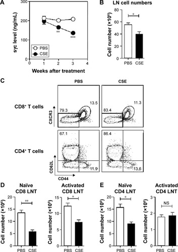
To investigate the effects of sγc in a COPD animal model induced by CSE, we first analyzed the sγc level in sera from CSE-treated mice. Interestingly, after 14 days of CSE exposure, the sγc level started to significantly decrease, with further descending tendencies at later time points (). Because T cells are a major source of sγc, we specifically analyzed the T cell profile and response in the peripheral lymph node (LN) of CSE or control mice. CSE significantly reduced the total number of LN cells (), and we also observed that the numbers of naïve (CD44lowCXCR3−) and activated (CD44hiCXCR3+) CD8 T cells were decreased by CSE (). Next, we examined the population of CD4 T cells and found that there was a significant reduction in the number of naïve (CD44lowCD62Lhi) CD4 LNT cells, while the number of activated (CD44hiCD62Llow) CD4 LNT cells was unaffected by CSE (). This translated into increased percentages of activated CD4 T cells in the CSE group (), since the number of overall LN cells was decreased (). Since sγc production is enhanced in activated T cells,Citation22 these data indicate that a reduction in the sγc level by CSE results from a diminished cell number, specifically activated T cells.
Figure 1 sγc levels and T cell profiles in COPD-induced WT mice.
Abbreviations: sγc, soluble form of common gamma chain; WT, wild type; CSE, cigarette smoke extract; PBS, phosphate-buffered saline; SEM, standard error of the mean; LN, lymph node; LNT, lymph node T; NS, not significant.
LN cellularity is rescued by sγc overexpression from CSE
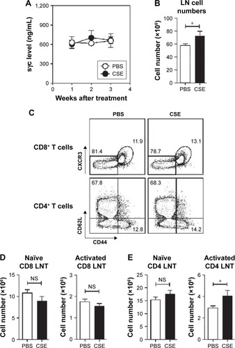
To assess the impact of decreased sγc expression in COPD mice, we analyzed sγc transgenic (sγcTg) mice, in which sγc is specifically overexpressed in T lineage cells under the control of a human CD2 promoter.Citation22 Contrary to wild-type (WT) mice with COPD, overexpressed sγc significantly promoted total LN cell numbers (), and we found that there was no decrease in the number of CD8 and CD4 T cells () and an increase in the number of activated CD4 T cells in sγcTg mice with COPD (). Collectively, although it is unclear why the sγc levels were reduced by CSE, decreased sγc expression may directly be related to reduced LN cell numbers, as the overexpression of sγc prevents the reduction in the number of LN cells.
Figure 2 T cell profiles in COPD-induced sγcTg mice.
Abbreviations: sγcTg, soluble form of common gamma chain transgenic; CSE, cigarette smoke extract; PBS, phosphate-buffered saline; SEM, standard error of the mean; LN, lymph node; LNT, lymph node T; sγc, soluble form of common gamma chain; NS, not significant.
sγc overexpression impairs LNT cell response in CSE
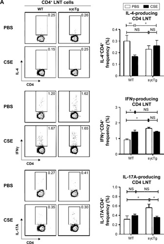
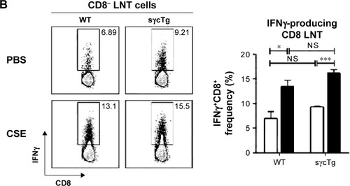
To investigate the functional effect of sγc in LNT cell in response to CSE, LN cells from CSE mice were stimulated with PMA (phorbol 12-myristate 13-acetate)/ionomycin, and the cytokine profiles were analyzed. The percentage of IL-4+ CD4 T cells was reduced, but IFNγ was increased in CD4 LNT cells from WT mice with COPD compared with the PBS control mice, suggesting that CSE acts as an inducer of Th1 (, top and middle). Th17 differentiation was not observed in LN under CSE treatment ( bottom). Th1 immunity skewed by CSE was not observed in sγcTg mice compared with CD4 T cell differentiation in WT mice with COPD, implying that sγc overexpression impairs CD4 LNT cell response to CSE. Consequently, CD4 T cells in CSE-treated WT LN were more skewed to Th1 compared to those in PBS-treated WT LN, while Th1 differentiation and Th2 differentiation in PBS- or CSE-treated LNs of sγcTg mice were comparable. Interestingly, reduction in Th17 differentiation was observed in CSE-treated LNs of sγcTg mice. Next, we tested CD8 LNT cell response and found that IFNγ-producing CD8 LNT cells were increased in both WT and sγcTg mice by CSE (). These data suggest that a downregulation of sγc expression in the COPD animal model may be one mechanism to specifically dampen T cell response in LN.
Figure 3 Cytokine profiles in COPD-induced WT and sγcTg LNT cells.
Abbreviations: WT, wild type; sγcTg, soluble form of common gamma chain transgenic; LNT, lymph node T; CSE, cigarette smoke extract; PBS, phosphate-buffered saline; SEM, standard error of the mean; NS, not significant; PMA, phorbol 12-myristate 13-acetate.
sγc overexpression enhances inflammatory response in respiratory tracts
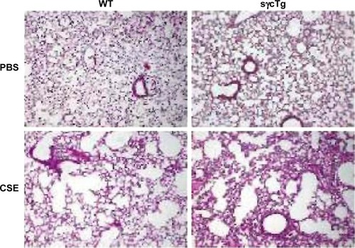
To further determine the biological function of decreased sγc expression in CSE, we assessed the infiltration of inflammatory immune cells into the lung tissue of COPD. We found that there was a significant increase in immune cell infiltration in sγcTg mice (). The numbers of infiltrating TCR+ cells were not shown to be different between WT and sγcTg mice with COPD, while the TCR− cells in sγcTg mice were more infiltrated by CSE compared with WT mice (). To address whether in vivo sγc upregulation had any effect on the infiltrated T cells into the respiratory tract, we stimulated the infiltrated cells with PMA/ionomycin and examined the ex vivo cytokine production profiles of CD8+ and CD4+ T cells. Interestingly, sγcTg CD4+ T cells were more skewed to Th1 and Th17 than were WT CD4+ T cells in CSE (); whereas IFNγ and IL-17 productions of CD8 T cells were not affected (). Accordingly, decreased sγc level in COPD results from the prevention of a Th1- and Th17-cell-prone proinflammatory environment. Indeed, the lung alveolar sections of CSE-exposed WT and sγcTg mice were stained by H&E, and the data showed that sγcTg mice exhibited more severe interstitial edema, alveolar wall thickening, and inflammatory cell infiltration compared with WT mice, while there were no significant histological differences between PBS-exposed WT versus sγcTg mice (). Collectively, these data implicate that IL-17 environment with increased Th17 may recruit more non-T cells such as neutrophils, eosinophils, and macrophagesCitation29,Citation30 and it may induce more severe immunopathogenesis of COPD.
Figure 4 Cytokine profiles in COPD-induced WT and sγcTg BALF cells.
Notes: (A) Infiltrating cell numbers in BALF from WT and sγcTg mice exposed to CSE for 3 weeks. Data are presented as the mean and SEM of four WT and five sγcTg mice. (B) Infiltrating pattern of T or non-T cells in BALF from WT and sγcTg mice exposed to CSE for 3 weeks. Data are presented as the mean and SEM of four WT and five sγcTg mice. (C) BALF CD8 (top) or CD4 (bottom) T cells from CSE or PBS-treated WT and sγcTg mice were stimulated for 3 h with PMA and ionomycin and assessed for IFN-γ and IL-17A expression by intracellular staining. Dot blots are representative of four to five mice per group. (D) Bar graph shows percent (%) IFN-γ (left)- or IL-17A (right)-producing CD8 BALF T cells. Error bars represent the mean and SEM of four to five mice per group. (E) Bar graph shows percent (%) IFN-γ (left)- or IL-17A (right)-producing CD4 BALF T cells. Error bars represent the mean and SEM of four to five mice per group. *P<0.05, **P<0.01, and ***P<0.001.
Abbreviations: WT, wild type; sγcTg, soluble form of common gamma chain transgenic; BALF, bronchoalveolar lavage fluid; CSE, cigarette smoke extract; SEM, standard error of the mean; PBS, phosphate-buffered saline; NS, not significant; PMA, phorbol 12-myristate 13-acetate.
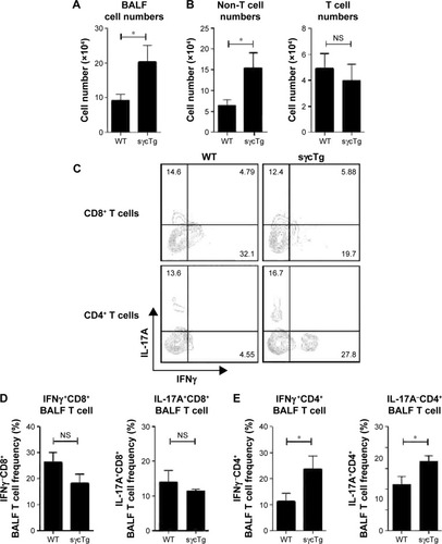
Figure 5 Histopathological evaluation from COPD-induced WT and sγcTg lung.
Abbreviations: WT, wild type; sγcTg, soluble form of common gamma chain transgenic; PBS, phosphate-buffered saline; CSE, cigarette smoke extract.
Discussion
COPD is characterized by septal tissue damage, remodeling of small airways, airway obstruction, and subsequent decline in lung function.Citation1 It has been conventionally known that inflammatory responses by innate and adaptive inflammatory immune cells contribute to lung tissue damage.Citation1,Citation31,Citation32 Previous studies have reported that the levels of Th1- and Th17-related cytokines in lung tissues of COPD patients are increased and play a pivotal role in the progression of COPD.Citation33,Citation34 Since sγc was identified as regulators of Th1 and Th17 differentiation,Citation22 it is logical that sγc may closely be related to the immunopathogenesis of COPD. Here, we assessed the pattern of sγc expression on COPD development and found that the expression of sγc was dramatically reduced in a CSE-induced COPD mouse model. To elucidate the biological role of its reduction, we analyzed sγcTg mice and showed that an increased sγc production more skewed CD4 T cells to Th1 and Th17, which resulted in more severe lung tissue damage in COPD mice. These findings propose a new regulatory mechanism in COPD immunopathogenesis that the downregulation of sγc expression is one of the defense mechanisms from excessive inflammation by CSE. Inflammatory immune response is more induced in the sγcTg animal model in which the regulatory mechanism in sγc expression is compromised. Collectively, this study reports a new regulatory role for sγc in enhancing the progression of COPD, and it implicates that sγc can be a novel target to control COPD immunopathogenesis caused by CS.
CS changes a broad range of immunological functions, including innate and adaptive immune responses.Citation24 It has been surmised that many of the health consequences of CS are due to its negative effects on the immune system. One of the adverse effects is an autoreactive CD8 T cell response.Citation35 CS triggers tissue cell death and the release of self-antigens. These events induce DC maturation and generate self-antigen specific cytotoxic CD8 T cells that induces more deterioration of tissue injury.Citation24 Thus, regulatory mechanisms are operated to control self-reactive T cells at the thymicCitation36 and peripheral levels.Citation37,Citation38 The autoreactive T cells are negatively selected in the thymus and controlled by Treg cells in the periphery. IL-2, which plays a critical role in the development and homeostasis of Treg cells,Citation39 is elevated in COPD patients who show disease stability,Citation40 inducing dominant upregulation of Treg cells in smokers with preserved lung function compared with COPD patients.Citation41 As our previous studies demonstrated that sγc inhibits IL-2 signaling,Citation22 a high level of sγc leads to impaired IL-2 signaling, resulting in the inhibition of Treg cell function and survival.Citation39 This suggests that the low level of sγc in a CSE animal model may result in the prevention of COPD progression by restricting excessive T cell response with IL-2-induced Treg cells. On the other hand, Treg development depends on IL-2 and TGFβ, which is also linked to Th17 differentiation.Citation42,Citation43 The function of Th17 in COPD is less known; an enhancement of Th17 differentiation was observed in peripheral blood and bronchial biopsies of COPD patients compared to those of healthy control.Citation44,Citation45 We found that CD4 T cells in sγcTg mice were much more skewed to Th17 compared to WT mice with CSE treatment, indicating that more IL-17 production from increased Th17 cell differentiation may recruit more inflammatory responsible cells in COPD. Indeed, IL-17 may induce chemokine secretionCitation46–Citation48 and promote the infiltration of neutrophils and macrophages to peripheral airway.Citation29,Citation30 It may partially explain the reason for the increased numbers of non-T cells in BALF from sγcTg mice with CSE treatment in our study. If the expression of sγc is regulated and maintained at low levels in smokers, CS-mediated disease stability may be maintained over a long period or it is possible for disease severity to be ameliorated. Whether this is indeed the case remains to be tested.
COPD is a typical disease induced by chronic inflammation.Citation24 It has been known that Th1 and Th17 cells act as helper T cells to robustly induce the recruitment of inflammatory cells and tissue damage in chronic inflammation. As shown in sγcTg mice with COPD that exhibited the enhanced Th1 and Th17 differentiation, sγc may play a critical role in chronic inflammation. Although the downregulation of sγc expression as part of physiological defense mechanism is induced to prevent severe COPD progress, Th1 and Th17 differentiation is induced in a COPD animal model. Therefore, we speculate that a complete block of sγc expression or function may inhibit the progression of COPD or maintain disease stability. In sum, the regulatory role of sγc on the progression and exacerbation of COPD in sγcTg mice put forward a model of inflammation regulatory mechanism that requires the integration of a role of sγc in controlling inflammation.
Author contributions
All authors contributed toward data analysis, drafting and critically revising the paper and agree to be accountable for all aspects of the work.
Acknowledgments
This work was supported by a 2-year research grant of Pusan National University.
Disclosure
The authors report no conflicts of interest in this work.
References
- PauwelsRABuistASMaPJenkinsCRHurdSSGlobal strategy for the diagnosis, management, and prevention of chronic obstructive pulmonary disease: National Heart, Lung, and Blood Institute and World Health Organization Global Initiative for Chronic Obstructive Lung Disease (GOLD): executive summaryRespir Care200146879882511463370
- BarnesPJChronic obstructive pulmonary diseaseN Engl J Med2000343426928010911010
- SaettaMTuratoGMaestrelliPMappCEFabbriLMCellular and structural bases of chronic obstructive pulmonary diseaseAm J Respir Crit Care Med200116361304130911371392
- ByronKAVarigosGAWoottonAMIL-4 production is increased in cigarette smokersClin Exp Immunol19949523333368306509
- de HeensGLvan der VeldenULoosBGCigarette smoking enhances T cell activation and a Th2 immune response; an aspect of the pathophysiology in periodontal diseaseCytokine200947315716119616447
- NakamuraYMiyataMOhbaTCigarette smoke extract induces thymic stromal lymphopoietin expression, leading to T(H)2-type immune responses and airway inflammationJ Allergy Clin Immunol200812261208121418926564
- SmelterDFSathishVThompsonMAPabelickCMVassalloRPrakashYSThymic stromal lymphopoietin in cigarette smoke-exposed human airway smooth muscleJ Immunol201018553035304020660708
- LiuYJSoumelisVWatanabeNTSLP: an epithelial cell cytokine that regulates T cell differentiation by conditioning dendritic cell maturationAnnu Rev Immunol20072519321917129180
- VassalloRTamadaKLauJSKroeningPRChenLCigarette smoke extract suppresses human dendritic cell function leading to preferential induction of Th-2 primingJ Immunol200517542684269116081845
- Di StefanoACaramoriGCapelliASTAT4 activation in smokers and patients with chronic obstructive pulmonary diseaseEur Respir J2004241788515293608
- SzaboSJKimSTCostaGLZhangXFathmanCGGlimcherLHA novel transcription factor, T-bet, directs Th1 lineage commitmentCell2000100665566910761931
- AgnelloDLankfordCSBreamJCytokines and transcription factors that regulate T helper cell differentiation: new players and new insightsJ Clin Immunol200323314716112797537
- MajoJGhezzoHCosioMGLymphocyte population and apoptosis in the lungs of smokers and their relation to emphysemaEur Respir J200117594695311488331
- O’ShaughnessyTCAnsariTWBarnesNCJefferyPKInflammation in bronchial biopsies of subjects with chronic bronchitis: inverse relationship of CD8+ T lymphocytes with FEV1Am J Respir Crit Care Med199715538528579117016
- SaettaMBaraldoSCorbinoLCD8+ve cells in the lungs of smokers with chronic obstructive pulmonary diseaseAm J Respir Crit Care Med1999160271171710430750
- SaettaMDi StefanoATuratoGCD8+ T-lymphocytes in peripheral airways of smokers with chronic obstructive pulmonary diseaseAm J Respir Crit Care Med19981573 pt 18228269517597
- SteinmanLState of the art. Four easy pieces: interconnections between tissue injury, intermediary metabolism, autoimmunity, and chronic degenerationProc Am Thorac Soc20063648448616921119
- ChrysofakisGTzanakisNKyriakoyDPerforin expression and cytotoxic activity of sputum CD8+ lymphocytes in patients with COPDChest20041251717614718423
- LiebermanJThe ABCs of granule-mediated cytotoxicity: new weapons in the arsenalNat Rev Immunol20033536137012766758
- VernooyJHMollerGMvan SuylenRJIncreased granzyme A expression in type II pneumocytes of patients with severe chronic obstructive pulmonary diseaseAm J Respir Crit Care Med2007175546447217138956
- RochmanYSpolskiRLeonardWJNew insights into the regulation of T cells by gamma(c) family cytokinesNat Rev Immunol20099748049019543225
- HongCLuckeyMALigonsDLActivated T cells secrete an alternatively spliced form of common gamma-chain that inhibits cytokine signaling and exacerbates inflammationImmunity201440691092324909888
- ParkJYJoYKoESoluble gammac cytokine receptor suppresses IL-15 signaling and impairs iNKT cell development in the thymusSci Rep201663696227833166
- CosioMGSaettaMAgustiAImmunologic aspects of chronic obstructive pulmonary diseaseN Engl J Med2009360232445245419494220
- van der ToornMSlebosDJde BruinHGCigarette smoke-induced blockade of the mitochondrial respiratory chain switches lung epithelial cell apoptosis into necrosisAm J Physiol Lung Cell Mol Physiol20072925L1211L121817209140
- HalbowerACMasonRJAbmanSHTuderRMAgarose infiltration improves morphology of cryostat sections of lungLab Invest19947111491537518881
- DeVossJJAndersonMSLessons on immune tolerance from the mono-genic disease APS1Curr Opin Genet Dev200717319320017466510
- ChenJPetrusMBryantBRInduction of the IL-9 gene by HTLV-I Tax stimulates the spontaneous proliferation of primary adult T-cell leukemia cells by a paracrine mechanismBlood2008111105163517218339896
- CurtisJLFreemanCMHoggJCThe immunopathogenesis of chronic obstructive pulmonary disease: insights from recent researchProc Am Thorac Soc20074751252117878463
- HoshinoHLaanMSjöstrandMLötvallJSkooghBELindenAIncreased elastase and myeloperoxidase activity associated with neutrophil recruitment by IL-17 in airways in vivoJ Allergy Clin Immunol20001051 pt 114314910629464
- ParkerLCPrinceLRSabroeITranslational mini-review series on Toll-like receptors: networks regulated by Toll-like receptors mediate innate and adaptive immunityClin Exp Immunol2007147219920717223959
- PasareCMedzhitovRToll-like receptors and acquired immunitySemin Immunol2004161232614751760
- ShanMChengHFSongLZLung myeloid dendritic cells coordinately induce TH1 and TH17 responses in human emphysemaSci Transl Med2009144ra10
- ZivadinovRWeinstock-GuttmanBHashmiKSmoking is associated with increased lesion volumes and brain atrophy in multiple sclerosisNeurology200973750451019687451
- LeeSHGoswamiSGrudoAAntielastin autoimmunity in tobacco smoking-induced emphysemaNat Med200713556756917450149
- AndersonGLanePJJenkinsonEJGenerating intrathymic microenvironments to establish T-cell toleranceNat Rev Immunol200771295496317992179
- OuyangWBeckettOFlavellRALiMOAn essential role of the Forkhead-box transcription factor Foxo1 in control of T cell homeostasis and toleranceImmunity200930335837119285438
- TakahashiTKuniyasuYTodaMImmunologic self-tolerance maintained by CD25+CD4+ naturally anergic and suppressive T cells: induction of autoimmune disease by breaking their anergic/suppressive stateInt Immunol19981012196919809885918
- FontenotJDRasmussenJPGavinMARudenskyAYA function for interleukin 2 in Foxp3-expressing regulatory T cellsNat Immunol20056111142115116227984
- D’ArmientoJMScharfSMRothMDEosinophil and T cell markers predict functional decline in COPD patientsRespir Res20091011319925666
- BarcelóBPonsJFerrerJMSauledaJFusterAAgustíAGPhenotypic characterisation of T-lymphocytes in COPD: abnormal CD4+CD25+ regulatory T-lymphocyte response to tobacco smokingEur Respir J200831355556218057064
- WeaverCTHattonRDInterplay between the TH17 and TReg cell lineages: a (co-)evolutionary perspectiveNat Rev Immunol200991288388919935807
- LeeYKMukasaRHattonRDWeaverCTDevelopmental plasticity of Th17 and Treg cellsCurr Opin Immunol200921327428019524429
- Vargas-RojasMIRamírez-VenegasALimón-CamachoLOchoaLHernández-ZentenoRSansoresRHIncrease of Th17 cells in peripheral blood of patients with chronic obstructive pulmonary diseaseRespir Med2011105111648165421763119
- Di StefanoACaramoriGGnemmiIT helper type 17-related cytokine expression is increased in the bronchial mucosa of stable chronic obstructive pulmonary disease patientsClin Exp Immunol2009157231632419604272
- KornTBettelliEOukkaMKuchrooVKIL-17 and Th17 cellsAnnu Rev Immunol20092748551719132915
- LanckackerEARobaysLJJoosGFVermaelenKYA new danger in the air: how pulmonary innate immunity copes with man-made airborne xenobioticsJ Innate Immun2010229610620375628
- RuddyMJShenFSmithJBSharmaAGaffenSLInterleukin-17 regulates expression of the CXC chemokine LIX/CXCL5 in osteoblasts: implications for inflammation and neutrophil recruitmentJ Leukoc Biol200476113514415107456