Abstract
The angiosome principle was first described by Jan Taylor in 1987 in the plastic reconstructive surgery field, providing useful information on the vascular anatomy of the human body. Specifically concerning foot and ankle pathology, it may help the clinician to select better vascular access and specific strategies for revascularization. This knowledge may be particularly beneficial when treating diabetic neuroischemic foot wounds associated with particularly aggressive atherosclerotic disease and a poor collateral circulation. The implementation of angiosome-based strategies in diabetic infragenicular vascular reconstruction may afford encouraging wound healing and limb preservation rates using both bypass and endovascular techniques. The minimal invasiveness of these novel strategies enables us to perform more specific and more distal tibial and/or foot arterial reconstructions, in one or multiple targeted vessels. This paper reviews the available literature on this revascularization strategy and focuses on the potential benefit of angiosome-guided primary angioplasty for diabetic ischemic foot ulcers.
Introduction
Although surgical bypass still plays a key role in revascularization of critical limb ischemia (CLI),Citation1 increasing clinical experience over the past two decades shows encouraging results for primary endovascular strategies, with acceptable feasibility, low complication rates,Citation2–Citation4 and limb salvage rates comparable with surgery.Citation2–Citation5 Diabetes mellitus is becoming increasingly common in CLI presentations featuring pro-longed foot inflammation or tissue necrosis. This currently involves frail patients at high perioperative risk and affected by numerous vascular comorbidities.Citation1,Citation2 This specific patient population, with systemic atherosclerosis, neuroischemic limb ulcers, gangrene, and sepsis (the so-called “diabetic foot syndrome”) is prone to a higher rate of periprocedural surgical complications.Citation1,Citation2,Citation5 Therefore, endovascular techniques may have many applications in this fieldCitation2 because of their low invasiveness, absence of scarring, and lack of need for venous conduits.Citation2,Citation6,Citation7 These strategies seem to have the advantages of enabling simultaneous multiple vessel recanalization with high reproducibility if necessary,Citation6,Citation7 resulting in shorter hospital stays and health care expenditure.Citation1–Citation7
Common revascularization issues in diabetic patients
The diabetic ischemic foot, because of its multifaceted pathology, presents well recognized challenges for the vascular interventionist. Chronically and critically oxygen-deprived tissues in these frail patients often develop ulcers, extended sepsis, and limb edema. Heavily calcified and occluded calf and foot arteries make all types of vascular reconstruction more difficult.Citation1,Citation2,Citation6 Not surprisingly, the main infragenicular arterial axis that supplies the distal wound territory is frequently affected by severe atherosclerotic disease.Citation4,Citation8–Citation10 Current critical limb ischemia data suggest that bypass using vein grafts achieves 79%–90% limb salvage rates at 1 year,Citation8 while below-knee angioplasty affords technically successful recanalization rates of up to 80%, with comparable rates of limb preservation.Citation1,Citation5,Citation8 However, these series have a heterogeneous Rutherford grade II–III component and class 4–6 presentation.Citation1,Citation8 It is generally accepted for distal bypass and endovascular reconstructions that the outflow vessel based on angiographic details modulates the strategy for vascular reconstruction and enables technical success and good postoperative patency.Citation1,Citation8 From this perspective, the aim is to reirrigate the distal ischemic wound territory directly, but is often achieved indirectly via collaterals surrounding the diseased zone. The decision-making process often focuses on the most suitable artery to be reopened or a patent distal vessel to anchor the bypass.Citation1,Citation2,Citation6–Citation8
In the last two decades, some authors have proposed complementary topographic orientations in critical limb ischemia revascularization, because the choice of the most appropriate target vessel starts from a clinical point of view, ie, the wound location.Citation9–Citation11 They have described particular tissue sectors of the human body depending on specific arterial and venous irrigation sources, named “angiosomes”.Citation9–Citation14 The distribution of these angiosomes was eventually sought to harmonize already used arterial reconstruction techniques.Citation10–Citation12
Angiosomes and limb salvage
Parallel efforts to decrease diabetic limb losses have unfolded in the field of plastic reconstructive surgery, by promoting the angiosome model of perfusion.Citation9–Citation14 This concept is based on the anatomical studies of Taylor et alCitation9,Citation12 and Attinger et al,Citation10,Citation11,Citation13,Citation14 who pioneered preferential strategies for surgical access, tissue reconstruction, and amputation, following specific three-dimensional tissue sectors of the body, ie, angiosomes in specific arteriovenous bundles.
Practical implementation of the angiosome concept in daily limb salvage bypass or transcatheter techniques started only a few years ago. Although few results have been published as yet, and mainly in the vascular interventional field, retrospective studies seem to favor this hypothesis.Citation14–Citation17 In a retrospective surgical series of 48 patients, Neville et alCitation15 found a 91% healing rate with a 9% amputation rate in the angiosome-guided group vs a 62% healing rate and 38% amputation rate in the nonangiosome-guided group. In 203 consecutive limbs with critical ischemia and ischemic ulcerations undergoing endovascular reconstruction, Iida et alCitation18 reported limb preservation in 86% of the angiosome-related group compared with 69% in the nonspecific treatment group. Consistent with these reports, in a similar series of 76 ischemic ulcers treated by both bypass and endovascular therapies, Varela et alCitation19 noted significant better results for wound healing (92% vs 73%) and limb salvage (93% vs 72%) in the angiosome-guided cohort of patients. We have previously published our experience in 98 diabetic patients with ischemic foot ulcers treated by angiosome-oriented primary angioplasty, with similarly encouraging results for limb preservation (91%, 88%, and 84% at 12, 24, and 36 months, respectively) and wound healing (85%, 81%, and 73% at the same time intervals).Citation17
In practical terms, the angiosome concept delineates the human body into three-dimensional tissue blocks irrigated by specific arteriovenous sources.Citation9–Citation12 Adjacent angiosomes are interconnected by numerous “choke vessels” that create an integrated compensatory system between different territories.Citation9–Citation14 Although not part of everyday practice, this model already has some contemporary clinical applications, including specific myocardial revascularization, selective visceral embolization, characteristic types of covering flaps,Citation9,Citation12 incisions or amputationsCitation10,Citation14 in plastic reconstructive surgery,Citation9–Citation14 and, more recently, targeted bypass or endovascular revascularization for limb salvage in critical limb ischemia.Citation11,Citation13–Citation19 In the lower limb, five angiosomes were initially describedCitation9,Citation12,Citation13 in the foot and lower ankleCitation9–Citation11,Citation13 emerging from the three main tibial trunks, ie, the anterior and posterior tibial arteries joined to the peroneal vessels.
Schema for foot and lower ankle angiosomes
Schematically, the distribution of angiosomesCitation12–Citation17 in the foot encompasses: the medial calcaneal, medial plantar, and lateral plantar artery angiosomes derived from the posterior tibial artery supplying the entire plantar heel and the medial and lateral plantar surface beyond the toes; the dorsalis pedis angiosome, which extends from the anterior tibial artery, supplying the dorsum of the foot and toes, and also upper anterior perimalleolar vascularization; and the lateral calcaneal artery angiosome (optionally adding the lateral perimalleolar artery angiosome) derived from the peroneal artery, covering the lateral and plantar heel and succeeding the anterior peroneal perforating branch, that connects the latter to the anterior tibial territory (). Going up to the superior ankle and distal calf zones, other angiosome territories have been identified, including the anterior perforator artery angiosome (from the peroneal artery) and the lateral malleolar angiosomes with the corresponding medial malleolar angiosomes from the anterior-tibial artery.Citation9,Citation11,Citation12 Some of these perimalleolar angiosomes have been additionally considered by recent reports of revascularization procedures.Citation15,Citation16,Citation18 A few examples of current angiosome-guided endovascular interventions for diabetic neuroischemic foot wounds are shown in –.
Figure 1 A simplified illustration of previously suggested angiosomes of the foot and lower ankle.
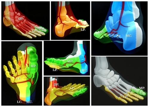
Figure 2 Selective revascularization of the posterior tibial and lateral plantar artery angiosome: (a–f) Staged angioplasties in the posterior tibial artery. (g–i) Selective angioplasties in the lateral plantar artery and its appended angiosome.
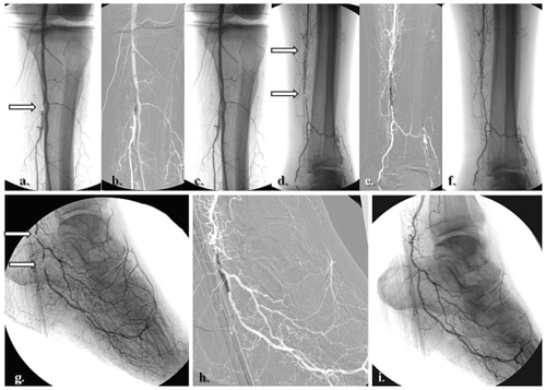
Figure 3 Clinical correspondence of the angiographic pattern showed in . A neuroischemic plantar ulcer in an acute diabetic foot presentation: (a) Initial clinical aspect featuring a lateral plantar artery hypoperfusion and sole forefoot abscess. (b) and (c) Abscess drainage and debridement. (d) and (e) Clinical evolution at weeks 3 and 5.
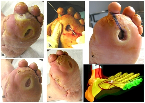
Figure 4 Selective revascularization of the distal posterior tibial artery and its appended medial plantar artery angiosome. (a) and (b) Selective angioplasty in the posterior tibial artery, and (c) and (d) specific revascularization of the medial plantar artery and its angiosome.
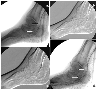
Figure 5 A right hallux neuroischemic sole ulceration matching the angiographic features showed in . There is medial plantar artery hypoperfusion in an end-artery occlusive disease pattern for the first toe. (a) Initial presentation and (b) clinical evolution at 1 month after angioplasty. (c) Results 2 months later.
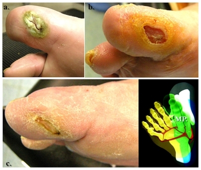
Figure 6 Global medial and lateral plantar artery critical ischemia and acute diabetic foot syndrome matching staged occlusions in the posterior tibial artery (with end-artery occlusive disease model to the sole). (a) Prime posterior tibial artery staged subocclusive lesions. (b) and (c) The reestablished flow in the posterior tibial and both right plantar arteries. (d) The initial clinical aspect. (e) Subsequent evolution at 3 weeks. (f) Clinical results after 5 months of team surveillance.
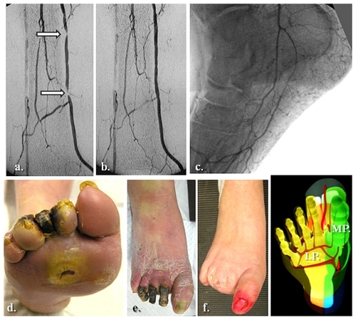
Figure 7 Selective anterior tibial and related dorsalis pedis artery angiosome: (a–c) Primary staged anterior tibial angioplasty, (d) initial dorsal foot ulceration, (e) clinical results at 1 month following (f–h) associated dorsalis pedis selective angioplasty.
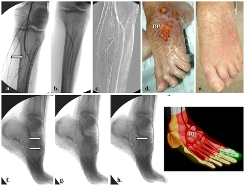
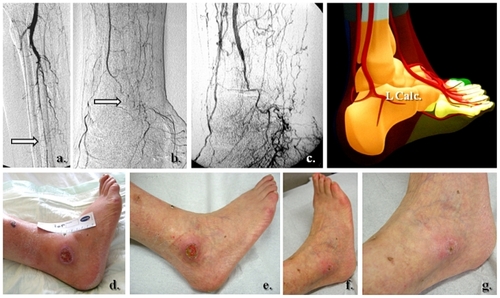
Figure 8 Specific posterior tibial and adjacent medial calcaneal artery angiosome revascularization in heel related wound. (a) Tight stenosis in the distal posterior tibial artery, above emergence of the medial calcaneal branch, (b) Angiographic result after selective angioplasty, (c) initial presentation of heel ulcer, and (d) clinical results after 5 weeks of team surveillance.
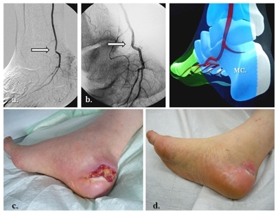
Figure 9 Lateral calcaneal artery and peroneal main flow-related angiosome ulceration. (a, b) Initial pattern of perfusion featuring the peroneal artery as single and severely diseased (end-artery occlusive model) calf vessel, (c) re-established flow in the peroneal territory, (d) prime aspect of lateral calcaneal and inframalleolar tissue defect, and (e–g) subsequent clinical evolution at weeks 1, 5, and 6.
The diabetic foot is a preferential application for topographic revascularization. Availability of the angiosome strategy for infragenicular revascularization seems to represent a particular interest in diabetic patients for several reasons. It has been suggested that the neuroischemic diabetic foot is associated with more distal and aggressive atherosclerotic macroangiopathy, as well as functional microcirculatory impairment induced by both neuropathy and local sepsis.Citation20,Citation21 In this specific context, featuring multiple blockades of large and medium-sized foot arteries,Citation20 O’Neal has outlined the concept of diabetic end-artery occlusive disease.Citation21 This hypothesis considers critical irrigation to the foot, whereby medium-sized “patchy atherosclerosis” is associated with acute septic thrombosis and loss of small collaterals with surrounding inflammation.Citation21 The end-artery occlusive disease theoryCitation21 probably explains better why irrigation from a few millimeters of skin to the entire diabetic foot or legCitation11,Citation16,Citation17,Citation21 relies on specific nourishing vessels, although solely hinged to one specific dominant angiosome-dependent artery.Citation9–Citation12 Consequently, it might be emphasized that in these subjects, the more distal and specific the revascularization, the higher the probability of re-establishing an adequate blood supply in a specific amount of threatened tissue.
The association between the end-artery occlusive disease theoryCitation21 and the broader angiosome concept may reveal promising results in diabetic wound regeneration,Citation13,Citation15–Citation19,Citation22 particularly if specific pedal and/or plantar revascularizations are added to those of the tibial trunkCitation14,Citation17,Citation18,Citation23 in single interventions. This also underscores the advantage of endovascular strategies to allow multiple below-knee and below-ankle vessel reconstructions, in addition to surgery. On the complex background of a diabetic foot, the angiosome concept may equally allow vascular interventionists to select more specific wound-oriented vascular reconstruction with probably a better prognosis in terms of tissue healing, as suggested in recent reports by Setacci et al,Citation16 Clemens and Attinger,Citation22 and Bazan.Citation24 Beyond the initial clinical orientation (focusing on the specific location of foot ulcers, –), other laboratory examinations may help the clinician to choose the appropriate angiosome for revascularization, ie, Doppler assessment of the dominant foot arch,Citation11–Citation13 angiographic computed tomographic or magnetic resonance evaluation of homolateral and contralateral limb arterial patterns,Citation17 and transcutaneous oxygen pressure monitoring stratification in adjacent angiosomes.Citation17
Challenging endovascular procedures in diabetic infragenicular trunks
, , , and show the main categories of targeted angiosome revascularization and indicate the presence of specific calcifications in the tibial, dorsal foot, or plantar diabetic arteries. These calcific deposits represent one of the major technical concerns for the vascular interventionist.Citation2,Citation6,Citation17 Inasmuch as nondiabetic subjects seem to develop intimal, eccentric, and patchy arterial wall calcific deposits (ie, type I calcifications),Citation25 diabetic infrapopliteal atherosclerosis affects the medial layer, with mostly concentric continuous wall calcifications (“Mönckeberg sclerosis” or type II calcification).Citation25 Although the precise etiology is unknown, their severity and spread have been related to the duration of diabetes, concomitant autonomic neuropathy, and specific alterations in the vasa vasorum.Citation10,Citation25 Type II calcifications undoubtedly present challenges for revascularization using endoluminal or surgical techniques in these rigid conduits with a diameter of only 2 or 3 millimeters.Citation3,Citation17,Citation23,Citation26 This observation becomes even more manifest when choosing an angiosome orientation for tibial revascularization,Citation17,Citation19,Citation26 acknowledging that the appropriate angiosome-dependent artery may not necessarily be the simplest vessel to treat.Citation15–Citation17 It has been shown that severely ischemic territories with tissue loss more often include long segments of chronic and calcific arterial occlusions in the corresponding angiosome-related vessels.Citation16–Citation19
Another challenging factor to be faced by the vascular specialist when planning treatment of a diabetic ulcer is the control of local neuropathy. Beyond chronic exposure of the foot with sensory loss to microtrauma, skin tears, and deformation, neuropathy adds characteristic functional microcirculatory impairmentCitation16 by autonomic denervation.Citation16,Citation20,Citation21 The latter strongly affects residual perfusion of the skin by capillary steal, impairing the local healing processCitation16,Citation21 and consequently the distal arterial runoff to the foot (an important consideration in any vascular reconstruction).Citation16,Citation20 An additional element of the diabetic environment is local sepsis. This component of the diabetic foot puzzle commonly causes purulent collections, swelling, and adjacent hyperpressure in the form of foot compartment syndromes.Citation20,Citation21 A consequent neural (peripheral entrapment) and remote arterial collateral depletion has been described.Citation20,Citation21,Citation26 Therefore, the compensatory capillary network of angiosomes (ie, “choke” vessels)Citation9–Citation14,Citation16,Citation26 seems strongly determined by the duration of diabetes and chronic inflammation,Citation20,Citation21,Citation26 indicating the need to treat ischemic foot areas by more distal and more specific vascular reconstructions.Citation15–Citation17 These specific diabetic foot challenges seem to increase the postoperative risk in terms of tissue recoveryCitation6,Citation14,Citation16,Citation17,Citation20,Citation26 which may still jeopardize limb salvage after successful revascularization.Citation14,Citation16,Citation21,Citation26 Because the angiosome strategy seems to be advantageous when performing revascularization,Citation14–Citation16,Citation18,Citation24 with particular utility in the diabetic foot ischemic model,Citation13,Citation17,Citation22 it seems reasonable to predict an increasing role for this conceptCitation16,Citation17,Citation19,Citation24 in an era of expanding endovascular applications, including the “first approach”Citation26–Citation29 and hybrid surgical and transcatheter procedures.Citation27,Citation28
Limitations and challenges of angiosome-based strategies
Our group has encountered two main challenges while using the angiosome strategy for infragenicular angioplasty. First, the angiosome concept of revascularization has shifted our indications from “which vessel is most suitable for revascularization” to a multidisciplinary clinical consideration of “which region of perfusion governed by which artery should be treated?” This policy has engaged our vascular group in more laborious procedures, acknowledging that the most suitable angiosome-dependent artery to treat may not necessarily be the simplest vessel to recanalize.Citation15–Citation19 As emphasized earlier, extended calcifications in long segment chronic occlusions or tight stenoses are commonly encountered during angiosome-targeted revascularization. Following local learning curves and evolving skills, our initially observed global technical failure rate of 20%Citation17 seems to match similar critical limb ischemia angioplasty feasibility reports, ranging from 69%–85% for occlusions and 77%–95% for severe stenoses.Citation3–Citation7,Citation30,Citation31 In an attempt to address these technical limitations, we have favored antegrade approaches, long (50–70 cm) sheaths reinforced by stiff catheters (Lumax/Cook, UK or 5-French Pier, Cordis, New York, NY) and a stiff shaft with 0.018 in over-a-wire balloons (ReeKross, Clear Stream Technologies Ltd, Wexford, Ireland). Occasionally, in heavily calcified vessels, cutting balloons (Boston Inc, Fall River, MA) or blunt microdissection devices (Frontrunner, Cordis) have been used to improve access ( and ). New endovascular techniques (eg, tibial retrograde approaches) or recanalization devices based on rotational endarterectomy, excimen laser, or ultrasound debulking therapy, have seldom been used by our team, but may offer promise for mandatory topographic recanalization.Citation23,Citation26,Citation31,Citation33
The second limitation relates to occasional anatomical variation of the main angiosome boundaries between patients. Although constant in number,Citation10–Citation14 each of the main arterial bundles in the ischemic foot and ankle remains strongly dependent on the collateral supply.Citation10–Citation13,Citation18,Citation19 The end-artery occlusive disease theoryCitation21 referred to earlier emphasizes the deficiency of medium-sized and small-sized collateral networksCitation9,Citation14 in the diabetic foot syndrome, the angiosome equivalents of which being the choke vessels. Unlikely nondiabetic critical limb ischemia presentations, diabetic neuroischemic wound territories show major collateral deterioration,Citation20–Citation22,Citation32 with no opportunity for a compensatory collateral circulation.Citation21,Citation32 The amount of tissue depending on one specific arterial source to be reopened varies from a few square millimeters of skin, to the entire foot or leg.Citation21 In our experience, in presentations showing complex overlapping wounds with adjacent angiosomes with questionable collateral resources, we orient procedures toward tandem or multiple angiosome reopening, if technically feasible.
Recent reports of infragenicular-targeted revascularizations
In a recent review of our group experience based on multidisciplinary follow-up, we compared the efficacy of below-knee angioplasty in two consecutive cohorts of diabetic patients with and without the angiosome concept.Citation32 Although not reaching statistical significance for primary (P = 0.813) and secondary patency (P = 0.511), we found a significant difference in wound healing (hazards ratio [HR] 2.19, P = 0.025) and limb preservation (HR: 2.32, P = 0.035) between these consecutive groups, with better results for angiosome-guided revascularization.Citation32 In the same retrospective analysis,Citation32 although the number of initial technical failures in the angiosome-guided primary angioplasties was marginally higher (21% vs 18% for more challenging lesions), we observed no significant difference (P < 0.05) in terms of need for reintervention or perioperative morbidity and mortality between angiosome-oriented vs nonangiosome-oriented angioplasties.Citation32 Other published reports have found parallel healing/nonhealing and limb salvage rates, ie, 91% vs 62%Citation15 compared with 85% vs 76% in our groupCitation32 and 86% vs 69%,Citation18 analogous to 90% vs 84% in our registry data.Citation32 In summary, there has been equal benefit for both surgery and endovascular strategies using angiosome-guided approaches.Citation15,Citation19,Citation32
The BASIL (Bypass versus Angioplasty in Severe Ischaemia of the Leg) trialCitation5 reports similar early amputation-free survival rates for surgery and endovascular techniques in nontopographic below-knee revascularizations, although less than one third of patients had diabetes. The same prospective study did not detect a significant decrease in survival rates for diabetic subjects using both therapeutic options.Citation5,Citation31 A recent analysis conducted by O’Brien-Irr et alCitation33 studied the clinical outcome of 106 cases divided in Rutherford category 4 and 5 chronic limb ischemia presentations and following infrainguinal endovascular treatment, comprising 32% and 58% of diabetic patients, respectively. The authors found that for Rutherford category 5 cases, comparable limb salvage rates were 82%, with early wound healing occurring in only 21% of cases without target extremity revascularization. They concluded that refining patient selection and clinical indications may substantially improve the outcome of percutaneous transluminal angioplasty.Citation33 Hoping to provide more accurate information upon the tibial runoff and the collateral reserve in each critical limb ischemia pattern of presentation, Mustapha et alCitation34 recently proposed an original classification and interventional protocol for AM-guided below-knee interventions. They graded from 0–3 different types of ischemic crural patterns, adding a three-level collateral scoring system that may help the interventionist to better choose the target artery and consequent chronic total occlusion (CTO) strategy to be deployed.Citation34
Angiosome concept as a component of diabetic team work
Contemporary clinical experience suggests that using the aforementioned new technologies and principles of treatment, like the angiosome concept, a linear correlation between successful revascularization and unreserved tissue healing is difficult to ascertainCitation20,Citation26,Citation27 unless appropriate multidisciplinary surveillance is undertaken. This seems particularly true for diabetic neuroischemic foot wounds.Citation14,Citation16,Citation17,Citation20,Citation22,Citation26–Citation29 It is also generally accepted that people with diabetes should be offered specific education about preventive foot care.Citation20,Citation29,Citation35 The effectiveness of any educational program is critically linked to the availability of complementary clinical services.Citation36 Trying to merge primary (the patient) with secondary (the medical team) prevention in the diabetic foot group at our institution, we have included patients and their general practitioners as members of the team, with their active participation in all therapeutic decisions.
Conclusion
Implementation of angiosome-based strategies in infragenicular interventions may improve wound healing and limb preservation rates using bypass and endovascular techniques. It may also offer the opportunity in primary angioplasty to orient pedal/plantar revascularizations more specifically and more distally. Incorporating the angiosome model in contemporary below-knee team strategies for revascularization might be useful, although further comparative and prospective data are needed to evaluate this concept fully.
Acknowledgments
We would like to acknowledge the members of our institutional radiology, endocrine, and diabetic foot teams for their support in generating the data and figures included in this paper.
Disclosure
The authors report no conflicts of interest in this work.
References
- Norgreen L Hiatt WR Dormandy JA Inter-Society Consensus for the management of peripheral arterial disease (TASC II) Eur J Vasc Endovasc Surg 2007 33 Suppl 1 S32 S55
- Blevins WA Schneider PA Endovascular management of critical limb ischemia Eur J Vasc Endovasc Surg 2010 39 756 761 20299245
- Conrad MF Kang J Cambria RP Infrapopliteal balloon angioplasty for the treatment of chronic occlusive disease J Vasc Surg 2009 50 799 805 19786239
- Romiti M Albers M Brochado-Neto FC Meta-analysis of infrapopliteal angioplasty for chronic critical limb ischemia J Vasc Surg 2008 47 975 981 18372148
- Adam DJ Beard JD Cleveland T BASIL trial participants Bypass versus angioplasty in severe ischemia of the leg (BASIL): Multicentre, randomized controlled trial Lancet 2005 366 1925 1934 16325694
- Markose G Bolia A Below the knee angioplasty among diabetic patients J Cardiovasc Surg (Torino) 2009 50 323 329
- Faglia E Mantero M Caminiti M Extensive use of peripheral angioplasty, particularly infrapopliteal, in the treatment of ischemic diabetic foot ulcers: Clinical results of a multicentric study of 221 consecutive diabetic subjects J Intern Med 2002 252 225 232 12270002
- Simms M Peripheral vascular disease and reconstruction The Foot in Diabetes 4th ed Chichester, UK J Wiley and Sons Ltd 2007
- Taylor GI Palmer JH The vascular territories (angiosomes) of the body: Experimental studies and clinical applications Br J Plast Surg 1987 40 113 141 3567445
- Attinger CE Cooper P Blume P The safest surgical incision and amputations applying the angiosomes principle and using the Doppler to assess the arterial-arterial connections of the foot and ankle Foot Ankle Clin North Am 2001 6 745 801
- Attinger CE Evans KK Bulan E Angiosomes of the foot and ankle and clinical implications for limb salvage: Reconstruction, incisions and revascularization Plast Reconstr Surg 2006 117 7 Suppl 261S 293S 16799395
- Taylor GI Pan WR Angiosomes of the leg: Anatomic study and clinical implications Plast Reconstr Surg 1997 4 183 198
- Attinger CE Evans KK Mesbahi A Angiosomes of the foot and angiosome-dependent healing Diabetic Foot, Lower Extremity Arterial Disease and Limb Salvage Philadelphia, PA Lippincott Williams and Wilkins 2006
- Attinger CE Cooper P Blume P Vascular anatomy of the foot and ankle Op Tech Plast Reconstr Surg 1997 4 183 198
- Neville RF Attinger CE Bulan EJ Revascularization of a specific angiosome for limb salvage: Does the target artery matter? Ann Vasc Surg 2009 23 367 373 19179041
- Setacci C De Donato G Setacci F Ischemic foot: Definition, etiology and angiosome concept J Cardiovasc Surg (Torino) 2010 51 223 231
- Alexandrescu V Hubermont G Philips Y Selective angioplasty following an angiosome model of reperfusion in the treatment of Wagner 1–4 diabetic foot lesions: Practice in a multidisciplinary diabetic limb service J Endovasc Ther 2008 15 580 593 18840046
- Iida O Nanto S Uematsu M Importance of the angiosome concept for endovascular therapy in patients with critical limb ischemia Catheter Cardiovasc Interv 2010 75 830 836 20306500
- Varela C Aci NF Haro JD The role of foot collateral vessels on ulcer healing and limb salvage after successful endovascular and surgical distal procedures according to an angiosome model Vasc Endovasc Surg 2010 44 654 660
- Boulton AJM Armstrong DG The diabetic foot Clinical Diabetes, Translating Research into Practice Philadelphia, PA Saunders Elsevier 2006
- O’Neal LW Surgical pathology of the foot and clinicopathologic correlations Levin and O’Neal’s The Diabetic Foot Philadelphia, PA Mosby Elsevier 2008
- Clemens MW Attinger CE Angiosomes and wound care in the diabetic foot Foot Ankle Clin 2010 15 439 464 20682415
- Zhu YQ Zhao JG Liu F Subintimal angioplasty for below-the-ankle arterial occlusions in diabetic patients with chronic critical limb ischemia J Endovasc Ther 2009 16 604 612 19842730
- Bazan HA Think of the angiosome concept when revascularizing the patient with critical limb ischemia Catheter Cardiovasc Interv 2010 75 837 20432387
- Irvin CL Guzman RJ Matrix metalloproteinases in medial arterial calcification: Potential mechanisms and actions Vascular 2009 17 Suppl 1 S40 S44 19426608
- Alexandrescu V Below-the-ankle subintimal angioplasty: How far can we push this application for lower limb preservation in diabetic patients? J Endovasc Ther 2009 16 617 618 19842732
- Alexandrescu V Ngongang C Vincent G Ledent G Hubermont G Deep calf veins arterialization for inferior limb preservation in diabetic patients with extended ischaemic wounds, unfit for direct arterial reconstruction: Preliminary results according to an angiosome model of perfusion Cardiovasc Revasc Med 2011 12 10 19 21241966
- Norgren L Hiatt WR Dormandy JA The next 10 years in the management of peripheral artery disease: Perspectives from the PAD 2009 conference Eur J Vasc Endovasc Surg 2010 40 375 380 20554459
- Edmonds M A natural history and framework for managing diabetic foot ulcers Br J Nurs 2008 17 S24 S29
- Lyden SP Smouse HB TASC II and the endovascular management of infrainguinal disease J Endovasc Ther 2009 16 Suppl II 5 18
- Ihnat DM Mills JL Current assessment of endovascular therapy for infrainguinal arterial occlusive disease in patients with diabetes J Vasc Surg 2010 52 92S 95S 20804939
- Alexandrescu V Vincent G Azdad K A reliable approach to diabetic neuroischemic foot wounds: Below-the-knee angiosome-oriented angioplasty J Endovasc Ther 2011 18 376 387 21679080
- O’Brien-Irr MS Dosluoglu HH Harris L Outcomes after endovascular intervention for chronic critical limb ischemia J Vasc Surg 2011 53 1575 1581 21514777
- Mustapha JA Heaney CM A new approach to diagnosing and treating CLI Endovasc Today 2010 9 41 50
- Apelqvist J Elgzyri T Larsson J Factors related to outcome of neuroischemic/ischemic foot ulcer in diabetic patients J Vasc Surg 2011 53 1582 1588 21515021
- Radford K Chipchase S Jeffcoate W Education in the management of the foot in diabètes The Foot in Diabetes 4th ed Chichester, UK J Wiley and Sons Ltd 2007