

Abstract
Neurons in early visual cortical areas encode the local properties of a stimulus in a number of different feature dimensions such as color, orientation, and motion. It has been shown, however, that stimuli presented well beyond the confines of the classical receptive field can augment these responses in a way that emphasizes these local attributes within the greater context of the visual scene. This mechanism imparts global information to cells that are otherwise considered local feature detectors and can potentially serve as an important foundation for surface segmentation, texture representation, and figure–ground segregation. The role of early visual cortex toward these functions remains somewhat of an enigma, as it is unclear how surface segmentation cues are integrated from multiple feature dimensions. We examined the impact of orientation- and motion-defined surface segmentation cues in V1 and V2 neurons using a stimulus in which the two features are completely separable. We find that, although some cells are modulated in a cue-invariant manner, many cells are influenced by only one cue or the other. Furthermore, cells that are modulated by both cues tend to be more strongly affected when both cues are presented together than when presented individually. These results demonstrate two mechanisms by which cue combinations can enhance salience. We find that feature-specific populations are more frequently encountered in V1, while cue additivity is more prominent in V2. These results highlight how two strongly interconnected areas at different stages in the cortical hierarchy can potentially contribute to scene segmentation.
Supplementary material
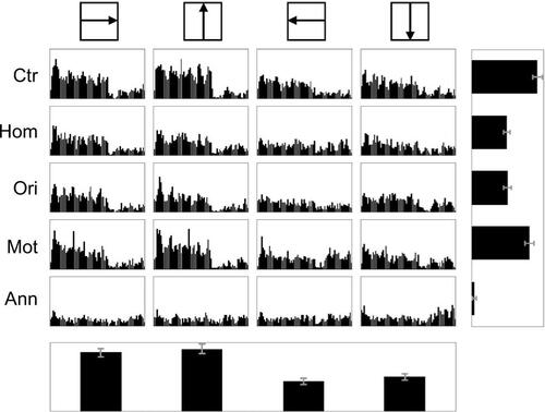
Figure S1 Response of a feature-specific cell in V1.
Notes: PSTHs depict spikes for all trials of the condition specified on the left of each row with a center direction specified at the top of each column. Bar orientation within the central region was 45 degrees for all conditions. The histogram on the right represents the mean spike rates (blank subtracted) summed over all directions. The histogram at the bottom represents the mean spike rates of the Ctr condition only, illustrating the direction-tuning properties of the cell. Mot stimuli evoked increases in firing rates in comparison to the Hom stimulus for all four center directions tested.
Abbreviations: Ctr, center-alone; Hom, homogeneous; Ori, orientation contrast; Mot, motion contrast; Ann, annulus; PSTHs, peri-stimulus time histograms.
Acknowledgments
The authors thank Dorothy Joiner and Sandra McGillis for technical support and Anthony Norcia for helpful comments on the manuscript.
Author contributions
Both MDZ and DYT designed and performed the experiments; MDZ analyzed the data and drafted the manuscript; both the authors revised the manuscript and approved the final version.
Disclosure
The authors report no conflicts of interest in this work.