

Abstract
Introduction
The nucleobase 2-amino-6-chloropurine-modified polyamidoamine (AP-PAMAM) was used as a carrier for p53 gene delivery to achieve the antitumor effects.
Methods and materials
The condensation of p53 plasmid was studied through gel retardation assay, and the transfection efficiency was evaluated through the transfection assay of pEGFP-N3 and pGL-3 plasmids. Using human cervical carcinoma cell line HeLa as a model, the inhibition of cell proliferation and migration was studied through flow cytometry, wound healing and Transwell migration assays, respectively. The p53 expression level was detected through quantitative polymerase chain reaction and Western blotting analyses.
Results
The carrier could condense p53 plasmid into stable nanoparticles at N/P ratios of 2.0, and higher transfection efficiency than polyamidoamine (PAMAM) could be obtained at all the N/P ratios studied. AP-PAMAM-mediated p53 delivery could achieve stronger antiproliferative effect than PAMAM/p53. The antiproliferative effect was identified to be triggered by the induction of cell apoptosis (apoptotic ratio of 26.17%) and cell cycle arrest at S phase. Additionally, AP-PAMAM/p53 transfection has been found to suppress the cell migration and invasion of cancer cells. Finally, the enhanced p53 expression level could be detected after p53 transfection at mRNA and protein levels.
Conclusion
The PAMAM derivative-mediated p53 delivery could be a promising strategy for achieving tumor gene therapy.
Supplementary materials
Figure S1 The in vitro transfection efficiency (A) and the quantitative assay through flow cytometry (B) using pEGFP-N3 in HeLa cells as a model: (a) control; (b) free pEGFP-N3; (c–g) PAMAM/pEGFP-N3 transfection at N/P ratios of 8, 12, 16, 20, and 24, respectively; and (h–l) AP-PAMAM/pEGFP-N3 transfection at N/P ratios of 8, 12, 16, 20, and 24, respectively.
Notes: The scale bar is 400 μm. **P<0.01.
Abbreviations: AP-PAMAM, 2-amino-6-chloropurine-modified PAMAM; PAMAM, polyamidoamine.
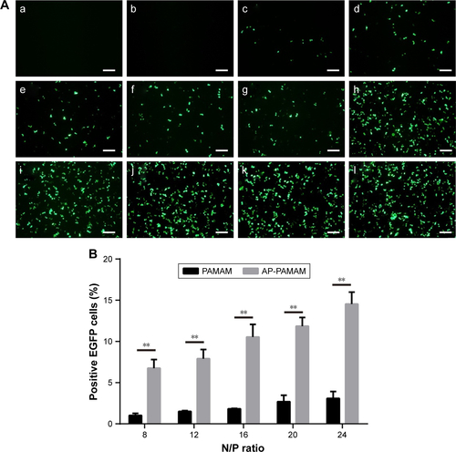
Figure S2 The quantitative assay of in vitro transfection efficiency of PAMAM and AP-PAMAM at different N/P ratios, using the transfection of pGL-3 in HeLa cells as a model.
Note: **P<0.01.
Abbreviations: AP-PAMAM, 2-amino-6-chloropurine-modified PAMAM; PAMAM, polyamidoamine.
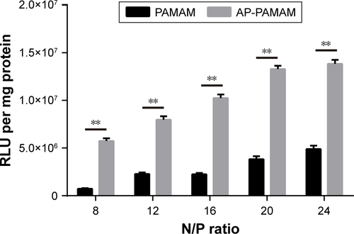
Figure S3 Flow cytometric analysis of the endocytosis of FITC-labeled PAMAM or AP-PAMAM after 2 h.
Abbreviations: AP-PAMAM, 2-amino-6-chloropurine-modified PAMAM; FITC, fluoresceine isothiocyanate; PAMAM, polyamidoamine.
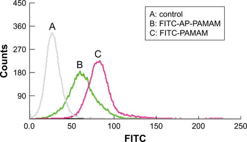
Figure S4 The confocal laser scanning microscope images of the intracellular distribution of FITC-labeled AP-PAMAM (A) and PAMAM (B) after 4 h.
Notes: Blue, nuclei (DAPI); red, lysosome (Lyso-Tracker Red); green, carrier (FITC labeled). The scale bar is 20 μm.
Abbreviations: AP-PAMAM, 2-amino-6-chloropurine-modified PAMAM; DAPI, 4,6-diamidino-2-phenylindole; PAMAM, polyamidoamine.
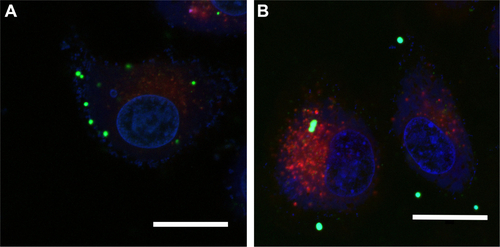
Figure S5 Cytotoxicity analysis of free nucleobase 2-amino-6-chloropurine using HeLa cell as a model.
Note: Data are expressed as mean ± SD of three experiments.
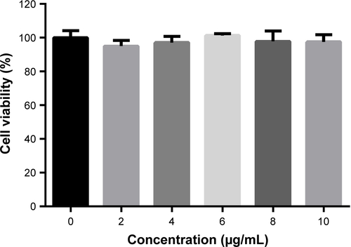
Figure S6 Inhibition of the formation of cell colony by p53 transfection mediated by different carriers: (A) control; (B) AP-PAMAM; (C) PAMAM/p53; and (D) AP-PAMAM/p53.
Note: The scale bar is 400 μm.
Abbreviations: AP-PAMAM, 2-amino-6-chloropurine-modified PAMAM; PAMAM, polyamidoamine.
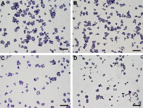
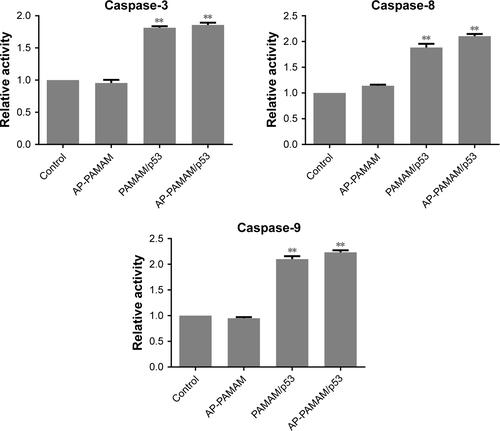
Figure S7 Relative activities of caspase-3, -8, and -9 after p53 transfection mediated by different carriers.
Note: **P<0.01.
Abbreviations: AP-PAMAM, 2-amino-6-chloropurine-modified PAMAM; PAMAM, polyamidoamine.
Acknowledgments
The authors gratefully acknowledge the financial supports from Natural Science Foundation of China (nos 81373344, 81473142, and 81673502), Province-University Cooperation Project of Jilin Province (SXGJQY2017-4), and Graduate Innovation Program of Jilin University (2016149 and 2017058).
Disclosure
The authors report no conflicts of interest in this work.