
Abstract
Background
Exposure to titanium dioxide nanoparticles (TiO2 NPs) that are widely used in food, medicine, sunscreen products and cosmetics is reported to cause ovarian damage and lower fertility in animals. However, the potential effects of TiO2 NPs application on premature ovarian failure (POF) have rarely been evaluated to date.
Methods
In this study, female mice were continuously exposed to TiO2 NPs at doses of 2.5, 5 or 10 mg/kg via gavage instillation for 30 days, and investigated the serum hormones and autoimmunity markers associated with POF.
Results
Exposure to TiO2 NPs resulted in POF, reductions in the levels of estradiol, progesterone and inhibin B and increases in luteinizing hormone, follicle-stimulating hormone, follicle-stimulating hormone/luteinizing hormone ratio, anti-Müllerian hormone, thyroid-stimulating hormone, free triiodothyronine, free tetraiodothyronine, anti-nuclear antibody and anti-thyroid peroxidase antibody levels in serum.
Conclusion
Exposure to TiO2 NPs induced POF triggered by alterations in hormones and autoimmunity markers. Our findings highlight the necessity for significant caution in handling and usage of TiO2 NPs by female consumers.
Introduction
Titanium dioxide nanoparticles (TiO2 NPs) are increasingly used as non-toxic, chemically inert and biocompatible pigment products or photocatalysts owing to their high surface area to particle mass ratio and high reactivity and are commercially manufactured for use in industry, comprehensive ecological improvement, food, medical, diagnostic and cosmetic applications in preference to bulk TiO2.Citation1,Citation2 However, the increased use of TiO2 NPs in various applications is significantly associated with potential human toxicity.
Recent published data support the toxicity of NPs in the reproductive system.Citation3,Citation4 For example, Yoshida et alCitation5 showed that exposure to black carbon NPs leads to adverse effects on male reproductive function in mice, characterized by increased serum testosterone and partial vacuolation of seminiferous tubules. Bai et alCitation6 observed that exposure to water-soluble multiwalled carbon nanotubes leads to oxidative stress in mouse testis. Amorphous nanosilica particles can cross the blood–testis barrier and nuclear membranes of spermatocytes in mouse testis.Citation7 In particular, it has been reported that TiO2 NPs exposure damages follicles, reduces follicular survival and inhibits development and oocyte maturation of rat preantral follicles.Citation8 Di Virgilio et alCitation9 demonstrated genotoxicity and cytotoxicity following TiO2 or aluminum oxide (Al2O3) NPs exposure in Chinese hamster ovary (CHO-K1) cells. Moreover, TiO2 NPs exposure is associated with reduced sperm number and motility or elevated abnormal sperm number and germ cell apoptosis in mouse testis.Citation10 Previous studies indicate that TiO2 NPs cross the blood–testis barrier to reach the testis and accumulate within, causing testicular lesions, sperm malformations and alterations in hormone levels and gene expression profiles in male mice.Citation11–Citation14 Importantly, we additionally showed that TiO2 NPs can translocate to and accumulate in the ovary, leading to reductions in body weight (BW), ovarian indices and fertility, inflammation, follicular atresia and necrosis as well as alterations in gene expression in female mice.Citation15–Citation17 Liu et alCitation18 suggested that nano-zinc oxide (ZnO) exposure affects ovarian development through regulating the number of neuroendocrine cells in the ovary and expression of neuronal factors. This study was conducted to examine the hypothesis that TiO2 NP-mediated suppression of fertility is also associated with premature ovarian failure (POF) in female mice.
POF is a primary ovarian defect characterized by absent menarche (primary amenorrhea) or premature depletion of ovarian follicles before the age of 40 years. POF is considered an immunological disorder that is potentially associated with several factors, including autoimmune disease,Citation19 ovarian lymphocytic infiltration,Citation20 presence of anti-ovarian antibodiesCitation21 and post-immunotherapy reversibility,Citation22 as well as toxics and drugs in women.Citation23 The defect is biochemically typified by low levels of gonadal hormones (estrogens and inhibins) and high levels of gonadotropins (luteinizing hormone [LH] and follicle-stimulating hormone [FSH])Citation24 and often associated with non-organ-specific autoantibodies, such as anti-nuclear antibodies (ANAs)Citation25 and anti-thyroid peroxidase antibodies (TPO-Abs),Citation26 indicative of altered autoimmunity in these patients. However, the issue of whether TiO2 NPs exposure triggers alterations in autoimmunity remains unclear at present.
Here, female mice were continuously exposed to TiO2 NPs at doses of 2.5, 5 or 10 mg/kg via gavage instillation for 30 days. Changes in serum parameters were assessed with the aim of establishing whether TiO2 NPs exposure is linked to POF and the underlying mechanisms in mice.
Methods
Chemicals
Nanoscale TiO2 (anatase, TiO2 content > 99.5%) was kindly provided by Professor Yang Ping (Chemical College of Soochow University, Suzhou, China). TiO2 NPs were characterized according to the procedures in the previous studies.Citation27,Citation28 The average particle size of TiO2 NPs suspended in 0.5% (w/v) hydroxypropyl methylcellulose (HPMC) solvent after 24 h incubation ranged from 5 to 6 nm based on X-ray diffraction patterns (Mercury CCD Corporation, Japan) and Tecnai G220 transmission electron microscopy (TEM) (FEI Corporation, USA) data, and hydrodynamic diameters of TiO2 NPs in suspension ranged from 208 to 330 nm (mainly ∼294 nm) as determined by dynamic light scattering (Brookhaven Instruments Corporation, USA). The surface area of TiO2 NPs was determined to be 174.8 m2/g based on Brunauer–Emmett–Teller adsorption measurements on a Micromeritics ASAP 2020M+ C instrument (Micromeritics Corporation, USA) and ζ potential was determined to be 9.28 mV using the Zeta PALS+BI-90 Plus analyzer (Brookhaven Instruments Corp.).Citation27,Citation28
Ethics approval
All animal experiments were conducted during the light phase and approved by the Animal Experimental Committee of Soochow University (ethical approval number: 2111270). Procedures were performed in accordance with the National Institutes of Health Guidelines for the Care and Use of Laboratory Animals.
Animals and treatment
Female specific pathogen-free (SPF) mice aged 4 weeks were used for this study. In total, 200 SPF female and 40 SPF male mice (18 ± 2 g) were purchased from the Animal Center of Soochow University (China). All mice were housed in stainless steel cages in a ventilated animal room. The room temperature of the housing facility was maintained at 24°C ± 2°C and a relative humidity of 60% ± 10% under a 12-h light/dark cycle. Distilled water and sterilized food for mice were available ad libitum. After acclimatization to laboratory conditions for 1 week, mice were fasted for 3 h prior to TiO2 NPs administration.
TiO2 NP powder was dispersed onto the surface of 0.5% w/v HPMC, and the suspension containing TiO2 NPs was treated ultrasonically for 30 min and mechanically vibrated for 5 min. Mice were randomly divided into four groups (n = 50 each), including a control group treated with 0.5% w/v HPMC and three experimental groups administered 2.5, 5 or 10 mg/kg of TiO2 NPs. Next, mice were weighed, and the volume of fresh TiO2 NPs suspensions required for each animal was calculated. Fresh TiO2 NPs suspensions were administered to mice via a gavage needle every day for 30 days. For appropriate dose selection, we referred to a report of the World Health Organization in 1969, according to which LD50 of TiO2 for rats is >12 g/kg BW after oral administration. We additionally referred to previous studies,Citation15,Citation16 which showed that the National Institute for Occupational Safety and Health (NIOSH) recommends exposure limits of 2–3 mg/m3 for fine TiO2 and 0.3–1 mg/m3 for ultrafine (including nanoscale engineered) TiO2.Citation29 The doses of TiO2 NPs selected for the study (2.5, 5 and 10 mg/kg) were equivalent to ∼0.15–0.7 g exposure for humans with 60–70 kg BW, representative of relatively safe doses.
Mating of animals
To evaluate the effects of TiO2 NPs on fertility, we treated three groups of female mice (10 per mating group). In total, 10 male and 10 control or treated female mice from each group were placed in a common cage for mating after 30 days of TiO2 NPs administration. Mating success was confirmed based on the formation of a white suppository at the mouth of the vagina tracked via vaginal smearing under an optical microscope (U-III Multi-point Sensor System; Nikon Corporation, Tokyo, Japan). Mating success was assessed by counting the number of pregnant mice.
BW and ovary weight
After 30 days, all mice were weighed. Blood samples were collected from the eye vein following rapid removal of the eyeball. After collecting blood, all mice were sacrificed via cervical dislocation, and ovaries of all animals were quickly removed, weighed, placed on ice and dissected and frozen at −80°C (with the exception of 40 ovaries that were used for histopathological examination). Serum was collected by centrifuging blood at 1,200× g for 10 min.
Serum parameter analysis
Plasma concentrations of anti-Müllerian hormone (AMH), inhibin B, estradiol (E2), progesterone (P), LH, FSH, prolactin (PRL), thyroid-stimulating hormone (TSH), free triiodothyronine (fT3) and free tetraiodothyronine (fT4) were detected with the aid of commercial kits (Bühlmann Laboratories AG, Switzerland). The levels of ANAs and anti-TPO-Abs were measured using enzyme-linked immunosorbent assay (ELISA; R&D Systems, Inc., Minneapolis, MN, USA). All biochemical assays were performed using an automated clinical chemistry analyzer (Type 7170A; Hitachi Ltd., Tokyo, Japan).
Histopathological evaluation of ovary
All histopathological examinations were performed using standard laboratory procedures. Five sets of ovarian tissues from mice were embedded in paraffin blocks, sliced into 5 μm sections and placed on separate glass slides (five slices from each kidney). After hematoxylin–eosin staining, sections were evaluated by a histopathologist blinded to the treatments under an optical microscope (U-III Multi-point Sensor System; Nikon Corporation).
Statistical analysis
Data are presented as mean ± SD. Statistical analyses were performed using SPSS 19.0 software (IBM Corporation, Armonk, NY, USA) and compared via one-way analysis of variance (ANOVA) followed by Tukey’s HSD post-hoc test. Differences were considered statistically significant at p-values < 0.05.
Results
Body and ovarian weights
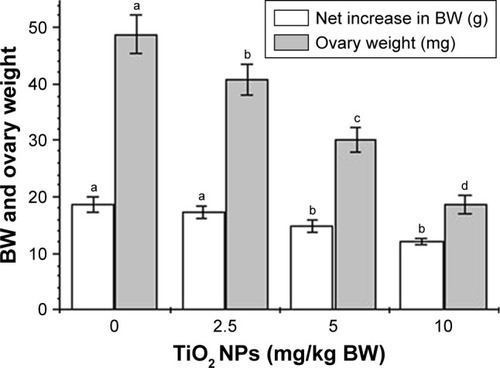
depicts the effects of TiO2 NPs exposure on net increase in body and ovarian weights. TiO2 NPs treatment induced a significant reduction in the net increase in BW (−7.81% to −35.87%) and ovarian weight (−16.32% to −61.44%), compared with the control group (p < 0.05). The decrease in ovarian weight caused by TiO2 NPs is suggestive of ovarian atrophy and may be related to tissue injury or POF, as confirmed by histopathological observation of mouse ovaries.
Figure 1 Effects of TiO2 NPs on net increase in BW and ovarian weight after gavage administration for 30 days.
Notes: Different letters within the same parameter indicate significant differences between groups (p < 0.05). Values represent mean ± SD (n = 10).
Abbreviations: BW, body weight; TiO2 NPs, titanium dioxide nanoparticles.
Reproduction
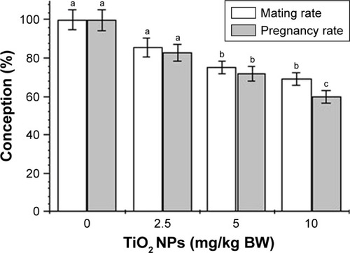
In response to increasing TiO2 NP doses, the mating and pregnancy rates of mice were significantly decreased by 14%–31% and 17%–40%, respectively, as shown in (p < 0.05). The decreased pregnancy rate may be associated with POF triggered by TiO2 NPs exposure.
Figure 2 Effect of TiO2 NPs on conception in female mice after gavage administration for 30 days.
Notes: Different letters within the same parameter indicate significant differences between groups (p < 0.05). Values represent mean ± SD (n = 10).
Abbreviations: BW, body weight; TiO2 NPs, titanium dioxide nanoparticles.
Histopathological evaluation
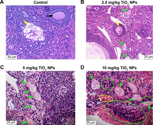
The histological changes in mouse ovary are presented in . Normal development of primary follicles from control ovary was observed (). However, samples from the 2.5 mg/kg TiO2 NP-exposed group showed not only atrophic secondary follicles but also a large number of primary atretic follicles and disposed disorder or apoptosis of granule cells. Similarly, the 5 mg/kg TiO2 NP-exposed group exhibited a large increase in primary atretic follicle number and disposed disorder or apoptosis of granule cells, and the 10 mg/kg TiO2 NP-exposed group showed severe inflammatory cell infiltration along with increased primary atretic follicle content and disposed disorder or apoptosis of granule cells in the ovary (). Our collective results support the theory that TiO2 NPs exposure induces POF in female mice.
Figure 3 Histopathological examination of ovary of mice following gavage administration of TiO2 NPs for 30 days.
Notes: (A) Control groups (unexposed mice) present normal development of primary follicles (black arrow) and secondary follicles (yellow arrow); (B) the 2.5 mg/kg TiO2 NP-exposed group shows atrophic secondary follicle (yellow arrow), primary follicle atresia (green arrow) and apoptosis of granule cells (blue arrow); (C) the 5 mg/kg TiO2 NP-exposed group shows large primary follicle atresia (green arrow) and granule cell apoptosis (blue arrow), and (D) the 10 mg/kg TiO2 NP-exposed group shows severe inflammatory cell infiltration (green circle), congestion (yellow circle), significant primary follicle atresia (green arrow) and disposed disorder or apoptosis of granule cells (blue arrow).
Abbreviation: TiO2 NPs, titanium dioxide nanoparticles.
Serum parameters
As shown in , serum FSH, LH, FSH/LH ratio, AMH, PRL, TSH, fT3 and fT4 levels were significantly increased, whereas inhibin B, E2 and P levels were markedly reduced with increasing TiO2 NP doses (p < 0.05). Notably, the levels of serum ANAs and TPO-Abs involved in the autoimmune response were gradually increased with exposure to increasing concentrations of TiO2 NPs in female mice (, p < 0.05).
Table 1 Changes in serum parameters involved in autoimmune response in female mice caused by gavage administration of TiO2 NPs for 30 days
Discussion
To determine the potential effects of TiO2 NPs exposure on POF of female mice, we focused on histopathological changes in ovary, fertility, and hormone and anti-ovarian antibody levels in serum in the present study. Exposure to 2.5, 5 and 10 mg/kg BW TiO2 NPs for 30 days led to significant reductions in body and ovarian weights () and mating and pregnancy rates (), severe inflammation, and follicle atresia as well as apoptosis of granule cells in the ovary (), consistent with our previous findings.Citation15–Citation17 These characteristics contribute to POF following TiO2 NP-induced toxicity, which may be attributable to TiO2 NP accumulation in ovaryCitation15 or biokinetics in vivo after oral application.Citation30–Citation32 Furthermore, TiO2 NPs induced a decrease in inhibin B, E2 and P levels and an increase in FSH, LH, FSH/LH ratio, AMH, PRL, TSH, fT3, fT4, ANA and TPO-Ab levels in mouse sera (), as discussed subsequently.
Gonadotrophin-releasing hormone (GnRH), FSH and LH regulate ovarian function through a sensitive feedback system. Elevated serum FSH in the early follicular phase, a consequence of reduced ovarian function, is predictive of impaired pregnancy outcomes after infertility treatment,Citation33 and a combination of low FSH and high LH levels can be utilized to predict pregnancy outcomes.Citation34 The FSH:LH ratio has been successfully applied as a predictor of pregnancy outcomes in infertile women.Citation23 Elevated serum FSH, LH and FSH:LH ratio observed in the present study are clearly suggestive of impaired ovarian function due to TiO2 NPs exposure in mice.
AMH is a dimeric glycoprotein generated by granulosa cells from pre-antral and antral follicles that mainly function to inhibit follicular development from the primordial to primary follicular stage. During the menstrual cycle, serum AMH levels are relatively stable.Citation35 Additionally, AMH is reported to be a better predictor of pregnancy outcome than other hormonal parameters.Citation36,Citation37 Inhibin B, produced by granulosa cells in antral follicles, has been proposed as an effective marker of follicular growth. Low serum inhibin B levels are associated with elevated FSH levels, leading to reduction in oocyte quality and fertility.Citation35 Therefore, increased AMH and decreased inhibin B levels in serum induced by TiO2 NPs exposure may be closely linked to POF generation in mice.
Estrogen plays important roles in oocyte maturation, embryo quality and fertilization.Citation38,Citation39 The most important estrogen is E2 in serum,Citation10 which, in combination with FSH and age, is predictive of pregnancy outcomes after in vitro fertilization treatment.Citation40 Progesterone is the most important hormone for endometrial development, implantation and maintenance of pregnancy,Citation41 and its activity during the luteal phase can increase pregnancy as well as live birth rates after infertility treatment.Citation42,Citation43 In the present study, lower serum E2 and P levels were associated with reduced mating and pregnancy rates in TiO2 NP-exposed female mice.
Thyroid hormones influence ovarian function directly and indirectly through elevation of PRL levels and alterations in GnRH secretion. Ovarian insufficiencies often occur in women with thyroid dysfunction.Citation44 Increased serum PRL levels are related to menstrual disorders because of their restraining effect on pulsatile GnRH secretion as well as inhibition of FSH and LH release.Citation45 Importantly, hyperprolactinemia is suggested to be associated with ovulatory dysfunction,Citation46 and both hypothyroidism and hyperprolactinemia are linked to infertility. However, the specific association between pregnancy outcome and serum TSH levels is a subject of controversy at present. For example, Cramer and co-workers suggested that infertile women with increased serum TSH levels have lower pregnancy rates than those with normal TSH levels.Citation47 Arojoki et al reported low serum TSH levels in women with infrequent menstruation and female infertility and high serum TSH levels in women with unexplained infertility and ovulatory dysfunction.Citation48 In our study, PRL, TSH, fT3 and fT4 levels were significantly increased in TiO2 NP-exposed female mice, suggesting that these factors are correlated with ovulatory dysfunction and low fertility due to POF.
Earlier reports indicate that 10.5% infertile women are positive for antithyroid antibodies (ATAs),Citation48 and TPO-Abs are associated with the TSH levels.Citation49 For instance, TPO-Ab-positive women have significantly elevated TSH levels, whereas a number of women with normal TSH levels are positive for TPO-Abs.Citation50,Citation51 The presence of ATAs in euthyroid women may be related to fertility problems, including increased abortion rate and incidence of infertility.Citation24 In addition, the incidence of ANAs in patients with POF was demonstrated to be higher than that in age-matched hypogonadotropic patients with secondary amenorrhea, refuting a potential role of hypoestrogenism in the formation of ANAs.Citation24 Data from this study collectively suggest that exposure to TiO2 NPs induces significant elevation of serum ANA and TPO-Ab levels in female mice, which may also be associated with altered autoimmunity, leading to POF.
Conclusion
Exposure of female mice to TiO2 NPs results in POF characterized by a reduction in reproductive ability along with decreased levels of inhibin B, E2 and P and increased FSH, LH, FSH/LH ratio, AMH, PRL, TSH, fT3, fT4, ANA and TPO-Ab levels in serum. Our results support the crucial need to improve public awareness of the hazards of oral exposure to TiO2 NPs for female consumers.
Acknowledgments
This work was supported by the National Natural Science Foundation of China (grant numbers 81473007, 31671033, 81273036 and 30901218), the National Natural Science Foundation of Jiangsu Province (grant number BK20161306) and the Top-notch Academic Programs Project of Jiangsu Higher Education Institutions (PPZY2015A018).
Disclosure
The authors report no conflicts of interest in this work.
References
- RocoMSBainbridgeWSSocietal Implications of Nanoscience and NanotechnologyNorwell, MANational Science Foundation, NSET Workshop Report Kluwer Academic Publishers2001
- JortnerJRaoCNRNanostructured advanced materials: perspectives and directionsPure Appl Chem200274914911506
- HongFSYuXHWuNProgress of in vivo studies on the systemic toxicities induced by titanium dioxide nanoparticlesToxicol Res20176115133
- AlaeeSIlaniMEffect of titanium dioxide nanoparticles on male and female reproductive systemsJ Adv Med Sci Appl Technol20173138
- YoshidaSHiyoshiKIchinoseTEffect of nanoparticles on the male reproductive system of miceInt J Androl200832433734218217983
- BaiYHZhangYZhangJPRepeated administrations of carbon nanotubes in male mice cause reversible testis damage without affecting fertilityNat Nanotechnol2010568368920693989
- MorishitaYYoshiokaYSatohHDistribution and histologic effects of intravenously administered amorphous nanosilica particles in the testes of miceBiochem Biophys Res Commun2012420229730122417826
- HouJWanXYWangFEffects of titanium dioxide nanoparticles on development and maturation of rat preantral follicle in vitroAcad J Second Mil Med Univ2009308869873 Chinese with English abstract
- Di VirgilioALReigosaMArnalPMFernández Lorenzo de MeleMComparative study of the cytotoxic and genotoxic effects of titanium oxide and aluminium oxide nanoparticles in Chinese hamster ovary (CHO-K1) cellsJ Hazard Mater20101771–371171820079968
- GuoLLLiuXHQinDXEffects of nanosized titanium dioxide on the reproductive system of male miceZhonghua Nan Ke Xue2009156517522 Chinese with English abstract19593991
- GaoGDZeYGZhaoXYTitanium dioxide nanoparticle-induced testicular damage, spermatogenesis suppression, and gene expression alterations in male miceJ Hazard Mater201325825913314323542321
- HongFSZhaoXYSiWHDecreased spermatogenesis led to alterations testis-specific gene expression in male mice following exposure to nano-TiO2J Hazard Mater201530071872826296075
- HongFSSiWHZhaoXYTiO2 nanoparticles exposure decreases spermatogenesis via biochemical dysfunctions in the testis of male miceJ Agric Food Chem201563317084709226145168
- HongFSWangYJZhouYJExposure to TiO2 nanoparticles induces immunological dysfunction in mouse testitisJ Agric Food Chem201664134635526720763
- GaoGDZeYGLiBThe ovarian dysfunction and its gene-expressed characteristics of female mice caused by long-term exposure to titanium dioxide nanoparticlesJ Hazard Mater2012243192723131501
- ZhaoXYZeYGGaoGDNanosized TiO2-induced reproductive system dysfunction and its mechanism in female micePLoS One201384e5937823565150
- HongFZhouYZhaoXShengLWangLMaternal exposure to nanosized titanium dioxide suppresses embryonic development in miceInt J Nanomedicine2017126197620428883729
- LiuXQZhangHFZhangWDRegulation of neuroendocrine cells and neuron factors in the ovary by zinc oxide nanoparticlesToxicol Lett2016256193227215404
- TungKSKLuCYImmunological basis of reproductive failureKransFTDanijanovIKaufmanNPathology of Reproductive FailureBaltimoreWilliams & Wilkins1991308333
- WheatcroftNJToogoodAALiTCDetection of antibodies to ovarian antigens in women with premature failureClin Exp Immunol19949611221288149656
- CorenblumBRoweTTaylorPJHigh doses, short term glucocorticoids for the treatment of infertility resulting from premature ovarian failureFertil Steril19935959889918486200
- Beck-PeccozPPersaniLPremature ovarian failureOrphanet J Rare Dis200611557566
- BroekmansFJKweeJHendriksDJMolBWLambalkCBA systematic review of tests predicting ovarian reserve and IVF outcomeHum Reprod Update200612668571816891297
- IshizukaBKudoYAmemiyaAYamadaHMatsudaTOgataTAnti-nuclear antibodies in patients with premature ovarian failureHum Reprod1999141707510374097
- McElduffAMorrisJThyroid function tests and thyroid autoantibodies in an unselected population of women undergoing first trimester screening for aneuploidyAust N Z J Obstet Gynaecol200848547848019032663
- Stagnaro-GreenARomanSHCobinRHel-HarazyEAlvarez-MarfanyMDaviesTFDetection of at-risk pregnancy by means of highly sensitive assays for thyroid autoantibodiesJAMA199026411142214252118190
- YangPLuCHuaNTitanium dioxide nanoparticles co-doped with Fe3+ and Eu3+ ions for photocatalysisMater Lett2002574794801
- HuRPZhengLZhangTMolecular mechanism of hippocampal apoptosis of mice following exposure to titanium dioxide nanoparticlesJ Hazard Mater20111911–3324021570177
- National Institute for Occupational Safety and Health (NIOSH)Outlines Guidance on Handling Titanium Dioxide (TiO2)AtlantaEmerald Group Publishing Limited2011
- KreylingWGHolzwarthUHaberlNQuantitative biokinetics of titanium dioxide nanoparticles after intravenous injection in rats: part 1Nanotoxicology201711443444228290717
- KreylingWGHolzwarthUHaberlNQuantitative biokinetics of titanium dioxide nanoparticles after oral application in rats: part 2Nanotoxicology201711444345328290734
- KreylingWGHolzwarthUHaberlNQuantitative biokinetics of titanium dioxide nanoparticles after intratracheal instillation in rats: part 3Nanotoxicology201711445446428290735
- BrodinTBerghTBerglundLHadziosmanovicNHolteJHigh basal LH levels in combination with low basal FSH levels are associated with high success rates at assisted reproductionHum Reprod200924112755275919617206
- SeckinBTurkcaparFOzaksitGElevated day 3 FSH/LH ratio: a marker to predict IVF outcome in young and older womenJ Assist Reprod Genet201129323123622183503
- BroekmansFJSoulesMRFauserBCOvarian aging: mechanisms and clinical consequencesEndocr Rev200930546549319589949
- WuCHChenYCWuHHSerum anti-mullerian hormone predicts ovarian response and cycle outcome in IVF patientsJ Assist Reprod Genet200926738338919768530
- BrodinTHadziosmanovicNBerglundLAntimullerian hormone levels are strongly associated with live-birth rates after assisted reproductionJ Clin Endocrinol Metab20139831107111423408576
- SundarrajanCLiaoWRoyACNgSCAssociation of oestrogen receptor gene polymorphisms with outcome of ovarian stimulation in patients undergoing IVFMol Hum Reprod19995979780210460216
- SundarrajanCLiaoWXRoyACNgSCAssociation between estrogen receptor-beta gene polymorphisms and ovulatory dysfunctions in patients with menstrual disordersJ Clin Endocrinol Metab200186113513911231990
- SroujiSSMarkALevineZBetenskyRAHornsteinMDGinsburgESPredicting in vitro fertilization live birth using stimulation day 6 estradiol, age, and follicle-stimulating hormoneFertil Steril200584379579716169429
- GrahamJDClarkeCLPhysiological action of progesterone in target tissuesEndocr Rev19971845025199267762
- BjurestenKLandgrenBMHovattaOStavreus-EversALuteal phase progesterone increases live birth rate after frozen embryo transferFertil Steril201195253453720579989
- ErdemAErdemMAtmacaSGulerIImpact of luteal phase support on pregnancy rates in intrauterine insemination cycles: a prospective randomized studyFertil Steril20099162508251318692788
- PoppeKVelkeniersBGlinoerDThyroid disease and female reproductionClin Endocrinol (Oxf)200766330932117302862
- KostrzakAWarenik-SzymankiewiczAMeczekalskiBThe role of serum PRL bioactivity evaluation in hyperprolactinaemic women with different menstrual disordersGynecol Endocrinol2009251279980619905999
- CramerDWSlussPMPowersRDSerum prolactin and TSH in an in vitro fertilization population: is there a link between fertilization and thyroid function?J Assist Reprod Genet200320621021512877251
- ArojokiMJokimaaVJuutiAKoskinenPIrjalaKAnttilaLHypothyroidism among infertile women in FinlandGynecol Endocrinol200014212713110836200
- RevelliACasanoSPianeLDA retrospective study on IVF outcome in euthyroid patients with anti-thyroid antibodies: effects of levothyroxine, acetyl-salicylic acid and prednisolone adjuvant treatmentsReprod Biol Endocrinol2009713719941670
- PrummelMFWiersingaWMThyroid peroxidase autoantibodies in euthyroid subjectsBest Pract Res Clin Endocrinol Metab200519111515826919
- DebièveFDulièreSBernardPHubinontCDe NayerPDaumerieCTo treat or not to treat euthyroid autoimmune disorder during pregnancy?Gynecol Obstet Invest200967317818219092255
- PoppeKGlinoerDThyroid autoimmunity and hypothyroidism before and during pregnancyHum Reprod Update20039214916112751777