
Abstract
Introduction
One of the main issues in the medical field and clinical practice is the development of novel and effective treatments against infections caused by antibiotic-resistant bacteria. One avenue that has been approached to develop effective antimicrobials is the use of silver nanoparticles (Ag-NPs), since they have been found to exhibit an efficient and wide spectrum of antimicrobial properties. Among the main drawbacks of using Ag-NPs are their potential cytotoxicity against eukaryotic cells and the latent environmental toxicity of their synthesis methods. Therefore, diverse green synthesis methods, which involve the use of environmentally friendly plant extracts as reductive and capping agents, have become attractive to synthesize Ag-NPs that exhibit antimicrobial effects against resistant bacteria at concentrations below toxicity thresholds for eukaryotic cells.
Purpose
In this study, we report a green one-pot synthesis method that uses Acacia rigidula extract as a reducing and capping agent, to produce Ag-NPs with applications as therapeutic agents to treat infections in vivo.
Materials and methods
The Ag-NPs were characterized using transmission electron microscopy (TEM), high-resolution TEM, selected area electron diffraction, energy-dispersive spectroscopy, ultraviolet–visible, and Fourier transform infrared.
Results
We show that Ag-NPs are spherical with a narrow size distribution. The Ag-NPs show antimicrobial activities in vitro against Gram-negative (Escherichia coli, Pseudomonas aeruginosa, and a clinical multidrug-resistant strain of P. aeruginosa) and Gram-positive (Bacillus subtilis) bacteria. Moreover, antimicrobial effects of the Ag-NPs, against a resistant P. aeruginosa clinical strain, were tested in a murine skin infection model. The results demonstrate that the Ag-NPs reported in this work are capable of eradicating pathogenic resistant bacteria in an infection in vivo. In addition, skin, liver, and kidney damage profiles were monitored in the murine infection model, and the results demonstrate that Ag-NPs can be used safely as therapeutic agents in animal models.
Conclusion
Together, these results suggest the potential use of Ag-NPs, synthesized by green chemistry methods, as therapeutic agents against infections caused by resistant and nonresistant strains.
Introduction
Over the past few decades, there has been a growing interest in exploring alternative strategies to design and develop new antimicrobials due to the increased incidence in microbial resistance against commonly used antibiotics.Citation1,Citation2 The field of nanotechnology offers a toolbox of synthesis and design strategies to develop antimicrobial nanotherapeutics with tailored antimicrobial properties to counterattack the problem of antimicrobial resistance. Several reports have shown that nanoparticles made up of different noble metals can be used to kill both resistant and nonresistant bacterial strains.Citation87–Citation89 The large surface area-to-volume ratio exhibited by these systems at the nanoscale has been hypothesized to be one of the bases of the antimicrobial effects against a broad spectrum of microorganisms.Citation1 Silver nanoparticles (Ag-NPs) have obtained considerable attention due to their attractive physicochemical propertiesCitation2,Citation3 and remarkable antimicrobial effects exhibited against Gram-negative and Gram-positive multidrug-resistant bacteria.Citation4 The bactericidal effect of silver in its various chemical forms has been exploited since ancient civilizations;Citation5 during the 18th century, silver was commonly used to treat infected ulcers, and more recently, in the early 1920s, Ag-NPs were included for wound treatment by the US Food and Drug AdministrationCitation6 due to their strong toxicity against a wide range of microorganisms.Citation7–Citation9 There are various mechanisms that have been linked to the toxicity of silver compounds against microorganisms, among the most common are direct disruptive interactions with the cell membrane and DNACitation2 and the trigger of biochemical cascades that lead to free radical production, altering cell membrane permeability.Citation10 In the case of Ag-NPs, it has been shown that there is a correlation between size and the exhibited antimicrobial effect.Citation2 Nevertheless, the use of Ag-NPs for antimicrobial therapies has two major drawbacks: the toxicity exhibited to eukaryotic and mammalian systems and the toxicity of the chemical synthesis to produce them. Therefore, there have been efforts toward the development of greener synthesis processes to produce Ag-NPsCitation11,Citation12 with tailored properties that reduce toxicity against eukaryotic systems.Citation13 Although there are various methods to synthesize Ag-NPs, including chemical reduction,Citation14 photochemical reduction,Citation15,Citation16 electrochemical reduction,Citation17 vaporization, and thermal synthesis,Citation18 there is a growing interest in developing a green synthesis method to produce nanoparticles using biological systemsCitation19,Citation20 and plant extracts.Citation21 Notwithstanding the diversity of green synthesis approaches, the use of plant extracts, as reductive media for nanoparticle synthesis, is one of the most convenient routes.Citation22,Citation23
Ag-NPs have been synthesized using extracts obtained from different plants such as Acorus calamus (rhizome), Boerhaavia diffusa (whole plant), tea extract (leaves), Tribulus terrestris (fruit), Aloe vera (leaves), Eucalyptus hybrid (peel), Memecylon edule (leaves), and Citrus sinensis (peel).Citation24 In fact, within the last decade, hundreds of plant extracts have been reported to be effective reducing agents in the synthesis of Ag-NPs.Citation25 These methods have been widely explored since they provide environmentally friendly and cost-effective routes to the synthesis of metallic nanostructures, in addition to being easily scalable production processes.Citation26–Citation28 In this study, we developed a green synthesis method to produce Ag-NPs using the extract of an Acacia rigidula plant. This plant is commonly known as blackbrush acacia, and it is a shrub that can be found in the regions of west and southwest Texas, as well as the Mexican northeastern region.Citation29 Although there is limited information on the chemical composition of the A. rigidula plant,Citation30 gums and condensed tannins are the most characterized parts in the literature.Citation31 Forty-four amines and alkaloids,Citation29 such as phenethylamines, have been identified in A. rigidula. These components have been associated with promotion of weight loss; therefore, both the A. rigidula plant and its extracts have been extensively used as dietary supplements.Citation30 Furthermore, forages from A. rigidula have been widely explored as a protein supplement to feed cattle.Citation32,Citation33 It was then interesting to explore, in this study, the potential use of the extract, obtained from waste parts of the A. rigidula plant (roots and stems), as a reducing and stabilizing agent in the synthesis of Ag-NPs. The successfully obtained Ag-NPs showed antimicrobial activity in vitro against Gram-negative bacteria (Escherichia coli, Pseudomonas aeruginosa, and a multidrug-resistant P. aeruginosa strain) and Gram-positive bacteria (Bacillus subtilis). Moreover, in vivo studies showed promising antimicrobial effects of Ag-NPs in a murine skin infection model (using a P. aeruginosa clinical resistant strain). In addition, an in vivo toxicological study was carried out to verify the safety of using Ag-NPs; the results demonstrate them to be a promising treatment for skin infections in the clinic. The combined results presented in this work contribute toward the development and synthesis of alternative antimicrobial agents, nanotherapeutics, to treat infections caused by clinical multidrug-resistant strains.
Materials and methods
Biological materials
Stems and roots from the A. rigidula plant were collected in Nuevo León, Mexico. Gram-negative bacteria used in the antimicrobial assays were E. coli American Type Culture Collection (ATCC)-11229, P. aeruginosa ATCC 27853, and a clinical multidrug-resistant P. aeruginosa isolated at the San Vicente Hospital in Monterrey, Nuevo León, Mexico. The Gram-positive bacterium used in the antimicrobial assays was B. subtilis ATCC 23857.
Bacteria cultures were grown for 20 h at 37°C and 150 rpm (Lab Companion IS-971). After incubation, cultures were diluted 1:250 in fresh medium until an optical density (OD600) of 0.2±0.02 was reached, adjusting with fresh medium if necessary to reach a cell concentration range between 107 and 108 cells/mL. The resulting suspension was then diluted (1:100) with fresh medium, obtaining the bacterial working solution. Absorbance was measured using an ultraviolet–visible (UV–Vis) spectrophotometer (OPTIZEN 2120 UV plus), and cell number was monitored by the plate-counting method, measured by serial dilutions.
Synthesis of Ag-NPs
A. rigidula extract preparation
To prepare the extract, 5 g of stems and roots from the A. rigidula plant were mixed in 50 mL of distilled water. The mixture was then heated in a hotplate (Lab Companion HP-3100), until boiling, for 20 min. During the extraction time, agitation was performed using a magnetic stirrer. Once the infusion cooled, the extract was transferred to a 50-mL corning tube and centrifuged (Sorvall™ Leyend™ XFR; Thermo Fisher Scientific, Waltham, MA, USA) at 9,000 rpm for 10 min. Then, the supernatant was separated and vacuum-filtered (WP6111560; Merck Millipore, Billerica, MA, USA) using filter paper (Ahlstrom, Stockholm, Sweden). The obtained filtrate was stored at 4°C (Nor-Lake Scientific, Hudson, WI, USA).
Ag-NPs preparation
A total of 10 mL of AgNO3 0.1 M and 3 mL of A. rigidula extract were mixed in a 50 mL corning tube and heated in a hotplate for 1 h at 60°C. The formation of the Ag-NPs was monitored and determined through a change of appearance (color) of the mix that changed from transparent to brownish. The resulting solution with the Ag-NPs was centrifuged at 9,000 rpm for 15 min. Then, the product was washed with ethanol and was stored at −75°C for 24 h in an ultralow temperature freezer (Sanyo MDF-U53VA). Then, the product was lyophilized (FreeZone 6; Labconco, Kansas City, MO, USA) for 24 h to obtain the purified Ag-NPs.
Ag-NPs stability experiments
UV–Vis spectra (wavelength range from 200 to 800 nm) of the A. rigidula extract and the Ag-NPs (solution of 1 mg/mL in distilled water as a dispersive medium) were measured using a Multiskan G0 UV–Vis spectrophotometer (Thermo Fisher Scientific). To test stability of the synthesized Ag-NPs, UV–Vis spectra were obtained from Ag-NPs’ solution at two different time points (first time point right after being synthesized and second time point after being stored in solution for 4 months).
Characterization techniques
Morphological and structural characteristics of the Ag-NPs were studied with conventional transmission electron microscopy (TEM) and high-resolution TEM (HRTEM) and selected area electron diffraction (SAED) using an FEI-TITAN 80–300 kV microscope operated at an accelerating voltage of 300 kV. The specimen for these studies was prepared by depositing and evaporating a drop of the colloidal solution (Ag-NPs 1 mg/mL), previously dispersed by means of an ultrasonic cleaner (Bransonic® Branson 2510MT; Bransonic, Dansbury, CT, USA), onto lacey carbon-coated copper grids. The elements present in the samples were determined using an energy-dispersive spectrometry (EDS) analyzer integrated in the transmission electron microscope. In relation to the particle size, TEM images were used to measure the diameter of Ag-NPs to estimate the size distribution of our nanoparticles using the image processing software ImageJ – National Institute of Health (NIH). The images obtained from the SAED patterns were interpreted with simulated patterns using the Web-based Electron Microscopy Application Software (Web-EMAPS).Citation34
Furthermore, UV–Vis spectra (wavelength range from 200 to 1,000 nm) of the Ag-NPs (solution of 1 mg/mL in distilled water as a dispersive medium) were obtained using a Multiskan G0 UV–Vis spectrophotometer. To obtain information related to the interaction between A. rigidula extract and Ag+ ions to produce the Ag-NPs and their capping, infrared spectra were obtained for the extract alone and for the Ag-NPs. The spectra were acquired using a Fourier transform infrared (FT-IR) spectrophotometer (Nicolet™ iS™ 50; Thermo Fisher Scientific). Samples from the extract solution and from a 1 mg/mL sample of Ag-NPs were freeze-dried with a lyophilizer (FreeZone 6) for 24 h to obtain the samples, which were diluted in KBr, using the attenuated total reflectance (ATR) method.
In vitro antibacterial test
Minimum inhibitory concentrations (MICs)
MIC assays were assessed in 96-well plates (Costar; Corning Incorporated, Corning, NY, USA) based on a modified methodology reported previouslyCitation35 and National Committee for Clinical Laboratory Standards.Citation36 Stocks of the Ag-NPs were prepared at a final concentration of 1 mg/mL using distilled water as the vehicle. To perform the MIC tests, Ag-NPs’ stock solution was added to reach a 0.5 mg/mL concentration within a final volume of 200 μL. Next, serial dilutions of the Ag-NPs were performed by taking 100 μL from the previous solution to every next well, with 100 μL of culture media, and discarding the last 100 μL from the last dilution so the tested concentrations were 0.25–4.88×10⁻⁴ mg/mL for Ag-NPs. Then, 100 μL of the bacteria working solution was added to each test well to achieve a final concentration of 105 cells/mL, followed by incubation at 37°C and 150 rpm. After 20 h of incubation at 37°C, the MIC was measured. The MIC for Ag-NPs was defined as the concentration at which no significant growth was observed by the naked eye. All tests, and their respective control samples, were performed in triplicate.
Minimum bactericidal concentration (MBC)
To assess the bactericidal activity of the synthesized Ag-NPs, 1, 2, and 4 times, the Ag-NPs’ MIC was tested against the bacterial strains. In a typical assay, an overnight culture of each strain was diluted 1:250 in 5 mL of fresh Luria Broth (LB) medium, followed by incubation at 37°C and 150 rpm, until it reached exponential phase at a critical OD (OD600 =0.18–0.22). A total of 20 μL of the exponential cultures were transferred to 96-well plates, which contained the Ag-NP treatments, to be incubated at 37°C and 150 rpm for 20 h. After incubation, the colony-forming units (CFUs) per milliliter were determined through the drop count method. Tenfold diluted aliquots were obtained, and then, three 10 μL drops of each of the diluted samples were placed on LB agar plates. The plates were then incubated at 37°C for 24 h, and viable bacterial colonies were counted. All tests and their respective control samples were performed in replicates of three.
Fluorescence microscopy imaging of treated and untreated bacteria
Fluorescence microscopy of the treated and untreated bacteria was carried out based on the methodology described by Vazquez-Rodriguez et al.Citation37 A total of 105 bacterial cells of the resistant P. aeruginosa clinical strain were treated with Ag-NPs, at 0, 0.5, 1, and 2 times the MIC, for 20 h at 37°C and 150 rpm. After the 20 h treatment, bacterial cells were stained with 5 μg/mL of propidium iodide (PI) for 1 h at 37°C and 150 rpm. A total of 20 μL of the treated samples and the control were placed and spread in glass slides, previously cleaned with 96% ethanol, allowing the samples to air dry and fix by adding 200 μL of methanol for 2 min. The methanol excess was decanted, and the slides were then washed twice with phosphate-buffered saline (PBS). Micrograph images were obtained under DM3000 (Leica Microsystems, Wetzlar, Germany) using the 100× objective with an Y3 ET filter system.
In vivo antibacterial test
Animals
Female adult Wistar rats (200–250 g) were used and maintained in stainless steel cages with a 12 h light/dark regime. All the experimental animals were handled ethically (refer the “Ethics approval and informed consent” section).
In vivo skin infection model
The in vivo experiment based on a tape stripping infection modelCitation38 was performed with some modifications. The rats were anesthetized with pentobarbital (Pentosedal; Laboratorios Maver, Ciudad de México, Mexico) injecting intraperitoneally 2.5 mL/kg of body weight (bw). Once anesthetized, the back of the rats was shaved in an area of 3 cm2 with a trimmer (274 Series A; Oster, Boca Raton, FL, USA). Next, an area of 2.5 cm2 of the hairless skin of the rats was tape stripped five times in succession using an elastic adhesive bandage (Sedasiva®; BSN Medical, Yumbo, Colombia). Damage on the skin was defined as the reddening and glistening of the skin with the absence of regular bleeding. This procedure was performed in all the animals used in the experiment. After that, to cause infection, bacterial inoculation was performed by placing, in the wound produced with the tape stripping process, 100 μL of a 107 cells/mL P. aeruginosa culture in log phase. The P. aeruginosa culture was obtained from an overnight culture grown as described previously in the MBC description of the Materials and methods section.
Experimental design
The rats were divided into three groups (three animals per experimental group [n=3]): the control group (with the wound on the skin without infection, and treated with 100 μL of saline solution every 24 h for 3 days in the damaged zone), the infected group (treated in the damaged zone with 100 μL of the clinical strain of P. aeruginosa culture), and the Ag-NP-treated group (treated with 100 μL of Ag-NPs solution at 200 parts per million [ppm] every 24 h for 3 days after infection with the clinical strain of P. aeruginosa in the damaged zone). During all the experiments, the rats were kept with food and water ad libitum at room temperature (24°C±1°C).
The rats were anesthetized and sacrificed 24 h after the third day of treatment, and 300 mg of the wound (~1 cm2) was removed and homogenized (Tissue-Tearor 398; BioSpec, Bartlesville, OK, USA) with 3 mL of phosphate buffer (100 mgwound/mLbuffer).
Finally, 10 μL of homogenates (to have a 1:100 dilution) were used to carry out a pour plate technique to determine the CFUs of living bacteria and to compare these results among the study groups.
In vivo toxicological study
Experimental design
To demonstrate the safety in the use of the Ag-NPs produced in this study, four groups of female adult Wistar rats (200–250 g) with three animals per group (n=3) were used to carry out the toxicological test. The four groups included the control group and three groups treated with the Ag-NPs at different concentrations. In this experiment, the rats were injured based on the tape stripping infection model described previously in the in vivo skin infection model methodology. Briefly, the pentobarbital was administered intraperitoneally to the rats (2.5 mL/kg bw) to anesthesize them. After that, a 3 cm2 area from the back of the rats was shaved using a trimmer in which 2.5 cm2 of the shaved part was tape stripped five times with an elastic adhesive bandage until the hairless skin reddened and glistened, but there was no bleeding observed. This procedure was done in all of the animals used in this experiment. To start the test, the animals in the control group were treated with 100 μL of saline solution on the wound produced by the tape stripping procedure, while the rats in the other three tested groups were treated with 100, 200, and 400 ppm of Ag-NPs, respectively. The treatment consisted of applying either saline solution or the Ag-NPs solution (at the respective concentration) directly on the wound until the solution was absorbed through the skin. All groups were treated in the damaged zone every 24 h for 3 days; the same way as the antibacterial assay was performed. Throughout the experiment, the rats were kept with food and water ad libitum at room temperature (24°C±1°C). Then, 24 h after the third day of treatment, urine samples were collected in vessels attached with metabolic cages (3700M020; Tecniplast, Buguggiate (VA), Italy) in which the rats were placed. Afterward, with the rats previously anesthetized, blood samples were also obtained and the rats were sacrificed.
Biochemical assays
In order to test kidney and liver functions of the rats exposed to Ag-NPs, the urine volume was measured, and with their respective methods, the concentration of creatinine,Citation39,Citation40 glucose,Citation41 and proteinsCitation42 in urine was also measured using a UV–Vis spectrophotometer (DMS 80; Varian, Palo Alto, CA, USA). For the case of blood plasma samples, the concentration of creatinine,Citation39,Citation40 albumin,Citation43 alanine aminotransferase (ALT),Citation44 and aspartate aminotransferase (AST)Citation45 was measured using a UV–Vis spectrophotometer (DMS 80).
Likewise, during the experiment, the appearance of the skin (wound) of the experimental groups exposed to Ag-NPs was observed and compared with the control group.
Statistical analysis
For the in vivo study (antibacterial and toxicological experiments), the significance of the differences between group averages was calculated with the two-tailed Student’s t-test for grouped data with an analysis of variance (ANOVA) posttest. The calculations and graphs were prepared using the Prism 4 software (GraphPad Software, Inc., La Jolla, CA, USA).
Ethics approval and informed consent
Animal care and experimentation practices at the Universidad Autónoma de Aguascalientes are constantly evaluated by the Animal Care and Use Committee, adhering to the official Mexican regulations (NOM-062-ZOO-1999). Mexican regulations are in strict accordance with the recommendations in the Guide for the Care and Use of Laboratory Animals of the NIH and The Weatherall Report of the USA to ensure compliance with established international regulations and guidelines. Experimental protocols were approved by the local Institutional Bioethical Committee. At the end of experiments, animals were sacrificed by using an excess of sodium pentobarbital anesthesia (40 mg/kg bw). Efforts were made to minimize suffering.
Results
Characterization of Ag-NPs
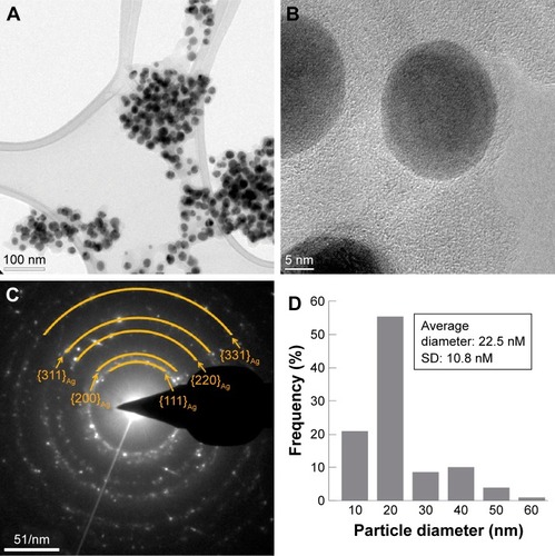
Ag-NPs were produced in an aqueous solution containing an extract from A. rigidula. As can be observed in the TEM and HRTEM micrographs (), the Ag-NPs display homogeneous spherical shapes (). An SAED pattern of the Ag-NPs showed spotty rings that can be attributed to the (111), (200), (220), (311), and (331) planes of a face-centered cubic (fcc) metallic Ag-NP (). The Ag-NPs exhibit a diameter size distribution that ranges from 8 to 66 nm with a mean of 22.46 nm and a standard deviation of 10.83 nm (). The histogram in shows that most of the Ag-NPs have a diameter size between 15 and 25 nm, which corresponds to a narrow homogeneous distribution.
Figure 1 Electron microscopic characterization of crystalline structure, size, and shape distributions of the Ag-NPs.
Notes: (A) Low-resolution TEM micrograph of the Ag-NPs. (B) High-resolution TEM micrograph of the Ag-NPs. (C) SAED pattern of the Ag-NPs with the rings labeled. (D) Histogram of the particle diameter size distribution of the Ag-NPs.
Abbreviations: Ag-NPs, silver nanoparticles; SAED, selected area electron diffraction; TEM, transmission electron microscopy.
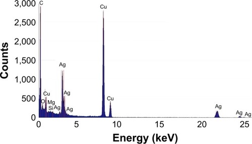
Further characterization of the nanoparticles by EDS analysis shows the presence of Ag, which confirms the presence of Ag-NPs (). In the EDS analysis, other elements such as C and Cu were detected, where C corresponds to the organic matrix and lacey carbon of the TEM grid and Cu is observed since the grid used for the TEM analysis is made up of Cu ().
Figure 2 EDS spectrum of the Ag-NPs with the peaks labeled.
Abbreviations: Ag-NPs, silver nanoparticles; EDS, energy-dispersive spectrometry.
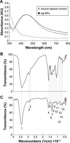
The UV–Vis spectra of the A. rigidula alone, and the Ag-NPs produced by reduction and capping using the A. rigidula extract, are shown in . The Ag-NPs exhibit a plasmon resonance spectrum with an absorption band between 430 and 480 nm. Furthermore, shows a dotted UV–Vis spectra that corresponds to the Ag-NPs’ sample that was stored in solution for 4 months. As can be observed, the spectra present minimal changes and keep their absorption peak between 430 and 480 nm. This result demonstrates the stability of the Ag-NPs after being stored for long time periods. FT-IR characterization of the extract alone () and the Ag-NPs synthesized with the extract () showed that both samples contained functional groups that have been previously reported to be present in an A. rigidula extract.Citation46–Citation51 Some of the characteristic peaks observed in both samples include () a prominent peak at 3,419 cm−1 due to the stretching of O–H in alcohols and phenols; two peaks at 2,929 and 2,854 cm−1 due to the stretching of CH3− and CH2− in aliphatic compounds; one peak at 1,614 cm−1 due to the bending of N−H in amines; one peak at 1,517 cm−1 corresponding to the stretching of C=C in aromatic compounds; another peak at 1,447 cm−1 attributed to −C−N groups; one peak at 1,384 cm−1 due to the symmetric deformation of CH3 in aromatic and aliphatic compounds; another peak at 1,285 cm−1 corresponding to the wagging of C−H in alkyl halides; a peak at 1,250 cm−1 due to the stretching of C=O groups; two peaks at 1,114 and 1,068 cm−1 due to the stretching of C−O in polysaccharides; three peaks at 894, 823, and 767 cm−1 due to the bending of =CH in aromatic hydrocarbons; and one peak at 689 cm−1 attributed to the bending of C−H in alkynes.
Figure 3 Physical characterization of the Ag-NPs.
Notes: (A) UV–Vis spectra of Acacia rigidula extract and Ag-NPs biosynthesized. (B) FT-IR spectra of A. rigidula extract. (C) FT-IR of Ag-NPs synthesized by the A. rigidula extract. The band and peaks are labeled as follows: 1) 3,419, 2) 2,929, 3) 2,854, 4) 1,614, 5) 1,517, 6) 1,447, 7) 1,384, 8) 1,285, 9) 1,250, 10) 1,114, 11) 1,068, 12) 894, 13) 823, 14) 767, and 15) 689 cm−1.
Abbreviations: Ag-NPs, silver nanoparticles; FT-IR, Fourier transform infrared; UV–Vis, ultraviolet–visible.
In vitro antimicrobial activity of Ag-NPs in Gram-negative and Gram-positive bacteria
MIC and MBC assays were performed under the same culture conditions for each of the bacteria tested in this work. B. subtilis had the lowest MIC with a value of 0.48 ppm. For P. aeruginosa, the obtained MIC was 15.6 ppm, twice the MIC obtained for the clinical multidrug resistant bacteria of the same strain. The MIC value obtained for E. coli was 62.5 ppm, the highest of all the MICs determined. The MIC values obtained in this experiment are summarized in .
Table 1 Nominal MICs of Ag-NPs for the four different strains tested in this work
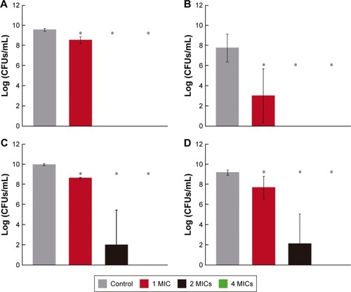
Bactericidal activity was tested through MBC assays using Ag-NPs at different treatment concentrations linked to the MICs reported. As can be observed in , E. coli and B. subtilis show the lowest MBC (two times the MIC). In addition, with a statistical significance (P<0.05), even at one MIC, the treatments show bactericidal activity. For P. aeruginosa strains, the MBCs were found at four times the MICs. However, as observed for E. coli and B. subtilis, bactericidal activity can be observed as low as the concentration corresponding to one MIC. Thus, the Ag-NPs produced by A. rigidula extract show bactericidal activity in both Gram-negative and Gram-positive bacteria.
Figure 4 MBC of Ag-NPs tested at 1, 2, and 4 times the MIC.
Notes: The image reports the MBCs of Ag-NPs against (A) Escherichia coli, (B) Bacillus subtilis, (C) Pseudomonas aeruginosa, and (D) a clinical resistant strain of P. aeruginosa. *P<0.05.
Abbreviations: Ag-NPs, silver nanoparticles; CFUs, colony-forming units; MBCs, minimum bactericidal concentrations; MICs, minimum inhibitory concentrations.
Morphological changes in P. aeruginosa after treatment with Ag-NPs
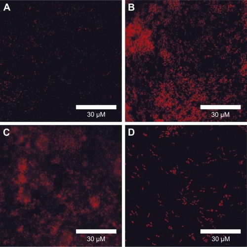
Since antimicrobial activity has been extensively linked to bacterial envelope damage, morphological changes were explored in a clinically relevant multidrug-resistant P. aeruginosa that was treated with Ag-NPs. Bacterial membrane disruption caused by the Ag-NPs treatments was tested through a fluorescence PI stain assay.Citation10 As can be observed in , when compared with the untreated bacteria (), the florescent micrographs () show a general qualitative increased fluorescence for all the treatments (0.5, one, and two times the MIC). These results suggest that bacterial membrane integrity is compromised either directly or as a consequence of the antimicrobial activity exhibited by the Ag-NPs.
Figure 5 Fluorescence microscopy imaging of clinical resistant strain of Pseudomonas aeruginosa.
Notes: Microscopic results show a PI assay of the clinical resistant strain of P. aeruginosa treated with Ag-NPs at the following concentrations: (A) 0, (B) 0.5 MIC, (C) 1 MIC, and (D) 2 MIC.
Abbreviations: Ag-NPs, silver nanoparticles; MICs, minimum inhibitory concentration; PI, propidium iodide.
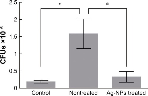
In vivo antimicrobial activity of Ag-NPs in a murine skin infection model triggered by a P. aeruginosa clinical strain
A murine skin infection model in rats was performed to test the ability of Ag-NPs to treat a topical infection caused by a resistant clinical strain (P. aeruginosa). As can be observed in , there is a statistically significant higher amount of CFUs present in the group infected with the clinical strain of P. aeruginosa with respect to the control group (P<0.05). Moreover, the group infected with P. aeruginosa and treated with Ag-NPs exhibits a number of CFUs statistically similar to the control group and statistically much lower than the group infected with P. aeruginosa and not treated with the Ag-NPs (P<0.05).
Figure 6 Antimicrobial effects of Ag-NPs in a murine skin infection model.
Notes: CFUs obtained from the in vivo antibacterial test. The mean values are expressed (±SEM) (*P<0.05).
Abbreviations: Ag-NPs, silver nanoparticles; CFUs, colony-forming units; SEM, standard error of the mean.
In vivo toxicological assay of the Ag-NPs
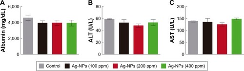
A murine toxicity infection model in rats was performed to test the toxicity of Ag-NPs during treatment of a topical infection caused by a resistant clinical strain (P. aeruginosa). For the toxicity study, different parameters of renal function were evaluated. These parameters included urinary volume, urinary concentrations of proteins, glucose and creatinine and blood concentrations of creatinine. The results shown in demonstrate that the control urine volumes (), as well as glucose (), total proteins (), and creatinine () concentration levels in the urine, are statistically similar (P<0.05) to the values obtained for all the treatments with different Ag-NP concentrations (100, 200, and 400 ppm). The same trend is observed for creatinine levels shown in the blood (). In addition, no statistically significant differences (P<0.05) were observed between the groups of rats treated with the Ag-NPs and the control groups in the parameters tested to evaluate hepatic function (albumin [], ALT [], and AST [] concentrations in blood plasma).
Figure 7 Parameters of renal function of Wistar rats treated with Ag-NPs.
Notes: Test was performed for different concentrations of Ag-NPs (100, 200, and 400 ppm), and different parameters of renal function are measured: (A) urinary volume, (B) urinary glucose concentration, (C) urinary total proteins concentration, (D) urinary creatinine concentration, and (E) blood plasma creatinine concentration. The mean values are expressed (±SEM).
Abbreviations: Ag-NPs, silver nanoparticles; SEM, standard error of the mean.
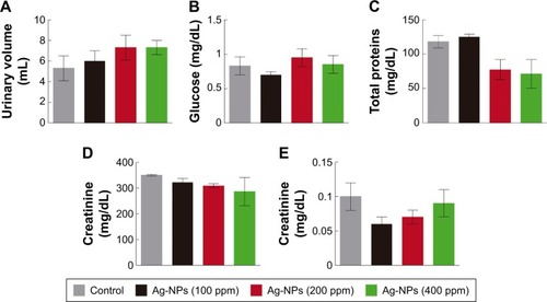
Figure 8 Parameters of hepatic function of Wistar rats treated with Ag-NPs.
Notes: Test was performed for different concentrations of Ag-NPs (100, 200, and 400 ppm), and different parameters of hepatic function are measured: (A) blood plasma concentration of albumin, (B) blood plasma concentration of ALT, and (C) blood plasma concentration of AST. The mean values are expressed (±SEM).
Abbreviations: Ag-NPs, silver nanoparticles; ALT, alanine aminotransferase; AST, aspartate aminotransferase; SEM, standard error of the mean.
Finally, no differences were observed in the appearance and characteristics of the skin between the treated animals at any of the concentrations of Ag-NPs.
Discussion
Synthesis and characterization of Ag-NPs
It is well-known that plant crude extracts contain secondary metabolites, such as phenolic acids, flavonoids, alkaloids, and terpenoids, that can trigger redox reactions, such as those that take part in the synthesis of Ag-NPs, which are involved in the reduction in silver ions into metallic silver atoms that further assemble into nanostructures.Citation52 Many species of the Acacia genus contain different types of secondary metabolites that can be used as reducing agents such as amines and alkaloids, nonprotein amino acids, cyanogenic glycosides, cyclitols, fatty acids and seed oils, fluoroacetate, gums, terpenes, diterpenes, phytosterols, triterpene genins and saponins, hydrolyzable tannins, condensed tannins, and flavonoids.Citation31 However, the polysaccharides or gums and complex phenolic compounds or condensed tannins are among the most studied and most recognized.Citation31 Thus, the effective reducing capability of the A. rigidula extract used in the synthesis of Ag-NPs can be attributed to the phenolic compounds, saponins, and flavonoids.
The homogeneity of the size distributions observed for the Ag-NPs reported in this work compares with other works in the literature that report Ag-NPs produced by green synthesis methods. Ag-NPs have been obtained with sizes between 50 and 250 nm using Carica papaya leaf extract;Citation53 with average sizes 23±12, 32±24 and 19±22 nm using Illicium verum (star anise) seed extracts,Citation54 and average size 73 nm using an aqueous extract of Tephrosia tinctoria.Citation55 Regarding spherical shape homogeneity, our synthesis method was compared with that of other works where spherical Ag-NPs are synthesized, such as in the case of other green synthesis methods that employ leaf extracts of Saraca indica,Citation56 Ficus carica,Citation57 and Cocos nucifera.Citation58
The chemical and physical characterization of the Ag-NP samples suggests that the Acacia extract is capable of not only reducing the silver ions but also capping the Ag-NPs and stabilizing their surface. This is a phenomenon not commonly observed, but it has been previously reported that when both reducing and capping effects are present in an extract, it allows a tighter control of the nanoparticle sizes obtained and prevents further aggregation of the samples in time.Citation52,Citation59
The well-defined spotty rings correspond to (111), (200), (220), (311), and (331) planes of fcc metallic silver (Joint Committee on Powder Diffraction Standards file no 87-0720), while EDS analysis demonstrates that the nanoparticles obtained are constituted of silver and makes evident their crystalline nature ( and ). Similar information from the EDS and SAED analyses of Ag-NPs has been reported when using I. verum (star anise) seedsCitation54 and extracts of Capsicum annuum var. aviculare (piquin) fruits.Citation12
The UV–Vis spectrum of both the A. rigidula extract and the Ag-NPs shows that for the extract, the absorbance was low, but in the case of Ag-NPs, it displays the maximum absorbance at 444 nm, indicating that the extinction band is a result of the surface plasmon resonance (SPR) phenomenon produced by Ag-NPs (). These results correlate with previous reports that show a maximum absorption peak at 480 nm, when Ag-NPs were synthesized using T. tinctoria extract as a reducing agent,Citation55 and an absorption peak at 420 nm when Ag-NPs were produced using Chrysopogon zizanioides aqueous extract.Citation59 Hence, these results agree with the Mie scattering theory about single spherical Ag-NPs well dispersed in water with different diameters.Citation54 In this regard, the width of the peak obtained in the UV–Vis spectrum is mainly attributed to the size distribution and differences in the shape of the Ag-NPs.Citation60 Furthermore, the UV–Vis spectra of a sample that was stored in solution for 4 months show a sustained maximum absorbance at ~450 nm (), strongly suggesting that Ag-NPs remain stable during considerable long periods of time. Thus, the organic matrix of A. rigidula extract acts as an effective stabilizing agent.
In the case of the FT-IR spectroscopy (), the different functional groups observed in the Ag-NPs spectrum also appear in the A. rigidula extract. In addition, the peaks observed are in accordance with the different components that the extract may contain, such as carbohydrates, flavonoids, and polyphenols, that can be carrying out the process of reduction in the Ag+ ions to produce Ag-NPs. Therefore, the characterization data strongly indicate the function of the extract as a capping and stabilizing agent of the Ag-NPs. Moreover, the results suggest the presence of the components of the extract in the surface of the Ag-NPs.
Antibacterial test: in vitro and in vivo study
For >2,000 years, silver has been recognized for its medicinal properties. Hence, silver has been used as an antimicrobial agent due to its effectiveness against some pathogenic microorganisms and its lower toxicity in animal cells in comparison with other metals.Citation24 Moreover, recently, there has been an increasing interest in the application of silver as nanoparticles, for medical applications, as an alternative antimicrobial agent. Taking into account the problem of multidrug resistance to the conventional antibiotics, both Gram-negative and Gram-positive bacteria have developed recently.Citation61 For this reason, there is a need to search and produce more efficient antimicrobial agents that can fight strains of multidrug-resistant bacteria.Citation62
The Ag-NPs produced by green synthesis with A. rigidula extract exhibited antimicrobial activity against Gram-negative bacteria (E. coli, P. aeruginosa, and the clinical strain of P. aeruginosa) and Gram-positive bacteria (B. subtilis) tested in our study ( and ). Interestingly, the MIC values observed in our study for P. aeruginosa are similar to the values obtained by Ag-NPs produced from an aqueous extract of Phyllanthus amarus against several P. aeruginosa clinical strains,Citation63 where MIC values varied from 6.25 to 12.5 ppm. MIC values obtained in this study for B. subtilis are lower than those previously reported, where a 12.5 ppm MIC value was observed with Ag-NPs obtained from an aqueous rhizome extract of Coptis chinensis.Citation64 However, the same study showed lower MIC values for E. coli, where an MIC value of 25 ppm was reported.Citation64 A similar MBC of the green synthesized Ag-NPs against the bacteria tested in this study was found using plant extracts as reducing and stabilizing agents.Citation65–Citation67 In this regard, it is possible to infer that Ag-NPs produced by an A. rigidula extract display effective antibacterial activity as bacteriostatic and bactericidal agents against both Gram-positive and Gram-negative bacteria.
We hypothesized that Ag-NPs might be interacting with both membrane and cell wall components, impeding bacterial growth. The PI assay results obtained by fluorescence microscopy () show an increased fluorescence in the bacteria treated with Ag-NPs compared to the untreated control sample. This indicates that the Ag-NPs induce bacterial cell permeability by the disruption of cell membrane integrity. Thus, this can be one potential antibacterial mechanism linked to the effective antimicrobial properties exhibited by Ag-NPs. In this context, alterations in permeability and membrane structure have been mentioned as one mechanism of antimicrobial activity of Ag-NPs.Citation68 Previous reports have shown that cell membrane damage of P. aeruginosa can be induced by Ag-NPs produced by green synthesis methods.Citation69
For the in vivo study, we confirmed that the Ag-NPs’ antibacterial activity involved a reduction of CFUs in the group treated with the Ag-NPs. In comparison, Ag-NPs elaborated by means of an aqueous extract of Caltropis procera fruit or leaves, administered to infant mice colonized with Vibrio cholerae or enterotoxic E. coli, significantly reduced from 75 to 100 times the colonization rates of these pathogenic bacteria.Citation70
Furthermore, nanobiocomposites of Ag-NPs synthetized with leaf extract of Syzygium cumini into a matrix of cellulose nanocrystals from Dendrocalamus hamiltonii and Bambusa bambos leaves showed antibacterial activity and were effective in obtaining an accelerated wound healing on, in vivo, skin of male, Swiss albino mice.Citation71 Moreover, flavonoid-loaded Ag-NPs produced by a Ricinus communis leaf extracted flavonoid mixture were efficient in curing a Staphylococcus aureus infection on a silkworm Bombyx mori larvae.Citation72 Therefore, molecules such as flavonoids have been found to be able to coat Ag-NPs,Citation73,Citation74 a similar to what we have seen in our study, and their presence in the surface could be a factor involved in the antimicrobial activity of these nanoparticles.
The main difference between Gram-negative and Gram-positive bacteria is the thickness of the cell wall. Gram-negative bacteria contain a thin peptidoglycan, 2–3 nm thick, between the outer membrane and the cytoplasmic or inner membrane,Citation75 while Gram-positive bacteria possess a thicker peptidoglycan layer of ~30 nm thick, without outer membrane.Citation76 In the case of B. subtilis (Gram-positive), growth inhibition can be attributed to interactions between Ag-NPs embedded in the cell wall, causing membrane permeability due to structural changes in the cell wall.Citation77 In contrast, on Gram-negative bacteria such as E. coli and P. aeruginosa, the outer membrane is composed mainly of lipids and proteins, which are the first structures encountered by the Ag-NPs. Therefore, Ag-NPs could have adhered to these structures, altering their properties and integrity-forming gaps within the membrane, increasing its permeability.Citation6 It is also well known that the membrane contains large amounts of sulfur-containing proteins and their inactivation can play a role in the deterioration of the membrane produced by Ag-NPs.Citation78,Citation79 Therefore, these kinds of proteins may be another target of the Ag-NPs, inactivating enzymes and contributing to the deterioration of the membrane.Citation78,Citation79 It has been reported that the antimicrobial activity of colloidal Ag-NPs is influenced by different particle characteristics such as size, shape, and capping agent.Citation24,Citation52,Citation80 In this context, small sized Ag-NPs (as the Ag-NPs obtained in this study) have been reported to present a better bactericidal activity due to a greater binding surface in comparison to larger Ag-NPs.Citation2 Moreover, the capping of the Ag-NPs produced in this study contains O−H functional groups () from polyphenol compounds present in the organic matrix. This functional group may produce damage and toxicity in the membrane and cell wall of the bacteria through oxidative stress by free radicals and reactive oxygen species (ROS).Citation81 However, Ag-NPs alone are capable of producing ROS causing a microbial growth inhibition.Citation82
Another mechanism that explains the toxicity of Ag-NPs against some bacteria refers to the release of Ag+ ions from the Ag-NPs. In fact, it has been reported that the toxicity of Ag-NPs with a size of 20–80 nm is predominantly due to the release of Ag+ ions.Citation2,Citation6 Since the Ag-NPs synthesized in this study (22.46 nm) are within the size range reported, it is possible that Ag+ ions release may also explain the growth inhibition shown here. This means that Ag+ ions could interact with membrane or cell wall components or participate in the generation of ROS. Hence, this mechanism can cause changes and damage in the permeability and physiology of bacteria. In addition, it has been shown that the release of Ag+ ions is the main antibacterial mechanism of toxicity of Ag-NPs.Citation83
In vivo toxicological study
In an organism, the suitable functioning of its organs is very important to achieve a healthy balance maintaining the composition of its internal environment. This is largely achieved with the correct functioning of organs such as the kidney and liver. In the kidneys, the basic structural and functional units are the nephrons. They carry out their function by the following three fundamental processes: glomerular filtration, tubular reabsorption, and tubular secretion producing the final excretion of substances in the urine.Citation84 In relation to the renal function parameters in urine assessed in this study, the kidneys regulate the volume (amount of body fluid), the concentration of glucose (associated with tubular function in nephrons), the concentration of total proteins (linked with glomerular filtration and general function of nephrons), and the concentration of creatinine both in plasma and urine (related with glomerular filtration rate in nephrons) in the organism. All the parameters measured in the treated samples were very similar to the values of the control group (). Thus, the Ag-NPs produced in our study did not produce any adverse or toxic effects in the nephrons and renal function of the animals treated.
Similarly, the liver is responsible for numerous vital functions for the proper functioning of higher order organisms, where the metabolization of chemical substances and biliary excretion occur.Citation84 Therefore, this work analyzed hepatic function parameters in blood plasma when rats were treated with the Ag-NPs. The concentration of ALT and AST (biomarker of hepatic cell necrosis) and the concentration of albumin (biochemical biomarker of damage and hepatic function) of the treated groups with Ag-NPs were similar to the values of control groups (). Thus, the Ag-NPs obtained in this study did not have any adverse or toxic effects in the hepatic function. These findings are in agreement with other results that show zero mortality, absence of abnormal reactions, and minimal effect on kidney and liver indices in male and female Sprague Dawley rats administered orally with 2,000 mg/kg bw of Ag-NPs produced by Sargassum siliquosum, indicating low toxicity of these nanoparticles.Citation85 Moreover, in the same study, it was observed that a pretreatment with Ag-NPs prevented liver cell damage caused by paracetamol at 100 and 200 mg/kg bw dose. Thus, Ag-NPs obtained by green synthesis provide a cheaper nontoxic method that ensures effectiveness and safety in the application of these nanoparticles in different fields of study.Citation86 Finally, in the skin directly exposed to Ag-NPs, no adverse effects or interactions between tissue and the nanoparticles were found, since there were no alterations or changes in the appearance and structure of the treated skin. Therefore, the Ag-NPs at the tested concentration are safe for use in topical medical applications as an alternative antimicrobial therapeutic.
Conclusion
The use of the extract, obtained from roots and stems of A. rigidula, as a reducing and stabilizing agent represents a suitable alternative to produce antimicrobial metal nanoparticles (green synthesis). The synthesis reported here provides many advantages over conventional synthesis methods, such as the use and production of less toxic reagent and products. This allows for greater care of the environment. Currently, in the field of pharmacology and toxicology, there is a great need for better nontoxic treatment options that are more effective and have better antimicrobial activity against infectious diseases caused by different types of bacteria, especially against multidrug-resistant clinical strains. The antimicrobial properties shown by the Ag-NPs reported here represent a great alternative to achieve this objective. The results reported in this work are relevant to the field since they represent a contribution toward the development of more efficient and nontoxic alternatives for pharmacological treatments, taking advantage of the tools developed by nanotechnology. Nevertheless, further research in this subject is required to elucidate completely the interaction mechanism between Ag-NPs and bacteria. Moreover, research in pharmacological and toxicological studies, especially in vivo, is much needed to develop and design future antimicrobial therapeutic agents.
Availability of data and material
All data generated or analyzed during this study are included in this published article.
Author contributions
CEE-G, JAG-C, AV-R, LZM-P, and JRM-R designed, performed, and analyzed the experimental data and wrote the article. EDBC and EMS-S designed and performed the TEM analysis and contributed to the discussion and format of the article. RMCM, DIRS, FMTG, JLCR, and VCR designed and performed the antimicrobial experiments in vivo. MTT-G helped with the format and methodology of the article. All authors contributed toward data analysis, drafting and revising the paper and agree to be accountable for all aspects of the work.
Acknowledgments
The authors thank the Universidad Autónoma de Nuevo León and CONACyT for financially supporting this work and Silvia Torres and JA Mercado Silva, who carried out some of the characterization studies reported herein, for providing technical support. This study was supported by Paicyt 2016–2017 Science Grant from the Universidad Autónoma de Nuevo León and by CONACyT Grants for Basic science (grant 221332), Fronteras de la Ciencia (grant 1502), and Infraestructura (grant 279957).
Disclosure
The authors report no conflicts of interest in this work.
References
- KhalilKAFouadHElsarnagawyTAlmajhdiFNPreparation and characterization of electrospun PLGA/silver composite nanofibers for biomedical applicationsInt J Electrochem Sci2013834833493
- MoronesJRElechiguerraJLCamachoAThe bactericidal effect of silver nanoparticlesNanotechnology20051610234620818017
- ElechiguerraJLBurtJLMoronesJRInteraction of silver nanoparticles with HIV-1J Nanobiotechnology200531615987516
- JhaDThiruveedulaPKPathakRMultifunctional biosynthesized silver nanoparticles exhibiting excellent antimicrobial potential against multi-drug resistant microbes along with remarkable anti-cancerous propertiesMater Sci Eng C Mater Biol Appl20178065966928866213
- Morones-RamirezJRPlata, metal precioso con amplio espectro de aplicacionesRev Cien Desarrollo2010362415662
- DuránNDuránMde JesusMBSeabraABFávaroWJNakazatoGSilver nanoparticles: a new view on mechanistic aspects on antimicrobial activityNanomedicine201612378979926724539
- LiauSReadDPughWFurrJRussellAInteraction of silver nitrate with readily identifiable groups: relationship to the antibacterialaction of silver ionsLett Appl Microbiol19972542792839351278
- GuptaASilverSMolecular genetics: silver as a biocide: will resistance become a problem?Nat Biotechnol199816108889788326
- NomiyaKYoshizawaATsukagoshiKKasugaNCHirakawaSWatanabeJSynthesis and structural characterization of silver (I), aluminium (III) and cobalt (II) complexes with 4-isopropyltropolone (hinokitiol) showing noteworthy biological activities. Action of silver (I)-oxygen bonding complexes on the antimicrobial activitiesJ Inorg Biochem2004981466014659632
- Morones-RamirezJRWinklerJASpinaCSCollinsJJSilver enhances antibiotic activity against Gram-negative bacteriaSci Transl Med20135190190ra81
- Mendoza-ReséndezRGómez-TreviñoABarriga-CastroEDNúñezNOLunaCSynthesis of antibacterial silver-based nanodisks and dendritic structures mediated by royal jellyRSC Adv20144416501658
- Mendoza-ReséndezRNúnezNOBarriga-CastroEDLunaCSynthesis of metallic silver nanoparticles and silver organometallic nanodisks mediated by extracts of Capsicum annuum var. aviculare (piquin) fruitsRSC Adv20133432076520771
- GeethalakshmiRSaradaDGold and silver nanoparticles from Trianthema decandra: synthesis, characterization, and antimicrobial propertiesInt J Nanomedicine20127537523091381
- WangYLiuWLiuWSynthesis of SnAgCu nanoparticles with low melting point by the chemical reduction methodMicroelectron Reliabil2017781724
- GabrielJSGonzagaVAPoliALSchmittCCPhotochemical synthesis of silver nanoparticles on chitosans/montmorillonite nanocomposite films and antibacterial activityCarbohydr Polym201717120221028578955
- dos SantosPLKaticVToledoKCBonacinJAPhotochemical one-pot synthesis of reduced graphene oxide/Prussian blue nanocomposite for simultaneous electrochemical detection of ascorbic acid, dopamine, and uric acidSens Actuat B Chem2018255324372447
- WiniarskiJPde BarrosMRMagossoHAJostCLElectrochemical reduction of sulfite based on gold nanoparticles/silsesquioxane-modified electrodeElectrochim Acta2017251522531
- LandgeSGhoshDAikenKSolvent-free synthesis of nanoparticlesTörökBDransfieldTGreen ChemistryBoston, MAElsevier2017609646
- AnjugamMVaseeharanBIswaryaADivyaMPrabhuNMSankaranarayananKBiological synthesis of silver nanoparticles using β-1, 3 glucan binding protein and their antibacterial, antibiofilm and cytotoxic potentialMicrob Pathog2018115314029208541
- FariqAKhanTYasminAMicrobial synthesis of nanoparticles and their potential applications in biomedicineJ Appl Biomed2017154241248
- ChahardoliAKarimiNFattahiANigella arvensis leaf extract mediated green synthesis of silver nanoparticles: their characteristic properties and biological efficacyAdv Powder Technol2018291202210
- MittalAKChistiYBanerjeeUCSynthesis of metallic nanoparticles using plant extractsBiotechnol Adv201331234635623318667
- MohanrajRAntimicrobial activities of metallic and metal oxide nanoparticles from plant extractsGrumezescuAMAntimicrobial NanoarchitectonicsBucharest, RomaniaElsevier201783100
- AhmedSAhmadMSwamiBLIkramSA review on plants extract mediated synthesis of silver nanoparticles for antimicrobial applications: a green expertiseJ Adv Res201671172826843966
- RajeshkumarSBharathLMechanism of plant-mediated synthesis of silver nanoparticles – a review on biomolecules involved, characterisation and antibacterial activityChem Biol Interact201727321922728647323
- ArunachalamKDAnnamalaiSKChrysopogon zizanioides aqueous extract mediated synthesis, characterization of crystalline silver and gold nanoparticles for biomedical applicationsInt J Nanomedicine20138237523861583
- LedezmaARomeroJHernándezMSíntesis biomimética de nanopartículas de plata utilizando extracto acuoso de nopal (Opuntia sp.) y su electrohilado poliméricoSuperf Vacío2014274133140
- SinghAKSrivastavaOOne-step green synthesis of gold nanoparticles using black cardamom and effect of pH on its synthesisNanoscale Res Lett2015101353
- ClementBAGoffCMForbesTDAToxic amines and alkaloids from Acacia rigidulaPhytochemistry199849513771380
- PawarRSGrundelEFardin-KiaARRaderJIDetermination of selected biogenic amines in Acacia rigidula plant materials and dietary supplements using LC–MS/MS methodsJ Pharm Biomed Anal20148845746624176750
- SeiglerDSPhytochemistry of Acacia – sensu latoBiochem Syst Ecol2003318845873
- RamírezRLaraJInfluence of native shrubs Acacia rigidula, Cercidium macrum and Acacia farnesiana on digestibility and nitrogen utilization by sheepSmall Rumin Res19982813945
- RamírezRLedezma-TorresRForage utilization from native shrubs Acacia rigidula and Acacia farnesiana by goats and sheepSmall Rumin Res19972514350
- ZuoJMabonJWeb-based electron microscopy application software: web-EMAPSMicro Microanal200410S021000
- AndrewsJMDetermination of minimum inhibitory concentrationsJ Antimicrob Chemother200148suppl 151611420333
- CavaleriJRankinDHarbeckJManual of antimicrobial susceptibility testingAm Soc Microbiol Seattle Washington2005125342
- Vazquez-RodriguezAVasto-AnzaldoXGBarboza PerezDMicrobial competition of Rhodotorula mucilaginosa UANL-001L and E. coli increase biosynthesis of non-toxic exopolysaccharide with applications as a wide-spectrum antimicrobialSci Rep20188179829335484
- KugelbergENorströmTPetersenTKDuvoldTAnderssonDIHughesDEstablishment of a superficial skin infection model in mice by using Staphylococcus aureus and Streptococcus pyogenesAntimicrob Agents Chemother20054983435344116048958
- RartelsHBöhmerMEine mikromethode 7air kreatininbestimmungClin Chim Acta197132181855096431
- FabinyDLErtingshausenGAutomated reaction-rate method for determination of serum creatinine with the CentrifiChemClin Chem19711786967005562281
- TrinderPDetermination of glucose in blood using glucose oxidase with an alternative oxygen acceptorAnn Clin Biochem1969612427
- GornallAGBardawillCJDavidMMDetermination of serum proteins by means of the biuret reactionJ Biol Chem1949177275176618110453
- DoumasBTWatsonWABiggsHGAlbumin standards and the measurement of serum albumin with bromcresol greenClin Chim Acta197131187965544065
- MurrayRAlanine aminotransferaseKaplanLAPesceAJClinical Chemistry: Theory, Analysis, and Correlation2nd ed Chap St LouisSt Louis, MOThe CV Mosby Company1989895898
- MurrayRAspartate aminotransferaseKaplanLAPesceAJClinical Chemistry Theory, Analysis and CorrelationSt Louis, MOCV Mosby Company198411051108
- SadiqMBHanpithakpongWTarningJAnalAKScreening of phytochemicals and in vitro evaluation of antibacterial and antioxidant activities of leaves, pods and bark extracts of Acacia nilotica (L.) DelInd Crops Prod201577873882
- D’AngeloJAZodrowELChemometric study of functional groups in different layers of Trigonocarpus grandis ovules (Pennsylvanian seed fern, Canada)Org Geochem201142910391054
- AadilKRBarapatreAMeenaASJhaHHydrogen peroxide sensing and cytotoxicity activity of Acacia lignin stabilized silver nanoparticlesInt J Biol Macromol201682394726434518
- KumarKMSinhaMMandalBKGhoshARKumarKSReddyPSGreen synthesis of silver nanoparticles using Terminalia chebula extract at room temperature and their antimicrobial studiesSpectrochim Acta A Mol Biomol Spectrosc20129122823322381795
- MuruganKSenthilkumarBSenbagamDAl-SohaibaniSBiosynthesis of silver nanoparticles using Acacia leucophloea extract and their antibacterial activityInt J Nanomedicine20149243124876776
- VietDQSon ThoVDStudy on characteristics of acacia wood by FTIR and thermogrametric analysisViet J Chem2017552259
- ChungI-MParkISeung-HyunKThiruvengadamMRajakumarGPlant-mediated synthesis of silver nanoparticles: their characteristic properties and therapeutic applicationsNanoscale Res Lett20161114026821160
- BanalaRRNagatiVBKarnatiPRGreen synthesis and characterization of Carica papaya leaf extract coated silver nanoparticles through X-ray diffraction, electron microscopy and evaluation of bactericidal propertiesSaudi J Biol Sci201522563764426288570
- LunaCChávezVBarriga-CastroEDNúñezNOMendoza-ReséndezRBiosynthesis of silver fine particles and particles decorated with nanoparticles using the extract of Illicium verum (star anise) seedsSpectrochim Acta A Mol Biomol Spectrosc2015141435025659741
- RajaramKAiswaryaDSureshkumarPGreen synthesis of silver nanoparticle using Tephrosia tinctoria and its antidiabetic activityMater Lett2015138251254
- PeruguSNagatiVBhanooriMGreen synthesis of silver nanoparticles using leaf extract of medicinally potent plant Saraca indicaAppl Nanosci201665747753
- BoraseHPSalunkheRBPatilCDInnovative approach for urease inhibition by Ficus carica extract-fabricated silver nanoparticles: an in vitro studyBiotechnol Appl Biochem201562678078425560197
- RoopanSMMadhumithaGRahumanAAKamarajCBharathiASurendraTLow-cost and eco-friendly phyto-synthesis of silver nanoparticles using Cocos nucifera coir extract and its larvicidal activityInd Crops Prod201343631635
- ArunachalamKDAnnamalaiSKHariSOne-step green synthesis and characterization of leaf extract-mediated biocompatible silver and gold nanoparticles from Memecylon umbellatumInt J Nanomedicine20138130723569372
- ZhangJLiXSunXLiYSurface enhanced Raman scattering effects of silver colloids with different shapesJ Phys Chem B200510925125441254816852551
- FrieriMKumarKBoutinAAntibiotic resistanceJ Infect Public Health201610436937827616769
- KapilAThe challenge of antibiotic resistance: need to contemplateIndian J Med Res200512128315756040
- SinghKPanghalMKadyanSChaudharyUYadavJPGreen silver nanoparticles of Phyllanthus amarus: as an antibacterial agent against multi drug resistant clinical isolates of Pseudomonas aeruginosaJ Nanobiotechnology20141214025271044
- AhmadAWeiYSyedFThe effects of bacteria-nanoparticles interface on the antibacterial activity of green synthesized silver nanoparticlesMicrob Pathog201710213314227916692
- BanasiukRKrychowiakMSwigonDCarnivorous plants used for green synthesis of silver nanoparticles with broad-spectrum antimicrobial activityArab J Chem201782332
- ShankarTKarthigaPSwarnalathaKRajkumarKGreen synthesis of silver nanoparticles using Capsicum frutescence and its intensified activity against E. coliRes Effic Technol201733303308
- PattanayakSMollickMMRMaityDButea monosperma bark extract mediated green synthesis of silver nanoparticles: characterization and biomedical applicationsJ Saudi Chem Soc2015216673684
- DakalTCKumarAMajumdarRSYadavVMechanistic basis of antimicrobial actions of silver nanoparticlesFront Microbiol20167183127899918
- KumarSSDHoureldNNKroukampEMAbrahamseHCellular imaging and bactericidal mechanism of green-synthesized silver nanoparticles against human pathogenic bacteriaJ Photochem Photobiol B201817825926929172133
- SalemWLeitnerDRZinglFGAntibacterial activity of silver and zinc nanoparticles against Vibrio cholerae and enterotoxic Escherichia coliInt J Med Microbiol20153051859525466205
- SinglaRSoniSKulurkarPMIn situ functionalized nanobiocomposites dressings of bamboo cellulose nanocrystals and silver nanoparticles for accelerated wound healingCarbohydr Polym201715515216227702499
- RajasekharreddyPRaniPUMattapallySBanerjeeSKUltra-small silver nanoparticles induced ROS activated toll-pathway against Staphylococcus aureus disease in silkworm modelMater Sci Eng C2017779901002
- AnjumSAbbasiBHThidiazuron-enhanced biosynthesis and antimicrobial efficacy of silver nanoparticles via improving phytochemical reducing potential in callus culture of Linum usitatissimum LInt J Nanomedicine20161171526955271
- HussainMRajaNIIqbalMAslamSApplications of plant flavonoids in the green synthesis of colloidal silver nanoparticles and impacts on human healthIran J Sci Technol Trans A Sci20172112
- MurrayRSteedPElsonHThe location of the mucopeptide in sections of the cell wall of Escherichia coli and other Gram-negative bacteriaCan J Microbiol196511354756014346132
- ShockmanGDBarrenJStructure, function, and assembly of cell walls of Gram-positive bacteriaAnnu Rev Microbiol19833715015276139058
- DibrovPDziobaJGosinkKKHäseCCChemiosmotic mechanism of antimicrobial activity of Ag+ in Vibrio choleraeAntimicrob Agents Chemother20024682668267012121953
- PalSTakYKSongJMDoes the antibacterial activity of silver nanoparticles depend on the shape of the nanoparticle? A study of the Gram-negative bacterium Escherichia coliAppl Environ Microbiol20077361712172017261510
- HoltKBBardAJInteraction of silver (I) ions with the respiratory chain of Escherichia coli: an electrochemical and scanning electrochemical microscopy study of the antimicrobial mechanism of micromolar Ag+Biochemistry20054439132141322316185089
- KaviyaSSanthanalakshmiJViswanathanBGreen synthesis of silver nanoparticles using Polyalthia longifolia leaf extract along with D-sorbitol: study of antibacterial activityJ Nanotechnol201120115
- DasBDashSKMandalDGreen synthesized silver nanoparticles destroy multidrug resistant bacteria via reactive oxygen species mediated membrane damageArab J Chem2017106862876
- KimJSKukEYuKNAntimicrobial effects of silver nanoparticlesNanomedicine2007319510117379174
- IvaskAElBadawyAKaweeteerawatCToxicity mechanisms in Escherichia coli vary for silver nanoparticles and differ from ionic silverACS Nano20138137438624341736
- KlaassenCDAmdurMOCasarett and Doull’s Toxicology: The Basic Science of PoisonsNew YorkMcGraw-Hill, Health Professions Division, USA1996
- VasquezRDApostolJGde LeonJDPolysaccharide-mediated green synthesis of silver nanoparticles from Sargassum siliquosum JG Agardh: assessment of toxicity and hepatoprotective activityOpenNano201611624
- RoyNGaurAJainABhattacharyaSRaniVGreen synthesis of silver nanoparticles: an approach to overcome toxicityEnviron Toxicol Pharmacol201336380781223958974
- ZhangXFLiuZGShenWGurunathanSSilver nanoparticles: synthesis, characterization, properties, applications, and therapeutic approachesInt J Mol Sci2016179 piiE153427649147
- ChiduralaSCKalagaddaVRTamburPAntimicrobial activity of pure Cu nano particles synthesized by surfactant varied chemical reduction methodEnvironmental Nanotechnology, Monitoring & Management201668894
- BindhuMUmadeviMAntibacterial activities of green synthesized gold nanoparticlesMaterials Letters2014120122125