

Abstract
Background
Trastuzumab plus docetaxel is a mainstay to treat HER2-positive breast cancers. However, developing nanoparticles could help to improve the efficacy/toxicity balance of this doublet by improving drug trafficking and delivery to tumors. This project aimed to develop an immunoliposome in breast cancer, combining docetaxel encapsulated in a stealth liposome engrafted with trastuzumab, and comparing its performances on human breast cancer cell lines with standard combination of docetaxel plus trastuzumab.
Methods
Several strategies to engraft trastuzumab to pegylated liposomes were tested. Immunoliposomes made of natural (antibody nanoconjugate-1 [ANC-1]) and synthetic lipids (ANC-2) were synthesized using standard thin film method and compared in size, morphology, docetaxel encapsulation, trastuzumab engraftment rates and stability. Antiproliferative activity was tested on human breast cancer models ranging from almost negative (MDA-MB-231), positive (MDA-MB-453) to overexpressing (SKBR3) HER2. Finally, cell uptake of ANC-1 was studied by electronic microscopy.
Results
ANC-1 showed a greater docetaxel encapsulation rate (73%±6% vs 53%±4%) and longer stability (up to 1 week) as compared with ANC-2. Both ANC presented particle size ≤150 nm and showed similar or higher in vitro antiproliferative activities than standard treatment, ANC-1 performing better than ANC-2. The IC50s for docetaxel combined to free trastuzumab were 8.7±4, 2±0.7 and 6±2 nM with MDA-MB-231, MDA-MB-453 and SKBR3, respectively. The IC50s for ANC-1 were 2.5±1, 1.8±0.6 and 3.4±0.8 nM and for ANC-2 were 1.8±0.3 nM, 2.8±0.8 nM and 6.8±1.8 nM with MDA-MB-231, MDA-MB-453 and SKBR3, respectively. Cellular uptake appeared to depend on HER2 expression, the higher the expression, the higher the uptake.
Conclusion
In vitro results suggest that higher antiproliferative efficacy and efficient drug delivery can be achieved in breast cancer models using nanoparticles.
Video abstract
Point your SmartPhone at the code above. If you have a QR code reader the video abstract will appear. Or use:
Supplementary materials
Figure S1 Representative SDS-PAGE gel, showing trastuzumab engraftment on liposome surface. Protein markers (A1), trastuzumab (A2), thiolated trastuzumab (A3), Liposome-1 (A4), ANC-1 engrafted with strategy B (A5), Liposome-1 engrafted with strategy B without maleimide function (A6), ANC-1 engrafted following strategy A (A7), Liposome-1 engrafted following strategy A without maleimide function (A8), protein markers (B1), trastuzumab (B2), ANC-1 engrafted following strategy B (B3), Liposome-1 engrafted following strategy B without maleimide function (B4), ANC-2 engrafted with strategy B (B5), Liposome-2 engrafted with strategy B without maleimide function (B6), ANC-1 engrafted with strategy B after 45 days (B7).
Abbreviations: SDS-PAGE, sodium dodecyl sulfate polyacrylamide gel electrophoresis; ANC, antibody nanoconjugate.
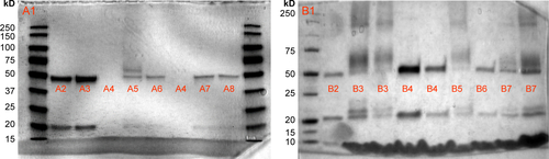
Figure S2 TEM observations of ANC-1 treated with osmium tetroxide (A and B) and uranyl acetate (C).
Abbreviations: TEM, transmission electron microscopy; ANC-1, antibody nanoconjugate-1.
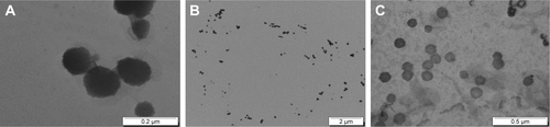
Figure S3 TEM observations of ANC-2 treated with osmium tetroxide.
Abbreviations: TEM, transmission electron microscopy; ANC-2, antibody nanoconjugate-2.
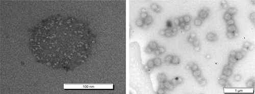
Figure S4 TEM observations of liposome-1 before extrusion.
Abbreviation: TEM, transmission electron microscopy.
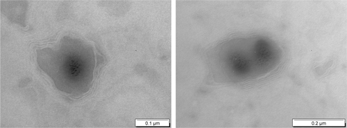
Figure S5 Monitoring size (nm) over time for ANC-1 following different storage conditions: diluted at −20°C [img], concentrated at 4°C [img], diluted at 4°C [img] and diluted at 25°C ([img]).a
Note: aValues are mean ± SEM of three or more experiments.
Abbreviation: ANC-1, antibody nanoconjugate-1.
![Figure S5 Monitoring size (nm) over time for ANC-1 following different storage conditions: diluted at −20°C [img], concentrated at 4°C [img], diluted at 4°C [img] and diluted at 25°C ([img]).aNote: aValues are mean ± SEM of three or more experiments.Abbreviation: ANC-1, antibody nanoconjugate-1.](/cms/asset/3b85d644-5dc8-4b6c-ba9f-5a519603f76e/dijn_a_12194050_sf0005_b.jpg)
Acknowledgments
The authors would like to thank the platform Amuticyt (VRCM) de la faculté de Pharmacie La Timone and the Service Commun de Microscopie Électronique de la Faculté de Médecine La Timone for their technical expertise. The authors would also like to thank the Ligue Contre le Cancer who generously provided a grant to AR. Additionally, we thank the French Institut Roche who partly supported this study and Genentech who kindly provided trastuzumab.
Disclosure
AS and FB are members of the Institut Roche, a joint-institute from Roche Laboratories that commercializes trastuzumab and has partly funded this study. JC and RF received fees as board members of Roche. The authors report no other conflicts of interest in this work.
Author contributions
AR, RF, JC, SG, JMB, HM, FC, EM and CO performed the bench experiments. AR, RF and JC performed statistical analyses. AR, RF, BL, AS, FB and JC wrote the manuscript. All authors contributed toward data analysis, drafting and revising the paper and agree to be accountable for all aspects of the work.