
Abstract
Infection, as a common postoperative complication of orthopedic surgery, is the main reason leading to implant failure. Silver nanoparticles (AgNPs) are considered as a promising antibacterial agent and always used to modify orthopedic implants to prevent infection. To optimize the implants in a reasonable manner, it is critical for us to know the specific antibacterial mechanism, which is still unclear. In this review, we analyzed the potential antibacterial mechanisms of AgNPs, and the influences of AgNPs on osteogenic-related cells, including cellular adhesion, proliferation, and differentiation, were also discussed. In addition, methods to enhance biocompatibility of AgNPs as well as advanced implants modifications technologies were also summarized.
Introduction
Implant-associated infection as a common postoperative complication of orthopedic surgery often results in patient suffering, financial burden, and even fatalities.Citation1,Citation2 Application of antibiotics is the most common approach to treat infection. However, the rise of resistant organisms has made routine antibiotic prophylaxis ineffective. What is worse, bacterium can rapidly form biofilm on the implant surface, making antibiotics unable to penetrate and develop antibacterial function.Citation3 Thus, it is urgent to find an antibacterial agent that can kill drug-resistant bacteria and modify the prosthesis to prevent biofilm formation.
Silver (Ag) has been demonstrated to possess effective antibacterial effect and has been vastly used in medicine.Citation4 Moreover, Ag can be manufactured into silver nanoparticles (AgNPs) through nanotechnology to have improved physical, chemical, and biological properties.Citation5–Citation7 Currently, application of AgNPs on orthopedic implant modification to prevent implant-associated infection has drawn more attention.Citation8,Citation9 In the field of orthopedic investigation, AgNP-coated external fixation pins, proximal femur or tibia mega-prostheses, and AgNPs containing bone cement have been innovated, and they show an infection inhibition trend.Citation10–Citation12 Since AgNP-coated orthopedic implants represent a promising approach to prevent infections, the specific antibacterial mechanism of AgNPs and its effect on osteogenic-related cells need to be understood.Citation13
Many research studies have investigated the antimicrobial activity of AgNPs, but the possible antibiotic mechanisms and potential hazard are still unclear.Citation14,Citation15 Castiglioni et al concluded that AgNPs cause cytotoxicity in various cell lines in a dose-dependent manner.Citation16 However, the effect of AgNPs on osteoblast and osteoclast, which are responsible for bone formation and resorption respectively, is not included. In addition, optimal dosage of AgNPs on osteogenic-related cells is still controversial. Hence, the effect of AgNPs on osteogenic-related cells needs to be considered in order to screen the optimal concentration. Although Prabhu et al summed up the potential toxicity of AgNPs, they did not propose how to reduce the toxicity effect.Citation17 Biocompatibility is a precondition for the application in medicine. Aiming to enhance the biocompatibility of AgNPs, biosynthesis process can be applied to change the morphology and surface characteristics of AgNPs.Citation18 Furthermore, recent studies have proven that surface modifications with biomolecules, polymers, or metal ions are effective strategies that can also fulfill this mission.Citation19–Citation21
In this review, we discuss the antibacterial mechanisms and antibiofilm activity of AgNPs, which are the fundamental of implants application. The disputed cellular effects of AgNPs on osteogenesis-related cells are discussed, and some advice for implants designed are provided. Methods such as biosynthesis, adjustments of physical properties, and combining with biomolecules to enhance the compatibility of AgNPs are highlighted. Finally, different modification methods of AgNP introduction into orthopedic implant are also summarized.
Antibacterial mechanisms and antibiofilm activity of AgNPs
As described above, AgNPs can destroy multiple drug-resistant strains and prevent biofilm formation, indicating significant potential in antibacterial application.Citation22 Although the antibacterial mechanisms of AgNPs have been discussed extensively, the exact effect of AgNPs on bacteria is still undefined. To our knowledge, two antibacterial mechanisms are widely accepted, namely contact killing and ion-mediated killing. In this section, specific antibacterial mechanisms and antibiofilm activity were described in detail.
Antibacterial mechanisms through direct contact with microorganisms
AgNPs have more outstanding physiochemical and biological properties beyond bulk silver. It has been reported that AgNPs can anchor to the bacterial cell wall and consequently infiltrate it. This action will cause physical changes in the bacterial membrane, like the membrane damage, which can lead to cellular contents leakage and bacterial death ().Citation23,Citation24 It was also demonstrated that the antibacterial effect of AgNPs on Gram-negative bacteria was stronger than Gram-positive bacteria. This phenomenon can be explained by the existing difference in the cell wall thickness between Gram-positive bacteria (30 nm) and Gram-negative bacteria (3–4 nm), which are mainly composed of peptidoglycan.Citation25 In addition, it has been proved that the cellular membrane of bacteria has a negative charge due to the presence of carboxyl, phosphate, and amino groups.Citation26 The positive charge confers electrostatic attraction between AgNPs and negatively charge cell membrane of the microorganisms, thereby facilitates AgNP attachment onto cell membranes.Citation27 Hence, enhanced antibacterial effects can be obtained by altering the surface charge of AgNPs to achieve stronger attractive force.Citation28
Figure 1 Antibacterial mechanisms of AgNPs. (A) The local enlarged picture shows that AgNPs can anchor to the bacterial cell wall and consequently infiltrate it. This action can lead to membrane damage and cellular content leakage. Furthermore, AgNPs or Ag+ can bind to the protein present in the cell membrane, which are involved in transmembrane ATP generation. (B) AgNPs can penetrate inside to microbial cell, and then AgNPs and the released Ag+ can interact with cellular structures and biomolecules such as proteins, enzymes, lipids, and DNA. The increased ROS lead to an apoptosis-like response, lipid peroxidation, and DNA damage. (C) AgNPs can sustainably release Ag+ in and out of bacteria, and Ag+ can interaction with proteins and enzymes.
Abbreviations: AgNP, silver nanoparticle; ROS, reactive oxygen species.
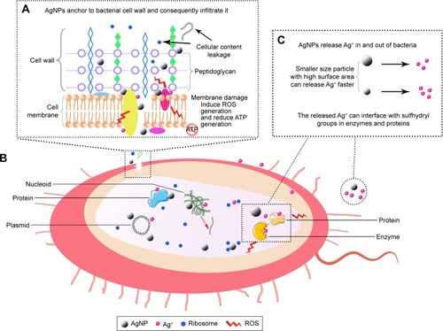
After adhesion to the bacterial wall, AgNPs can also penetrate the membrane and enter the bacteria. There is a size-dependent antibacterial effect, namely smaller nanoparticles has a large surface area in contact with the bacterial cells and can reach the cytoplasm more often than larger nanoparticles.Citation24 When AgNPs penetrate inside the microbial cell, it may interact with cellular structures and biomolecules such as proteins, lipids, and DNA. Interaction between AgNPs and cellular structures or biomolecules will lead to bacterial dysfunction and finally death. In particular, AgNP interaction with ribosomes lead to their denaturation causing inhibition of translation and protein synthesis (). It is also speculated that AgNPs interact effectively with the carboxyl and thiol groups of β-galactosidase, inhibit intracellular biological functions, and lead cell death.Citation29
Furthermore, the antibacterial mechanism of AgNPs is also due to their ability of producing high levels of reactive oxygen species (ROS) and free radical species such as hydrogen peroxide, superoxide anion, hydroxyl radical, hypochlorous acid, and singlet oxygen.Citation30–Citation32 Under normal circumstances, ROS generated in cells is limited and can be eliminated by antioxidant systems.Citation33 AgNPs exert antibacterial effect through inactivation of respiratory chain dehydrogenases and eventual excess ROS generation, which inhibited respiration and growth of cells.Citation34,Citation35 AgNPs can downregulate the expression of antioxidant enzyme such as glutathione (GSH), superoxide dismutase, and catalase, which can accelerate the accumulation of ROS.Citation22 Increased ROS lead to an apoptosis-like response, lipid peroxidation, depletion of GSH, and DNA damage.Citation36,Citation37 In addition, the antibacterial activity of AgNPs was also influenced by adenosine triphosphate (ATP)-associated metabolism and ROS.Citation38
Antibacterial mechanisms mediated by the release of silver ions
There are several evidences suggesting that the released silver ions (Ag+) from AgNPs are important in the antibacterial activity.Citation7,Citation39–Citation41 One of the important parameters of AgNPs against microbes is the surface area of the nanomaterials. AgNPs can sustainably release Ag+ in and out of bacteria. The highest concentration of released Ag+ was observed in the case of AgNPs with the highest surface area. The lowest concentration of Ag+ released was noted for AgNPs with the lowest surface area, resulting in weak antimicrobial property.Citation42 The mechanism of the antimicrobial action of Ag+ is closely associated with its interface with sulfhydryl groups in enzymes and proteins (). For instance, Ag+ can bind to proteins that are present in the cell membrane to form stable bonds resulting in protein deactivation. The proteins are involved in transmembrane ATP generation and mediate ion transport across cell membranes.Citation43 Besides, micromolar ranks of Ag+ have been described to uncouple respiratory electron transport from oxidative phosphorylation and limit respiratory chain enzymes or obstruct with the membrane penetrability to protons and phosphate.Citation44,Citation45 In addition, it has been found that Ag+ can form complex with nucleic acids, where it preferentially interact with the nucleosides. Ag+ intercalates between the purine and pyrimidine base pairs, disrupts the H-bonds between base pairs of the anti-parallel DNA strands, which prevent cell division and reproduction eventually.Citation46,Citation47
Ag+ as a heavy metal ion can cause the increase of cellular oxidative stress in microbes, which is another antibacterial mechanism. Long et alCitation48 prepared four different AgNPs by coating with different ligand and investigated the mechanism of AgNP-dependent antibacterial activity. The released Ag+ from AgNPs was suggested to interact with respiratory chain proteins on the membrane, interrupt intracellular O2 reduction, and induce ROS production.Citation48 The thioredoxin (Trx) system, which is composed of nicotinamide adenine dinucleotide phosphate, thioredoxin reductase (TrxR), and Trx, is one of the major disulfide reductase systems used by bacteria against oxidative stress.Citation49 It was demonstrated that Ag+ binds to the active sites of Staphylococcus aureus TrxR and Trx and leads to oligomerization and functional disruption of TrxR as well as Trx. Ag+ also depleted intracellular thiol levels in S. aureus, disrupting bacterial thiol-redox homeostasis, and the increased ROS induced bacteria death finally.Citation50 Although AgNPs can kill bacteria through the two different action modes mentioned above, the antibacterial mechanism is usually considered as the synergistic effect generated from AgNPs and Ag+.Citation32
With the wide application of AgNPs, researchers start to worry about the potential development of bacterial resistance to AgNPs. In a recent report, it was found that AgNPs enhanced bacterial resistance to antibiotics by promoting stress tolerance through induction of intracellular ROS.Citation51 In addition, Gram-negative bacteria Escherichia coli 013, Pseudomonas aeruginosa CCM 3955, and E. coli CCM 3954 can develop resistance to AgNPs after repeated exposure. This resistance was due to the production of flagellin, an adhesive protein of the bacterial flagellum, which caused the aggregation of AgNPs and thereby eliminated their antibacterial effect.Citation52 Indeed, bacterial resistance exists, and the mechanism is the aggregation of AgNPs. However, AgNPs are always incorporated into the implant surface in a dispersed state. Thus, further studies are needed to verify whether bacterial resistance develop in AgNP-coated implant surface.
Antibiofilm activity of AgNPs
Biofilms are communities of microorganisms attached to a solid surface. Once the biofilm is formed on the implant surface, it protects microorganisms from antibiotic treatment and causes serious consequences.Citation53,Citation54 The antibiofilm activity of AgNPs has been demonstrated in a number of studies. One pioneering study was performed to analyze the interactions of AgNPs with Pseudomonas putida biofilms. The results suggested that biofilms are impacted by the treatment with AgNPs.Citation54 Du et alCitation55 synthesized AgNPs by using benzoin gum extract and tested their antibiofilm effect by using E. coli. The AgNPs exhibited the antibiofilm activity at concentrations up to 10 μg/mL.Citation55 AgNPs were fabricated in situ and immobilized on the titanium surface. This modified surface can reduce bacterial biofilm formation in vitro by inhibiting bacterial adhesion and icaAD transcription. In addition, the antibiofilm activity of the immobilized AgNPs is independent of silver release, and AgNPs can defend several cycles of bacterial exposure in vitro and reduce implant-associated periprosthetic infection in vivo.Citation56 In another study, AgNPs were incorporated into porous titanium implants in the grown oxide layer and to create a micro-/nanoporous structure on the surface of the implants. Antimicrobial assays showed strong antimicrobial activity against methicillin-resistant S. aureus including released activity, surface antimicrobial activity, and prevention of biofilm formation.Citation57 These evidences showed that implant can be endowed with antibiofilm activity with AgNP incorporation.
Cellular effects of AgNPs on osteogenesis-related cells
Biocompatibility of AgNPs on osteogenesis-related cells, especially osteoblast, osteoclast, and mesenchymal stem cells (MSCs) should be concerned due to their key roles in bone regeneration.Citation58,Citation59 In this section, we discuss the influence of AgNPs on the abovementioned cell activity, adhesion, proliferation, and differentiation.
Effects of AgNPs on osteoblast and osteoclast
Bone metabolism is a critical factor during implants relative to bone integration, in which the osteoblast and osteoclast are responsible for bone formation and absorption during the integration, respectively.Citation59 AgNPs could be uptake into osteoblasts and could cause the first manifestation of cell injury through generation excessive nitric oxide, that is, swelling of the endoplasmic reticulum.Citation60 AgNPs were reported showing a cytotoxicity effect on osteoblasts in a dose-dependent manner and impaired cell viability at a concentration of 10 μg/g of AgNPs.Citation61 In addition, higher cytotoxicity concentrations of AgNPs were observed from other studies, which was 25 μg/mL and 50 μM, respectively.Citation16,Citation62 However, from the results mentioned above, the proper concentration of AgNPs was concluded to be 10 μg/mL aiming for medical application, possessing effective antibacterial and good biocompatibility simultaneously. Furthermore, size-dependent cytotoxicity effect was also observed. When several cell lines were treated with three different characteristic sized AgNPs, the smaller particles exhibited stronger cytotoxic effects on osteoblast, which is due to the size and surface area discrepancy release of Ag+ from AgNPs.Citation63,Citation64
Despite the side effect, AgNPs were demonstrated to possess the capacity of enhancing mineralization and alkaline phosphatase (ALP) expression in MC3T3-E1 cells at a concentration of 20 μg/mL. The underlying mechanisms were the miRNA regulation of expression of mothers against decapentaplegic (Smad) transcription factor 1 and 5, and Runt-related transcription factor 2 (Runx2), which were related to osteogenesis.Citation65 Furthermore, some results indicated that the incorporation of AgNPs into biomaterials might lead to decreased cytotoxicity by reducing the cellular uptake of AgNPs.Citation66 In addition, cell spreading is suggested to be beneficial to osteoblast differentiation and also results in better cell–cell communication, which is reported being critical to coordinate cell behavior.Citation67 When AgNPs were incorporated into TiO2 nanotube and cultured with MC3T3-E1 cells, some favorable effects on promoting cell spreading were observed from cell morphology assay after culturing for 3 days.Citation68 Another study indicated the same trend, and no significant cytotoxicity was observed when the osteoblast-like MG63 and MC3T3 cells were exposed to AgNPs, and the MG63 cell even promoted cellular proliferation.Citation69 Furthermore, nanostructure properties of implant surface were enhanced by the incorporation of AgNPs, which is a benefit for promoting osteogenesis with increased cell attachment, viability, and osteogenic gene expression (ALP, Runx2, and OCN).Citation70
Cellular effects of AgNPs on viability and differentiation of MSCs
MSCs act as multipotent precursors of various cells, which can differentiate into osteoblasts, adipocytes, and chondrocytes by a proper induction condition. Due to the key role of MSCs in bone regeneration, some researchers reported different effects of AgNPs on MSCs.
Similar to the effect on osteoblast and osteoclast, AgNPs exert cytotoxic effects on MSCs in a dose- and time-dependent manner. Greulich et alCitation71 investigated the biocompatibility of 100 nm AgNPs in human mesenchymal stem cells (hMSCs). The cytokine level of interleukin-8 (IL-8) was significantly higher than that of IL-6 and VEGF at concentrations of 5 μg/mL and above.Citation71 In addition, AgNPs can be absorbed into cells and then induce DNA damage, cell death, and functional impairment of MSCs. Distribution analysis showed that AgNPs were mainly located in the cytoplasm and the nucleus. Cytotoxic and genotoxic effects were positively correlated with dose. Interestingly, the migration ability of hMSCs was not impaired at subtoxic concentrations.Citation72 In terms of MSC proliferation, some studies have demonstrated that AgNPs can promote cell proliferation at nontoxic concentration. Jung et alCitation73 proved that AgNPs increased cell proliferation at subtoxic concentrations and decreased cell proliferation at concentrations above 10 μg/mL. In addition, hypoxia-inducible factor-1a acting as a transcription factor in regulating metabolism, development, proliferation, and pathology under hypoxic conditions plays a key factor in AgNPs-mediated cell proliferation in hMSCs.Citation73
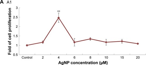
In the aspect of differentiation, Sengstock et al observed that AgNPs attenuate the adipogenic and osteogenic differentiation of hMSCs even at nontoxic concentrations, whereas chondrogenic was unaffected.Citation74 Conversely, Samberg et al exposed human adipose-derived stem cells in both undifferentiated and differentiated states to various concentrations from 0.1 to 100 μg/mL with 10 or 20 nm AgNPs, and no significant effect was observed on cell differentiation.Citation75 Qin et alCitation76 revealed that 4 μg/mL of AgNPs was safe to urine-derived stem cells (USCs). At this concentration, AgNPs can promote osteogenic differentiation of USCs, induce actin polymerization, increase cytoskeletal tension, and activate RhoA.Citation76 In another study, AgNPs promoted MSC differentiation even at a much higher concentration (4–20 μM). Furthermore, AgNPs were also considered to be able to promote the formation of fracture callus and induce early closure of the fracture gap in vivo (). The mechanisms may be via multiple routes: 1) chemoattraction of MSCs and fibroblasts to migrate to the fracture site; 2) induction of the proliferation of MSCs; 3) induction of osteogenic differentiation of MSCs via induction/activation of TGF-β/BMP signaling in MSCs.Citation77
Figure 2 (A) AgNPs increase MSC proliferation and do not reduce cell viability at low concentration. (A1) Cells were cultured with different concentrations of AgNPs for 2 days to test cell proliferation. (A2) Cell viability assay was performed to test the effect of AgNps on MSC. (B) ALP activity (B1) and Alizarin red staining (B2) indicated that AgNPs promote osteogenic differentiation of MSC in vitro. (C) AgNPs promote fracture healing in vivo. Plain X-ray radiograph of the fracture sites; broken line demarcates the unfilled fracture gap (C1). Hematoxylin and eosin staining of the middle section of the fracture site of each treatment group was shown. Broken lines indicate the two ends of the fracture femoral bone (C2). The areas of the facture gap of each treatment groups at different postoperative days were quantified and shown in (C3).
Note: Reprinted from Nanomedicine 11(8), Zhang R, Lee P, Lui V C, et al, Silver nanoparticles promote osteogenesis of mesenchymal stem cells and improve bone fracture healing in osteogenesis mechanism mouse model, 1949–1959, Copyright (2015) with permission from Elsevier.Citation77 *P<0.05, **P<0.01.
Abbreviations: AgNP, silver nanoparticle; ALP, alkaline phosphatase; MSC, mesenchymal stem cell; m, marrow; nc, new callus; gt, granulation tissue; cm, cartilage matrix.
As discussed above, these findings suggest that AgNPs could be used as a biocompatible agent within a particular dosage window. Maybe AgNPs ≤10 μg/mL is a safe dose window for osteogenesis-related cells. The cellular effects of AgNPs on osteogenesis-related cells with respect to size of particles, exposure doses, type of cell line, major outcome of each study, and their mechanisms were summarized in .
Table 1 Different size, dose of AgNPs, and their effect on osteogenesis-related cells and corresponding mechanisms
Methods to improve biocompatibility of AgNPs
Due to great application of AgNPs, their biocompatibility should be paid attention for medical use. In this part, we focus three methods to enhance the biocompatibility of AgNPs, namely biosynthesis process, adjustment of physical properties, and biomolecule combination.
Biosynthesis to improve biocompatibility
The traditional chemical and physical methods are widely used for AgNP synthesis. However, these methods always have associated risks, such as chemical precursor contamination, solvent toxicity, and hazardous byproduct formation, which make alternative synthetic methods imperative.Citation78 At present, bio-enthused synthesis of nanoparticles provides advantages over chemical and physical methods as it is environment-friendly, no need to use high-pressure, high temperature, and no toxic chemicals are needed in biological method.Citation79
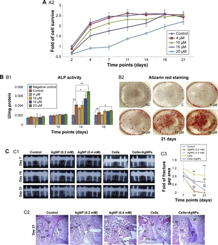
There are many resources such as bacteria, yeast, fungi, and various parts of plants can be used in the nanoparticle synthesis. Plant extracts frequently offer good manipulation and control over crystal growth and stabilization.Citation80 The major advantage of using plant extracts for biosynthesis is that they are easily available, safe, and nontoxic. In most cases, there are various metabolites that can be used to reduce Ag+.Citation17 Through adjustment reaction parameters, AgNPs can be achieved with better yield, controlled size, shape, greater particle stability, more biocompatibility, scalability, and applicability.Citation78,Citation81 For example, AgNPs can be green synthesized by using Artemisia tournefortiana Rchb ethanol extract; these AgNPs with spherical shape showed powerful antibacterial ability.Citation82 In addition, Mangifera indica inflorescence aqueous extract was used to reduce AgNO3 to produce green AgNPs. These particles with an average diameter of 40 nm showed powerful killing effect for Gram-negative (Klebsiella pneumoniae, P. aeruginosa, and E. coli) and Gram-positive (Staphylococcus mutans and S. aureus) strains. Importantly, AgNPs exhibited no significant toxic effect on HeLa cell line below 25 μg/mL ().Citation83 Kasithevar et al synthesized AgNPs by using aqueous leaf extract of Alysicarpus monilifer; these green synthesized AgNPs were biocompatible with normal Vero cell line and have high antibacterial activity.Citation84 In conclusion, biosynthesis approach is a promising method to produce AgNPs with both significant antibacterial effect and biocompatibility.
Figure 3 (A) Graphical presentation of green silver nanoparticles biosynthesis. (B) Characterization of green AgNPs: (a) ultraviolet-visible spectra indicated the synthesis of AgNPs were extremely stable. (b) Nanophox particles size analyzer graph showed the size of the particle was ranging between 30 and 70 nm with an average particle size of 40 nm. (c) Field-emission scanning electron microscope image. (d and e) Transmission electron microscope images. *P<0.05. (C) Visualization of the biofilm inhibition: (a and b) images display control and treated biofilm stained by crystal violet. (c and d) images show the difference in biofilm after treatment as visualized by the SEM, (e and f) are confocal laser scanning microscope images of the control and treated biofilms. Red arrows indicate the damage in the bacterial cells due to action of AgNPs. (D) Cell line (HeLa) toxicity assessment of the AgNPs.
Note: Reproduced from Qayyum S, Oves M, Khan AU. Obliteration of bacterial growth and biofilm through ROS generation by facilely synthesized green silver nanoparticles. PLoS One. 2017;12(8):e0181363.83
Abbreviations: AgNP, silver nanoparticle; SEM, scanning electron microscope.
Adjustment of physical properties
The fundamental characteristics of metallic nanoparticles strongly depend on their shapes, sizes, configurations, crystallinity, and structure whether they are in solid form or in hollow geometries. Thus, controlling such parameters can achieve the desired properties of the nanoparticles.Citation18 In this section, we discuss the role of different sizes and shapes of AgNPs, which are the two main properties in biomedical application.
Many studies have demonstrated that smaller AgNPs showed better antimicrobial activity. For instance, mono-dispersed AgNPs with sizes of 25, 35, 45, 60, and 70 nm were obtained, and cell viability test was performed by using human lung fibroblast. The smaller AgNPs can cause severe cell apoptosis and necrosis and generation of high levels of ROS.Citation85 Similar results were observed in other in vitro studies. Compared with larger AgNPs, increased apoptosis, activation of cytokines/chemokines IL-8, IL-1β, tumor necrosis factor, macrophage inhibitory protein, and ROS production were observed in macrophages treated with smaller AgNPs.Citation86
The shape of AgNPs can impact the degree of particle toxicity. In order to comprehensively investigate these facts, spherical, rod-shaped, truncated triangular AgNPs, and their skin penetration capabilities were studied. From the results, it was found that triangular AgNPs could be an ideal candidate for topical applications which can reduce systemic toxicity, compared to the rod-shaped and spherical AgNPs.Citation87 In another study, spherical, rectangular, penta, and hexagonal AgNPs of different dimensions were biosynthesized, and the results showed that the spherical AgNPs possessed more antimicrobial effect than other shaped AgNPs.Citation88 The electron charge transfer properties are also affected by different types of AgNPs. Regardless of the size required, narrow size distributions and more “spherical” shapes can improve sample quality.Citation89 In a word, the above studies have highlighted the role of size and shape in potentiating AgNP-based cellular effect.
Combination with biomolecules
Surface modification of nanomaterials is essential where the surface layer facilitates the reduction of surface energy. It can also provide a protective coating that prevents nanoparticles from agglomeration, thus increasing their long-term stability.Citation90 Many organic molecules can be used for surface functionalization. Examples include small molecules like lipids, vitamins, peptides, and sugar and larger ones such as natural polymers including proteins, enzymes, DNA, and RNA.Citation91 In this section, we discuss several biomolecules especially chitosan (CS), which is commonly used in surface modification.
CS is a linear polysaccharide, which has randomly distributed β-(1-4)-linked D-glucosamine (deacetylated unit) and N-acetyl-D-glucosamine (acetylated unit).Citation92 It is a highly biocompatible biodegradable material and shows antibacterial properties against pathogenic bacteria, with potential application as an antimicrobial agent.Citation93 These properties make CS suitable for surface modification of AgNPs. The hydrogels based on CS and modified with AgNPs exhibit toxicity in relation to S. aureus and did not exert any negative impact on the cells of the dermis.Citation94 AgNPs embedded in CS are capable to interact with amine and hydroxyl groups of the CS molecule, subsequently form complexes and produce stable nanostructured film. From the results, the CS/nAg nanocomposites showed enhanced antibacterial and biocompatibility properties.Citation93 Low-molecular-weight CS (LMWC) has better water solubility and biological activity than higher degree of polymerization of CS. Compared with AgNPs coated with high-molecular-weight CS, AgNPs coated with LMWC were more effective against methicillin-resistant S. aureus and showed better biocompatibility and lower body absorption characteristics.Citation95 In another study, an agarose composite embedded with CS-coated AgNPs was synthesized. This scaffold showed good swelling ratio, excellent hemocompatibility, and appreciable antibacterial activity. Moreover, it also showed good biocompatibility to several cell lines and benefits for cell sustained growth.Citation20 These studies suggest that CS is a good material for surface modification of AgNPs.
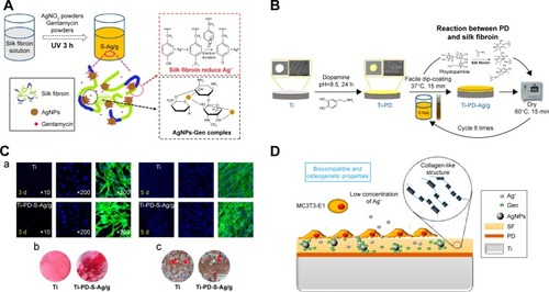
Except CS, there are many other biomolecules that can be used in combination with AgNPs for surface functionalization. Polyethylene glycol (PEG) can reduce osponization process in which nanoparticles are directed to liver through macrophages.Citation21 In addition, Kwon et al demonstrated that polyvinylpyrrolidone was a more suitable surfactant than PEG for AgNP capping, which could decrease aggregation and reduce cytotoxicity.Citation96 Furthermore, silk fibroin (SF) as a natural polymer shows the ability of reduction and osteogenic regeneration. It can reduce Ag+ to Ag0 by the Tyr residues with strong electron-donating property and also exposes to disperse and stabilize the produced AgNPs to maintain the stability of the SF-AgNPs composite solution. The SF-AgNPs composite showed good biocompatibility and improved osteogenic differentiation by decreasing the Ag+ release from AgNPs ().Citation97 Advantages of combination with biomolecules is that the surface chemistry properties of AgNPs can be easily controlled to realize multifunctionalization. Many biomolecules can meet our requirements to enhance the biocompatibility of AgNPs for medical use, which is worthy to investigate in vivo reaction in future study.
Figure 4 (A) Schematic of in situ preparation of SF/AgNPs/Gen composite solution and the possible reduction mechanism of Ag+ by SF as well as the interaction between AgNPs and gentamicin. (B) Illustrative diagram of fabrication process for PD-S-Ag/g coatings on titanium substrate, scheme is not in real scale. (C) SF-coated AgNPs shows good biocompatibility and improved osteogenic differentiation. (a) Fluorescent images for 3 and 5 days with actin stained with FITC (green) and nuclei stained with DAPI (blue). (b) Collagen secretion on different specimens for 28 days. (c) Calcium deposition (red arrow) on different specimens for 28 days. (D) Mechanism on the AgNPs/Gen-contained SF-based coatings.
Note: Reprinted with permission from Zhou W, Jia Z, Xiong P, et al. Bioinspired and biomimetic AgNPs/gentamicin-embedded silk fbroin coatings for robust antibacterial and osteogenetic applications. ACS Appl Mater Interfaces. 2017;9(31):25830–25846. Copyright (2017) American Chemical Society.Citation97
Abbreviations: AgNP, silver nanoparticle; PD, ploy-dopamine; SF, silk fibroin.
Modification of orthopedic implants by using AgNPs
Over the past decades, metallic implants have been widely used in orthopedic fields. However, it is biologically inert and cannot induce new bone regeneration or inhibit bacterial activity. AgNPs as an antibacterial agent are usually used to modify orthopedic implant surface to obtain antibacterial and potential osteointegration ability. In this section, we discuss various surface modification techniques for AgNP coating onto implants surface (), as well as their effects on antibacterial and bone formation.
Figure 5 Strategies and main techniques for the introduction of AgNPs onto implants surface.
Abbreviation: AgNP, silver nanoparticle.

Plasma immersion ion implantation (PIII)
PIIIs is proven to be economical and effective and have already been widely applied in the modification of semiconductors and biomedical industry. The process involved accelerating positive ion incorporation vertically into the negative potential surfaces in the electric field.Citation98 It can deposit different ions on many biomaterials, including metal, ceramics, and polymers.Citation99 Moreover, PIII can efficiently control the distribution and concentration of incorporated element in material by regulating the modification parameters.Citation100
AgNPs can be incorporated into the near-surface of implant by PIII process. This technique significantly improved the stability of surface coating with a negligible amount of Ag+ release. The immobilized AgNP implant surface not only inhibited bacterial adhesion and biofilm formation in vitro but also reduced implant-associated infection in vivo. While, the biocompatibility of titanium implants was also enhanced due to the change of implant surface physical topography.Citation101,Citation102 In addition, Ag-PIII-treated titanium accelerated the osteointegration of titanium implant by initiating the ERK1/2 signal.Citation102 In another study, similar results have been observed by activating the integrin-α5-orchestrated MAPK/ERK signal cascade.Citation103 Moreover, by increasing the PIII process time (1, 2, and 3 h), the diameter of AgNPs becomes larger, thus increasing implant surface roughness. The concentration of AgNPs can be effectively controlled within the safe range by changing the fabrication time.Citation104 Furthermore, two or more different metallic ions can be introduced on the implant surface through PIII process to obtain more biological functions.Citation105,Citation106 For instance, Zn and Ag were simultaneously and sequentially implanted into titanium by PIII. The advantages of Zn and Ag are preserved, the Zn/Ag microgalvanic couple formed on Zn/Ag dual-ion coimplanted titanium shows the best osseointegration as well as good antibacterial properties.Citation106
Magnetron sputtering
Magnetron sputtering constitutes one of the physical vapor deposition techniques that have been widely used in applications involving the metal-mechanic industry with great success. The sputtering technique has advantages of coating thinly, thickness uniformity, and high bonding strength to metal. Many stable bioactive coating can be deposited on various implant surface uniformly through magnetron sputtering.Citation107,Citation108 Magnetron sputtering requires simple equipment and thus is easy to control. In addition, it has low substrate temperature, high film forming rate, and strong film adhesion.Citation109
AgNPs coating on M2 tool steel and Si substrate by the sputtering technique, the antibacterial efficiency, and tribological properties of the substrate were significantly enhanced.Citation110,Citation111 By controlling magnetron sputtering time, coating morphological shapes could be controlled, as could the quantity of deposited structures.Citation112 In addition, the stable coating on the implant surface can achieve long-term antibacterial effect through magnetron sputtering technique. Furthermore, AgNPs incorporated on the implant surface by sputtering can increase water contact angle and improve hydrophobicity of the substrate surface.Citation113 It was found that with a decreased sputtering time, surface roughness and wettability were higher. An increasing roughness value is well known to be associated with increased osteoblast adhesion. However, it is also beneficial to bacterial adhesion.Citation114 In Liu et al study,Citation115 the homogeneous AgNP coating was uniformly distributed on the surface of the polyetheretherketone (PEEK) samples via magnetron sputtering. The AgNP-coated samples had a significant increase in surface roughness (P<0.05) as the thickness of their AgNP coating increased. Their antibacterial rates were above 99%, indicating that the AgNP-coated PEEK implant materials have strong antibacterial and bactericidal effects against S. mutans and S. aureus in the oral cavity.Citation115
In situ synthesis
In situ synthesis is a method through chemical reaction on the implant surface to incorporate different nanoparticles. This technology is simple, economical, and available for surface modification. However, adhesion between nanoparticles and substrate surface is not tight.
AgNPs have been obtained on different TiO2 substrates, through reduction in AgNO3 solutions. The amount of deposited nanoparticles depends on the concentration of the silver nitrate solution and reactive time.Citation116–Citation118 There is no doubt that the released Ag+ showed powerful antibacterial effect, but the key point is how to prolong the release time. To enhance the adhesion on the substrate and prolong antibacterial time, an initial layer on Ti surfaces using phase-transited lysozyme was established. Then, AgNPs were synthesized on CS and hyaluronic acid layer, and multilayer coatings were prepared on Ti surfaces via a layer-by-layer self-assembly technique. This system showed obvious antibacterial effect even in 14 days.Citation7 In another study, a mussel-inspired self-polymerized polydopamine anchor was employed, and the released time was up to 28 days.Citation119 A titanate nanowire film was produced on Ti substrate by an alkali hydrothermal reaction and subsequently doped by AgNPs through an ultraviolet light chemical reduction. The release of Ag+ was detrimental for the growth, proliferation, and differentiation of MC3T3. However, the additional CS will alleviate this cytotoxicity effect.Citation120 During bacterial infection, pH level around the peri-implant surface decreases as low as pH 5.5.Citation121 Dong et alCitation122 construct a pH-dependent AgNP releasing titanium implant to control peri-implant infection. Once the pH was doped to 5.5, the robust released AgNPs from implant system which efficiently controlled bacterial growth. Moreover, this system was biocompatible and showed osteoinductive properties.Citation122 In situ synthesis is a simple method, but the key point is how to design a safe and suitable system to achieve long-term antibacterial effect.
Plasma electrolytic oxidation (PEO)
PEO also known as micro-arc oxidation is a method that develops ceramic-like surfaces on the implant surface. The oxidation layer can also offer a wide variety of mechanical, biomedical, tribological, and antibacterial properties through the incorporation of several ions and particles.Citation123,Citation124 Nowadays, this method is widely used in the modification of implants to prepare bioactive coatings.
PEO can dope the surface of the implants with fully dispersed and firmly attached AgNPs all within the span of a few minutes. In addition, factors such as voltage and time were not affected significantly due to the addition of AgNPs in electrolyte.Citation125 AgNPs were immobilized in the oxide layer on titanium implant to create a micro-/nanoporous structure through PEO technique. The coating showed strong antimicrobial activity without any signs of cytotoxicity, because AgNPs are entrapped in an in-depth growing oxide layer which fully immobilizes them and prevents them from freely circulating through the blood stem.Citation57 Furthermore, the AgNPs were found to be very stable in biological fluids with material loss, as a result of dissolution, to be <0.07% for the silver nanocoatings after 24 h in a modified Krebs-Ringer bicarbonate buffer, which shows a stable coating with a significant effect for preventing bacterial growth.Citation126
Three-dimensional printing (3DP) silver-containing scaffolds
Additive manufacturing technology, also called 3DP, has emerged recently. It can precisely fabricate scaffolds with defined shape, size, porosity, and pore size distribution, which is beneficial for bone growth.Citation127,Citation128 In addition, more and more complex orthopedic diseases can be solved based on this technique.Citation129 In this section, we discuss the silver-containing 3D scaffolds in orthopedic field.
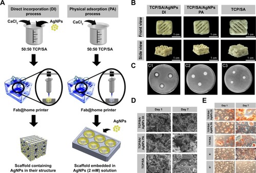
Many materials can be used to produce scaffold via this technology, such as metal, polymer, and inorganic materials. 3D scaffolds with a porosity structure is suitable for loading ions, nanoparticles, and biomolecules to achieve multifunctional application.Citation130 In Correia et al study,Citation131 the tricalcium phosphate (TCP)/sodium alginate (SA) scaffolds were produced by 3DP. Subsequently, AgNPs were incorporated into scaffolds through two different methods, direct incorporation and physical adsorption. Both of them present appropriate mechanical properties, biocompatibility, and bactericidal activity, which is suitable for being used in bone tissue regeneration ().Citation131 Similarly, graphene oxide and AgNPs nanocomposites were successfully modified on the β-TCP scaffolds by a simple soaking method to achieve biofunctions with antibacterial and osteogenic activity.Citation132 However, through soaking method, AgNP burst was released from the 3D scaffolds in short time, and this manner easily causes cytotoxicity. To control the release, a mixture of nanocomposites such as bioceramic and polyvinyl alcohol in different compartment can achieve different release kinetics.Citation133 Notably, a growing number of researchers focus on the bio-printing technology, which is a bio-printer that is used to dispense “bioinks,” consisting of cells, scaffolds, and biomolecules, in a spatially controlled manner.Citation134 Biological structures fabricated via bio-printing can encapsulate cells and bioactive agents directly, which include various biomimetic tissues such as the blood vessels, liver, heart, and tumors.Citation135 Furthermore, 3DP technology is a continuous development, and it will play a bigger role in regenerative medicine.
Figure 6 (A) Schematic representation of scaffold production by using the DI and PA processes. (B) Macroscopic images of the scaffolds produced by DI, PA, and without AgNPs (TCP/SA) (front and side view). (C) Characterization of the bactericidal activity of the produced scaffolds, DI process (C1), PA process (C2), TCP/SA scaffold (C3). (D) SEM images of osteoblast morphology in the presence of the scaffolds. (E) Qualitative evaluation of mineralization on the 3D scaffolds by human osteoblast through alizarin red staining.
Note: Reprinted from Int J Biol Macromol, 93, Correia T R, Figueira D R, De Sa K D, et al, 3D Printed scaffolds with bactericidal activity aimed for bone tissue regeneration, 1432–1445, Copyright (2016) with permission from Elsevier.Citation131
Abbreviations: AgNP, silver nanoparticle; TCP, tricalcium phosphate; SA, sodium alginate; SEM, scanning electron microscope; DI, direct incorporation; PA, physical adsorption.
Conclusion
Although orthopedic implants have made huge improvement for functional reconstruction of patients with bone fractures or defects, however, implant failure and revision surgeries are needed once the infection occurs. AgNPs with strong antibacterial efficacy are being used extensively in implant surface modification to prevent implant-related infection. The potential antibacterial mechanisms of the AgNPs were summarized, and their roles in optimization of orthopedic implants to provide some advice for implant designing were highlighted in this review. Future directions should focus on stability and long-term AgNP release, exploration of suitable size, shape, as well as the novel method of surface modification, such as 3DP technology.
Acknowledgments
Support from National Natural Science Foundation of China (Grant No 81772456, 51627805), Norman Bethune Program of Jilin University (Grant No 2012216), Science and Technology Development program of Jilin province (Grant No 20150414006GH, 20170520120JH), Special fund project of Jilin provincial industrial innovation (No 2016C037), Training Program of Outstanding Doctoral Student by Norman Bethune Health Science Center of Jilin University (No YB201501), Graduate Innovation Fund of Jilin University (No 2017089) is highly appreciated.
Disclosure
The authors report no conflicts of interest in this work.
References
- CochisAAzzimontiBDella ValleCThe effect of silver or gallium doped titanium against the multidrug resistant Acinetobacter baumanniiBiomaterials201680809526708086
- GaoAHangRHuangXThe effects of titania nanotubes with embedded silver oxide nanoparticles on bacteria and osteoblastsBiomaterials201435134223423524529392
- KuehlRBrunettoPSWoischnigAKPreventing implant-associated infections by silver coatingAntimicrob Agents Chemother20166042467247526883700
- BrennanSAFhoghlúCNDevittBO’MahonyFJBrabazonDWalshASilver nanoparticles and their orthopaedic applicationsBone Joint J201597558258925922449
- KelkawiAHAAbbasi KajaniABordbarAKGreen synthesis of silver nanoparticles using Mentha pulegium and investigation of their antibacterial, antifungal and anticancer activityIET Nanobiotechnol201711437037628530184
- LiYLinZZhaoMSilver nanoparticle based codelivery of oseltamivir to inhibit the activity of the H1N1 influenza virus through ROS-mediated signaling pathwaysACS Appl Mater Interfaces2016837243852439327588566
- ZhongXSongYYangPTitanium surface priming with phase-transited lysozyme to establish a silver nanoparticle-loaded chitosan/hyaluronic acid antibacterial multilayer via layer-by-layer self-assemblyPLoS One2016111e014695726783746
- XiangYLiJLiuXConstruction of poly(lactic-co-glycolic acid)/ZnO nanorods/Ag nanoparticles hybrid coating on Ti implants for enhanced antibacterial activity and biocompatibilityMater Sci Eng C Mater Biol Appl20177962963728629062
- NandiSKShivaramABoseSBandyopadhyayASilver nanoparticle deposited implants to treat osteomyelitisJ Biomed Mater Res B Appl Biomater201810631073108328508595
- HardesJHenrichsMPHauschildGNottrottMGuderWStreitbuergerASilver-coated megaprosthesis of the proximal tibia in patients with sarcomaJ Arthroplasty20173272208221328343825
- WickensDJWestGKellyPJAntimicrobial activity of nanocomposite zirconium nitride/silver coatings to combat external bone fixation pin infectionsInt J Artif Organs2012351081782523138705
- ProkopovichPLeechRCarmaltCJParlinIPPerniSA novel bone cement impregnated with silver–tiopronin nanoparticles: its antimicrobial, cytotoxic, and mechanical propertiesInt J Nanomedicine201382227223723818779
- WangJLiJQianSAntibacterial surface design of titanium-based biomaterials for enhanced bacteria-killing and cell-assisting functions against periprosthetic joint infectionACS Appl Mater Interfaces2016817111621117827054673
- DuránNDuránMde JesusMBSeabraABFávaroWJNakazatoGSilver nanoparticles: a new view on mechanistic aspects on antimicrobial activityNanomedicine201612378979926724539
- VelusamyPKumarGVJeyanthiVDasJPachaiappanRBio-inspired green nanoparticles: synthesis, mechanism, and antibacterial applicationToxicol Res20163229510227123159
- CastiglioniSCazzanigaALocatelliLMaierJAMSilver nanoparticles in orthopedic applications: new insights on their effects on osteogenic cellsNanomaterials (Basel)201776124
- PrabhuSPouloseEKSilver nanoparticles: mechanism of antimicrobial action, synthesis, medical applications, and toxicity effectsInt Nano Lett2012232112
- AhmedKBRNagyAMBrownRPZhangQMalghanSGGoeringPLSilver nanoparticles: significance of physicochemical properties and assay interference on the interpretation of in vitro cytotoxicity studiesToxicol In Vitro20173817919227816503
- ChengHXiongWFangZStrontium (Sr) and silver (Ag) loaded nanotubular structures with combined osteoinductive and antimicrobial activitiesActa Biomater20163138840026612413
- KumarNDesaganiDChandranGBiocompatible agarose-chitosan coated silver nanoparticle composite for soft tissue engineering applicationsArtif Cells Nanomed Biotechnol201846311328849679
- MuhammadZRazaAGhafoorSPEG capped methotrexate silver nanoparticles for efficient anticancer activity and biocompatibilityEur J Pharm Sci20169125125527132812
- YuanYGPengQLGurunathanSEffects of silver nanoparticles on multiple drug-resistant strains of Staphylococcus aureus and Pseudomonas aeruginosa from mastitis-infected goats: an alternative approach for antimicrobial therapyInt J Mol Sci2017183569
- SeongMLeeDGSilver nanoparticles against Salmonella enterica serotype typhimurium: role of inner membrane dysfunctionCurr Microbiol201774666167028321528
- KhalandiBAsadiNMilaniMA review on potential role of silver nanoparticles and possible mechanisms of their actions on bacteriaDrug Res (Stuttg)2017672707627824432
- ChatterjeeTChatterjeeBKMajumdarDChakrabartiPAntibacterial effect of silver nanoparticles and the modeling of bacterial growth kinetics using a modified Gompertz modelBiochim Biophys Acta20151850229930625450183
- Van Der WalANordeWZehnderAJBLyklemaJDetermination of the total charge in the cell walls of Gram-positive bacteriaColloids Surf B Biointerfaces199791–281100
- AbbaszadeganAGhahramaniYGholamiAThe effect of charge at the surface of silver nanoparticles on antimicrobial activity against gram-positive and gram-negative bacteria: a preliminary studyJ Nanomater201516153
- MandalDKumar DashSDasBBio-fabricated silver nanoparticles preferentially targets Gram positive depending on cell surface chargeBiomed Pharmacother20168354855827449536
- YouCHanCWangXThe progress of silver nanoparticles in the antibacterial mechanism, clinical application and cytotoxicityMol Biol Rep20123999193920122722996
- ZhaoRLvMLiYStable nanocomposite based on PEGylated and silver nanoparticles loaded graphene oxide for long-term antibacterial activityACS Appl Mater Interfaces2017918153281534128422486
- GomaaEZSilver nanoparticles as an antimicrobial agent: a case study on Staphylococcus aureus and Escherichia coli as models for Gram-positive and Gram-negative bacteriaJ Gen Appl Microbiol2017631364328123131
- SiritongsukPHongsingNThammawithanSTwo-phase bactericidal mechanism of silver nanoparticles against Burkholderia pseudomalleiPLoS One20161112e016809827977746
- RamalingamBParandhamanTDasSKAntibacterial effects of biosynthesized silver nanoparticles on surface ultrastructure and nanomechanical properties of gram-negative bacteria viz. Escherichia coli and Pseudomonas aeruginosaACS Appl Mater Interfaces2016874963497626829373
- QuinterosMACano AristizábalVDalmassoPRParajeMGPáezPLOxidative stress generation of silver nanoparticles in three bacterial genera and its relationship with the antimicrobial activityToxicol In Vitro20163621622327530963
- SuHLChouCCHungDJThe disruption of bacterial membrane integrity through ROS generation induced by nanohybrids of silver and clayBiomaterials200930305979598719656561
- KorshedPLiLLiuZWangTThe molecular mechanisms of the antibacterial effect of picosecond laser generated silver nanoparticles and their toxicity to human cellsPLoS One2016118e016007827575485
- LeeWKimKJLeeDGA novel mechanism for the antibacterial effect of silver nanoparticles on Escherichia coliBiometals20142761191120125104311
- HwangISHwangJHChoiHKimKJLeeDGSynergistic effects between silver nanoparticles and antibiotics and the mechanisms involvedJ Med Microbiol201261Pt 121719172622956753
- JinJCWuXJXuJWangBBJiangFLLiuYUltrasmall silver nanoclusters: highly efficient antibacterial activity and their mechanismsBiomater Sci20175224725727921105
- LombardoPCPoliALCastroLFPerussiJRSchmittCCPhotochemical deposition of silver nanoparticles on clays and exploring their antibacterial activityACS Appl Mater Interfaces2016833216402164727487246
- KimTBraunGBSheZGHussainSRouslahtiESailorMJComposite porous silicon-silver nanoparticles as theranostic antibacterial agentsACS Appl Mater Interfaces2016844304493045727754645
- ZawadzkaKKądziołaKFelczakASurface area or diameter – which factor really determines the antibacterial activity of silver nanoparticles grown on TiO 2 coatings?New J Chem201438732753281
- KluehUWagnerVKellySJohnsonABryersJDEfficacy of silver-coated fabric to prevent bacterial colonization and subsequent device-based biofilm formationJ Biomed Mater Res200053662163111074419
- HoltKBBardAJInteraction of silver(I) ions with the respiratory chain of Escherichia coli: an electrochemical and scanning electrochemical microscopy study of the antimicrobial mechanism of micromolar Ag+Biochemistry200544391321416185089
- SchreursWJRosenbergHEffect of silver ions on transport and retention of phosphate by Escherichia coliJ Bacteriol198215217136749823
- HatchettDWWhiteHSElectrochemistry of sulfur adlayers on the low-index faces of silverJ Phys Chem19961002398549859
- MonteiroDRGorupLFTakamiyaASde CamargoERFilhoACBarbosaDBSilver distribution and release from an antimicrobial denture base resin containing silver colloidal nanoparticlesJ Prosthodont201221171522050139
- LongYMHuLGYanXTSurface ligand controls silver ion release of nanosilver and its antibacterial activity against Escherichia coliInt J Nanomedicine2017123193320628458540
- GonSFaulknerMJBeckwithJIn vivo requirement for glutaredoxins and thioredoxins in the reduction of the ribonucleotide reductases of Escherichia coliAntioxid Redox Signal200685–673574216771665
- LiaoXYangFLiHTargeting the thioredoxin reductase–thioredoxin system from Staphylococcus aureus by silver ionsInorg Chem20175624148231483029182243
- KaweeteerawatCNa UbolPSangmuangSAueviriyavitSManiratanachoteRMechanisms of antibiotic resistance in bacteria mediated by silver nanoparticlesJ Toxicol Environ Health A20178023–241276128929020531
- PanáčekAKvítekLSmékalováMBacterial resistance to silver nanoparticles and how to overcome itNat Nanotechnol20181316529203912
- StoodleyPSauerKDaviesDGCostertonJWBiofilms as complex differentiated communitiesAnn Rev Microbiol200256118720912142477
- FranciGFalangaAGaldieroSSilver nanoparticles as potential antibacterial agentsMolecules20152058856887425993417
- DuJSinghHYiT-HAntibacterial, anti-biofilm and anticancer potentials of green synthesized silver nanoparticles using benzoin gum (Styrax benzoin) extractBioprocess Biosyst Eng201639121923193127495263
- QinHCaoHZhaoYIn vitro and in vivo anti-biofilm effects of silver nanoparticles immobilized on titaniumBiomaterials201435339114912525112937
- Van HengelIAJRioolMFratila-ApachiteiLESelective laser melting porous metallic implants with immobilized silver nanoparticles kill and prevent biofilm formation by methicillin-resistant Staphylococcus aureusBiomaterials201714011528622569
- TuanRSBolandGTuliRAdult mesenchymal stem cells and cell-based tissue engineeringArthritis Res Ther2002513212716446
- ShapiroFBone development and its relation to fracture repair. The role of mesenchymal osteoblasts and surface osteoblastsEur Cell Mater20081553e76
- ZielinskaETukajCRadomskiMWInkielewicz-StepnaikIMolecular mechanism of silver nanoparticles-induced human osteoblast cell death: protective effect of inducible nitric oxide synthase inhibitorPLoS One20161110e016413727716791
- PaukschLHartmannSRohnkeMBiocompatibility of silver nanoparticles and silver ions in primary human mesenchymal stem cells and osteoblastsActa Biomater201410143944924095782
- FloresCYMiñánAGGrilloCASalvarezzaRCVericatCSchilardiPLCitrate-capped silver nanoparticles showing good bactericidal effect against both planktonic and sessile bacteria and a low cytotoxicity to osteoblastic cellsACS Appl Mater Interfaces2013583149315923534883
- AlbersCEHofstetterWSiebenrockKALandmannRKlenkeFMIn vitro cytotoxicity of silver nanoparticles on osteoblasts and osteoclasts at antibacterial concentrationsNanotoxicology201371303622013878
- KimTHKimMParkHSShinUSGongMSKimHWSize-dependent cellular toxicity of silver nanoparticlesJ Biomed Mater Res A201210041033104322308013
- MahmoodMLiZCascianoDNanostructural materials increase mineralization in bone cells and affect gene expression through miRNA regulationJ Cell Mol Med201115112297230621143388
- CaoHLiuXMengFChuPKBiological actions of silver nanoparticles embedded in titanium controlled by micro-galvanic effectsBiomaterials201132369370520970183
- StainsJPCivitelliRCell-cell interactions in regulating osteogenesis and osteoblast functionBirth Defects Res C Embryo Today2005751728015838921
- GaoAHangRHuangXThe effects of titania nanotubes with embedded silver oxide nanoparticles on bacteria and osteoblastsBiomaterials201435134223423524529392
- CaoHQiaoYLiuXElectron storage mediated dark antibacterial action of bound silver nanoparticles: smaller is not always betterActa Biomater2013925100511023085265
- ZhengYLiJLiuXSunJAntimicrobial and osteogenic effect of Ag-implanted titanium with a nanostructured surfaceInt J Nanomedicine2012787588422393287
- GreulichCKittlerSEppleMMuhrGKöllerMStudies on the biocompatibility and the interaction of silver nanoparticles with human mesenchymal stem cells (hMSCs)Langenbecks Arch Surg2009394349550219280220
- HackenbergSScherzedAKesslerMSilver nanoparticles: evaluation of DNA damage, toxicity and functional impairment in human mesenchymal stem cellsToxicol Lett20112011273321145381
- JungSKKimJHKimHJJiYHKimJHSonSWSilver nanoparticle-induced hMSC proliferation is associated with HIF-1 [alpha]-mediated upregulation of IL-8 expressionJ Invest Dermatol201413412300324999595
- SengstockCDiendorfJEppleMSchildhauerTAKöllerMEffect of silver nanoparticles on human mesenchymal stem cell differentiationBeilstein J Nanotechnol201452058206925551033
- SambergMELoboaEGOldenburgSJMonteiro-RiviereNASilver nanoparticles do not influence stem cell differentiation but cause minimal toxicityNanomedicine2012781197120922583572
- QinHZhuCAnZSilver nanoparticles promote osteogenic differentiation of human urine-derived stem cells at noncytotoxic concentrationsInt J Nanomedicine201492469247824899804
- ZhangRLeePLuiVCSilver nanoparticles promote osteogenesis of mesenchymal stem cells and improve bone fracture healing in osteogenesis mechanism mouse modelNanomedicine20151181949195926282383
- RajanRChandranKHarperSLYunSIKalaichelvanPTPlant extract synthesized silver nanoparticles: an ongoing source of novel biocompatible materialsInd Crops Prod201570356373
- MittalAKBhaumikJKumarSBanerjeeUCBiosynthesis of silver nanoparticles: elucidation of prospective mechanism and therapeutic potentialJ Colloid Interface Sci2014415394724267328
- OvaisMKhalilATRazaAGreen synthesis of silver nanoparticles via plant extracts: beginning a new era in cancer theranosticsNanomedicine (Lond)2016122331573177
- PérezZEJMathiyalaganRMarkusJGinseng-berry-mediated gold and silver nanoparticle synthesis and evaluation of their in vitro antioxidant, antimicrobial, and cytotoxicity effects on human dermal fibroblast and murine melanoma skin cell linesInt J Nanomedicine20171270972328260881
- Baghbani-AraniFMovagharniaRSharifianASalehiSShandizSASPhotocatalytic, anti-bacterial, and anti-cancer properties of phyto-mediated synthesis of silver nanoparticles from Artemisia tournefortiana Rchb extractJ Photochem Photobiol B201717364064928711019
- QayyumSOvesMKhanAUObliteration of bacterial growth and biofilm through ROS generation by facilely synthesized green silver nanoparticlesPLoS One2017128e018136328771501
- KasithevarMSaravananMPrakashPGreen synthesis of silver nanoparticles using Alysicarpus monilifer leaf extract and its antibacterial activity against MRSA and CoNS isolates in HIV patientsJ Interdiscip Nanomed201722131141
- LiLSunJLiXControllable synthesis of monodispersed silver nanoparticles as standards for quantitative assessment of their cytotoxicityBiomaterials20123361714172122137123
- CarlsonCHussainSMSchrandAMUnique cellular interaction of silver nanoparticles: size-dependent generation of reactive oxygen speciesJ Phys Chem B200811243136081361918831567
- TakYKPalSNaogharePKRangasamySSongJMShape-dependent skin penetration of silver nanoparticles: does it really matter?Sci Rep201551690826584777
- KumariMPandeySGiriVPTailoring shape and size of biogenic silver nanoparticles to enhance antimicrobial efficacy against MDR bacteriaMicrob Pathog201710534635527889528
- SunBBarnardASThe impact of size and shape distributions on the electron charge transfer properties of silver nanoparticlesNanoscale2017934126981270828828432
- LabhasetwarVLeslie-PeleckyDLBiomedical Applications of Nanotechnology [M]Hoboken, NJJohn Wiley & Sons2007
- RavindranAChandranPKhanSSBiofunctionalized silver nanoparticles: advances and prospectsColloids Surf B Biointerfaces201310342352
- DivakarDDJastaniyahNTAltamimiHGEnhanced antimicrobial activity of naturally derived bioactive molecule chitosan conjugated silver nanoparticle against dental implant pathogensInt J Biol Macromol201810879079729102795
- Luna-HernándezECruz-SotoMPadilla-VacaFCombined antibacterial/tissue regeneration response in thermal burns promoted by functional chitosan/silver nanocompositesInt J Biol Macromol20171051241124928757422
- TyliszczakBDrabczykAKudłacik-KramarczykSBialik-WąsKKijkowskaRSobczak-KupiecAPreparation and cytotoxicity of chitosan-based hydrogels modified with silver nanoparticlesColloids Surf B Biointerfaces201716032533028950197
- PengYSongCYangCGuoQYaoMLow molecular weight chitosan-coated silver nanoparticles are effective for the treatment of MRSA-infected woundsGerodontology201712295304
- KwonTWooHJKimYHOptimizing hemocompatibility of surfactant-coated silver nanoparticles in human erythrocytesJ Nanosci Nanotechnol20121286168617522962723
- ZhouWJiaZXiongPBioinspired and biomimetic AgNPs/gentamicin-embedded silk fibroin coatings for robust antibacterial and osteogenetic applicationsACS Appl Mater Interfaces2017931258302584628731325
- LuTQiaoYLiuXSurface modification of biomaterials using plasma immersion ion implantation and depositionInterface Focus20122332533623741609
- AndersAFrom plasma immersion ion implantation to deposition: a historical perspective on principles and trendsSurf Coat Technol20021561312
- RautrayTRNarayananRKwonTYKimKHSurface modification of titanium and titanium alloys by ion implantationJ Biomed Mater Res B Appl Biomater201093258159120127988
- QiaoSCaoHZhaoXAg-plasma modification enhances bone apposition around titanium dental implants: an animal study in Labrador dogsInt J Nanomedicine20151065366425609967
- QinHCaoHZhaoYAntimicrobial and osteogenic properties of silver-ion-implanted stainless steelACS Appl Mater Interfaces2015720107851079425952114
- CaoHZhangWMengFOsteogenesis catalyzed by titanium-supported silver nanoparticlesACS Appl Mater Interfaces2017965149515728111942
- WangGJinWQasimAMAntibacterial effects of titanium embedded with silver nanoparticles based on electron-transfer-induced reactive oxygen speciesBiomaterials2017124253428182874
- ZhaoYCaoHQinHBalancing the osteogenic and antibacterial properties of titanium by codoping of Mg and Ag: an in vitro and in vivo studyACS Appl Mater Interfaces2015732178261783626202255
- JinGQinHCaoHZn/Ag micro-galvanic couples formed on titanium and osseointegration effects in the presence of S-aureusBiomaterials201565223126141835
- WanTAokiHHikawaJLeeJHRF-magnetron sputtering technique for producing hydroxyapatite coating film on various substratesBiomed Mater Eng200717529129717851171
- LiRQinYLiuGTantalum nitride coatings prepared by magnetron sputtering to improve the bioactivity and osteogenic activity for titanium alloy implantsRSC Adv20177875540855417
- SocolGMacoveiAMiroiuFHydroxyapatite thin films synthesized by pulsed laser deposition and magnetron sputtering on PMMA substrates for medical applicationsMater Sci Eng B20101691159168
- HsiehJTsengCChangYChangSYWuWAntibacterial behavior of TaN–Ag nanocomposite thin films with and without annealingSurf Coat Technol20082022255865589
- HsiehJHYehTHHungSYChangSYWuWLiCAntibacterial and tribological properties of TaN–Cu, TaN–Ag, and TaN–(Ag, Cu) nanocomposite thin filmsMater Res Bull2012471029993003
- ZawadzkaKKisielewskaAPiwonskiIMechanisms of antibacterial activity and stability of silver nanoparticles grown on magnetron sputtered TiO2 coatingsBull Mater Sci20163915768
- HuangH-LChangY-YChenH-JChouYLaiCChenMYCAntibacterial properties and cytocompatibility of tantalum oxide coatings with different silver contentJ Vac Sci Technol201432202B117
- UhmSHSongDHKwonJSLeeSBHanJGKimKNTailoring of antibacterial Ag nanostructures on TiO2 nanotube layers by magnetron sputteringJ Biomed Mater Res B Appl Biomater2014102359260324123999
- LiuXGanKLiuHSongXChenTLiuCAntibacterial properties of nano-silver coated PEEK preparedac through magnetron sputteringDent Mater2017339E348E36028734568
- KedzioraAKorzekwaKStrekWPawlakADoroszkiewiczWBugla-PloskonskaGSilver nanoforms as a therapeutic agent for killing escherichia coli and certain ESKAPE pathogensCurr Microbiol201673113914727086305
- ChenYDengYPuYTangBSuYTangJOne pot preparation of silver nanoparticles decorated TiO2 mesoporous microspheres with enhanced antibacterial activityMater Sci Eng C Mater Biol Appl201665273227157724
- KamarajKGeorgeRPAnandkumarBParvathavarthiniNKamachi MudaliUA silver nanoparticle loaded TiO2 nanoporous layer for visible light induced antimicrobial applicationsBioelectrochemistry2015106Pt B29029726205428
- JiaZXiuPLiMBioinspired anchoring AgNPs onto micro-nanoporous TiO2 orthopedic coatings: trap-killing of bacteria, surface-regulated osteoblast functions and host responsesBiomaterials20167520322226513414
- XuZLiMLiXAntibacterial activity of silver doped titanate nanowires on Ti implantsACS Appl Mater Interfaces2016826165841659427336202
- MaLLiuMLiuHChenJCuiDIn vitro cytotoxicity and drug release properties of pH-and temperature-sensitive core–shell hydrogel microspheresInt J Pharm20103851869119879345
- DongYYeHLiuYpH dependent silver nanoparticles releasing titanium implant: A novel therapeutic approach to control peri-implant infectionColloids Surf B Biointerfaces201715812713628688362
- Echeverry-RendónMGalvisOAguirreRRobledoSCastañoJGEcheverríaFModification of titanium alloys surface properties by plasma electrolytic oxidation (PEO) and influence on biological responseJ Mater Sci Mater Med2017281116928956201
- RizwanMAliasRZaidiUZMahmoodianRHamdiMSurface modification of valve metals using plasma electrolytic oxidation (PEO) for antibacterial applications: a reviewJ Biomed Mater Res A2018106259060528975693
- NeculaBSFratila-ApachiteiLEZaatSAApachiteiIDuszczykJIn vitro antibacterial activity of porous TiO 2–Ag composite layers against methicillin-resistant Staphylococcus aureusActa Biomater2009593573358019454327
- BesinisAHadiSDLeHRTredwinCHandyRDAntibacterial activity and biofilm inhibition by surface modified titanium alloy medical implants following application of silver, titanium dioxide and hydroxyapatite nanocoatingsACS Appl Mater Interfaces2017113327338
- ParkSHChoiYJMoonSWThree-dimensional bio-printed scaffold sleeves with mesenchymal stem cells for enhancement of tendon-to-bone healing in anterior cruciate ligament reconstruction using soft-tissue tendon graftArthroscopy201834116617928688825
- HolzapfelBMRudertMHutmacherDWGerüstträgerbasiertes knochen-tissue-engineering [Scaffold-based bone tissue engineering]Orthopade2017468701710 German28725934
- YusaKYamanochiHTakagiALinoMThree-dimensional printing model as a tool to assist in surgery for large mandibular tumour: a case reportJ Oral Maxillofac Res201782e4
- RitzUGerkeRGötzHSteinSRommensPMA new bone substitute developed from 3D-prints of polylactide (PLA) loaded with collagen I: an in vitro studyInt J Mol Sci201718122569
- CorreiaTRFigueiraDRDe SaKD3D printed scaffolds with bactericidal activity aimed for bone tissue regenerationInt J Biol Macromol2016931432144527267575
- ZhangYZhaiDXuM3D-printed bioceramic scaffolds with antibacterial and osteogenic activityBiofabrication20179202503728631614
- García-AlvarezRIzquierdo-BarbaIVallet-RegíM3D scaffold with effective multidrug sequential release against bacteria biofilmActa Biomater20174911312627845276
- JessopZMAl-SabahAGardinerMDCombellackEHawkinsKWhitakerIS3D bioprinting for reconstructive surgery: principles, applications and challengesJ Plast Reconstr Aesthet Surg20177091155117028734756
- LiY-CZhangYSAkpekAShinSRKhademhosseiniA4D bio-printing: the next-generation technology for biofabrication enabled by stimuli-responsive materialsBiofabrication20169101200127910820